Comparison of short pulse subthreshold (532 nm) and infrared micropulse (810 nm) macular laser for diabetic macular edema - Nature
←
→
Page content transcription
If your browser does not render page correctly, please read the page content below
www.nature.com/scientificreports
OPEN Comparison of short‑pulse
subthreshold (532 nm) and infrared
micropulse (810 nm) macular laser
for diabetic macular edema
Abdulrahman Al‑Barki1, Lamia Al‑Hijji1, Robin High2, Patrik Schatz1,3, Diana Do4,
Quan D. Nguyen4, Jeffrey K. Luttrull5 & Igor Kozak1,6*
The purpose of the study was to assess both anatomic and functional outcomes between short-
pulse continuous wavelength and infrared micropulse lasers in the treatment of DME. This was a
prospective interventional study from tertiary care eye hospital—King Khaled Eye Specialist Hospital
(Riyadh, Saudi Arabia). Patients with center-involving diabetic macular edema were treated with
subthreshold laser therapy. Patients in the micropulse group were treated with the 810-nm diode
micropulse scanning laser TxCell (IRIDEX Corporation, Mountain View, CA, USA) (subthreshold
micropulse—STMP group). Laser was applied according to recommendations for MicroPulse (125
microns spot size, 300 ms pulse duration and power adjustment following barely visible testing burn)
in a confluent mode (low intensity/high density) to the entire area of the macular edema. Patients
in the short-pulse group were treated with grid pattern laser with 20 ms pulse PASCAL laser 532 nm
(TopCon Medical Laser Systems, Tokyo, Japan) with EndPoint algorithm, which was either 30% or
50% of testing burn (EndPoint 30% and EndPoint 50% groups, respectively). Main outcome measures
included best-corrected visual acuity (BCVA in logMAR) and foveal thickness at baseline and the last
follow-up visit at 6 months. There were 44 eyes in the micropulse group, 54 eyes in the EndPoint 50%
group and 18 eyes in the EndPoint 30% group. BCVA for the whole cohort (logMAR) was 0.451 (Snellen
equivalent 20/56) at baseline, 0.495 (Snellen equivalent 20/62) (p = 0.053) at 3 months, and 0.494
(Snellen equivalent 20/62) at the last follow-up (p = 0.052). Foveal thickness for the whole cohort was
378.2 ± 51.7 microns at baseline, 347.2 ± 61.3 microns (p = 0.002) at 3 months, and 346.0 ± 24.6 microns
at the final follow-up (p = 0.027). As such the short-pulse system yields more temporary reduction
in edema. Comparison of BCVA between baseline and 6 months for EndPoint 30%, EndPoint 50%
and STMP groups was p = 0.88, p = 0.76 and p = 0.003, respectively. Comparison of foveal thickness
between baseline and 6 months for EndPoint 30%, EndPoint 50% and STMP groups was p = 0.38,
p = 0.22 and p = 0.14, respectively. We conclude that the infrared micropulse system seems to improve
functional outcomes. When applied according to previously published reports, short-pulse system may
yield more temporary reduction in edema while infrared micropulse system may yield slightly better
functional outcomes.
Diabetic macular edema (DME) is the leading cause of moderate vision loss in patients with d iabetes1. The role
of laser photocoagulation, previously the mainstay of treatment of D ME2, has undergone significant changes
after the introduction of anti-vascular endothelial growth factor (anti-VEGF) t herapy3–5. Three main areas of
current laser use in DME include management of extrafoveal leakage in non-center involving macular edema, an
adjunct management to intravitreal pharmacotherapy to reduce the frequency of required intravitreal injections
for vision improvement and/or vision stabilization, and the use of non-damaging/tissue sparing photocoagula-
tion techniques3,6–10.
1
King Khaled Eye Specialist Hospital, Riyadh, Saudi Arabia. 2Department of Biostatistics, College of Public Health,
University of Nebraska Medical Center, Omaha, NE, USA. 3Department of Clinical Sciences, Ophthalmology,
Lund University, Skane University Hospital, Lund, Sweden. 4Department of Ophthalmology, Byers Eye Institute,
Stanford University, Palo Alto, CA, USA. 5Retina Diagnostic Laboratory of Ventura County, Ventura, CA,
USA. 6Moorfields Eye Hospital Centre, Abu Dhabi, UAE. *email: igor.kozak@moorfields.ae
Scientific Reports | (2021) 11:14 | https://doi.org/10.1038/s41598-020-79699-9 1
Vol.:(0123456789)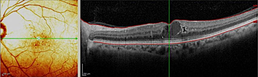
www.nature.com/scientificreports/
Figure 1. Graphic bar representation of central foveal thickness from baseline (pre-Tx), follow-up (3 months)
to the last follow-up (6 months) among three laser treatment groups. Bars represent standard deviations, circles
represent statistically significant difference among groups.
Laser Pre-treatment versus 3 months Pre-treatment versus 6 months 3 months versus 6 months
EndPoint 30% 0.18 0.38 0.99
EndPoint 50% 0.005 0.22 0.58
Micropulse 0.30 0.14 0.58
Table 1. Adjusted p-values for comparisons of foveal thickness in eyes with diabetic macular edema at
different time points among three treatment groups. Statistically significant value is in italics
Non-damaging retinal photocoagulation may be an effective treatment alternative to the modified Early
Treatment Diabetic Retinopathy Study (ETDRS) macular photocoagulation technique, but without causing
chorioretinal scarring or inducing visual field s cotoma10–12. Two approaches are commonly mentioned both
in clinical practice and in the scientific literature: short-pulse continuous wave (SPCW) and micropulsed laser
treatments, both of which limit heat spread to adjacent retinal layers. “Subthreshold” delivery indicates an invis-
ible laser application, and “micropulse” refers to a laser pulse in the microsecond range10,13. Published studies
have shown clinical efficacy of subthreshold short-pulse14,15 as well as subthreshold micropulse (STMP)16–21
laser for DME. In the case of STMP, treatment can be done which is not only subthreshold clinically, but “truly”
subthreshold, sublethal to the R PE9,13. However, there is no head-to-head comparison of patterned SPCW and
STMP according to manufacturer recommended treatment guidelines. To fill this gap and enhance our knowledge
and understanding of retina laser properties, we have conducted the following prospective study to compare
the efficacy of SPCW and STMP in center-involved DME. The aim of the study was to assess both anatomic and
functional outcomes in these two photocoagulation modalities.
Results
Patients. Overall, the patients included in this study are the ones who refused intravitreal injection therapy.
Out of 93 consented patients who started prospective study, 65 had complete data at the end of study period.
Fourteen patients had treatment in 1 eye and 51 patients had bilateral (116 eyes in total). Overall, there were
42 males and 23 females and 76 phakic, 38 pseudophakic and 2 aphakic eyes in the study (statistically signifi-
cant difference between aphakic group and other groups). All patients had type 2 diabetes mellitus. Glycemia
was poorly controlled (HbAC1 > 7.0%) in all patients (baseline: SPCW EndPoint 30% = 7.2%, SPCW EndPoint
50% = 7.3%, STMP = 7.2%; comparisons: EndPoint 30% vs EndPoint 50%: p = 0.22; EndPoint30% vs STMP:
p = 0.22; EndPoint 50% vs STMP: p = 0.22). There were 13 treatment naïve patients and 52 patients had been pre-
viously treated with bevacizumab monotherapy (no intravitreal steroids) with last injection received more than
6 months from study baseline. There were 27 patients (44 eyes) in the micropulse group, 26 patients (54 eyes)
in the EndPoint 50% group and 12 patients (18 eyes) in the EndPoint 30% group with mean ages of 59.4 ± 7.3,
63.4 ± 8.4 and 63.0 ± 6.3 years, respectively (no statistically significant difference). All treatments were performed
without any complications. Mean follow-up period for the whole group was 8.13 ± 3.93 months; for STMP, and
for SPCW EndPoint 50% and EndPoint 30% groups it was 9.5 ± 4.12, 6.97 ± 3.26 and 8.0 ± 4.37.0 months, respec-
tively.
Central foveal thickness. Foveal thickness for the entire cohort of all study subjects (116 eyes total) was
378.2 ± 51.7 microns at baseline; 347.2 ± 61.3 microns at 3 months (p = 0.002); and 346.0 ± 24.6 microns at the
final follow-up (p = 0.027). Figure 1 depicts changes in foveal thickness after treatment in each treatment group
(baseline: SPCW EndPoint 30% = 394.4 ± 201.5 microns, SPCW EndPoint 50% = 394.1 ± 128.5 microns and
STMP = 344 ± 105.8 microns; comparisons—EndPoint 30% vs 50%: p = 0.61; EndPoint 30% vs STMP: p = 0.08;
Scientific Reports | (2021) 11:14 | https://doi.org/10.1038/s41598-020-79699-9 2
Vol:.(1234567890)www.nature.com/scientificreports/
Figure 2. A pre-treatment spectral-domain optical coherence tomography scan of an eye with non-proliferative
diabetic retinopathy and diabetic macular edema with increased foveal thickness.
Figure 3. A post-treatment spectral-domain optical coherence tomography scan of the same eye following
micropulse macular laser demonstrates improvement in diabetic macular edema and reduction of foveal
thickness.
EndPoint 50% vs STMP: p = 0.09). Table 1 presents adjusted p-values for comparisons of foveal thickness at dif-
ferent time points in each treatment group. Foveal thickness was > 400 microns in 30 (25.8%) eyes before treat-
ment. In the subanalysis of eyes with foveal thickness < 400 microns the results were similar than in the whole
cohort (baseline: EndPoint 30% [273.3 ± 45.7 microns] vs EndPoint 50% [310.6 ± 55.8]: p = 0.06; EndPoint 30%
vs STMP[298.9 ± 43.1]: p = 0.27; EndPoint 50% vs STMP: p = 0.11; at 3 months EndPoint 30% vs 50%: p = 0.22;
EndPoint 30% vs STMP: p = 0.47; EndPoint 50% vs STMP: p = 0.77 and at 6 months EndPoint 30% vs 50%:
p = 0.25; EndPoint 30% vs STMP: p = 0.38; EndPoint 50% vs STMP: p = 0.22). Figures 2 and 3 show representative
images following STMP treatment.
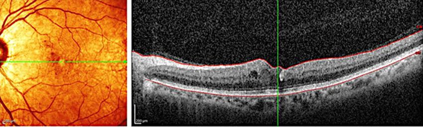
Best‑corrected visual acuity. BCVA for the whole cohort (logMAR) was 0.451 at baseline (Snellen equiv-
alent 20/56), 0.495 (Snellen equivalent 20/62) at 3 months (p = 0.053) and 0.494 (Snellen equivalent 20/62) at the
last follow-up (p = 0.052). Figure 4 depicts changes in BCVA after treatment in each treatment group (baseline:
SPCW EndPoint 30% = 0.43 ± 0.2 (Snellen equivalent 20/52), SPCW EndPoint 50% = 0.41 ± 0.2 (Snellen equiva-
lent 20/51) and STMP = 0.50 ± 0.2 (Snellen equivalent 20/61); comparisons—EndPoint 30% vs 50%: p = 0.3; End-
Point 30% vs STMP: p = 0.4; EndPoint 50% vs STMP: p = 0.4). Table 2 presents adjusted p-values for comparisons
of best-corrected visual acuity at different time points in each treatment group.
Laser parameters, rescue injections and laser spot imaging. The mean number of laser shots was
148.3 ± 106.8, 165.7 ± 99.3 and 140.7 ± 66.1 in the STMP, and SPCW EndPoint 50% and EndPoint 30% groups,
respectively. The mean laser power used was 340 ± 155 mW, 105 ± 48 mW and 125 ± 33 mW in the micropulse,
EndPoint 50% and EndPoint 30% groups, respectively. At the end of final follow-up period, rescue therapy
(intravitreal injections of antiangiogenic pharmacotherapeutic agents) were administered in 6 (13%), 14 (6%)
and 4 (22%) eyes in the STMP, and SPCW EndPoint 50% and EndPoint 30% groups, respectively. None of the
three pair-wise differences in the proportion of patients receiving rescue therapy were statistically significant.
Table 3 presents adjusted p-values for comparisons of laser applications, power and need for rescue therapy at
different time points in each treatment group. Table 4 presents data on presence of post-treatment laser scar in
each group as imaged by color fundus photography and fundus autofluorescence.
Discussion
Subthreshold macular laser treatment (thermal retinal photo-stimulation) is an appealing technique for treat-
ment of diabetic macular edema if the clinician wishes to avoid traditional complications of conventional
photocoagulation12. Its efficacy has been found comparable to conventional laser treatment of D
ME10–12. Our
Scientific Reports | (2021) 11:14 | https://doi.org/10.1038/s41598-020-79699-9 3
Vol.:(0123456789)www.nature.com/scientificreports/
Figure 4. Graphic bar representation of best-corrected visual acuity (BCVA) from baseline (pre-Tx), follow-up
(3 months) to the last follow-up (6 months) among three laser treatment groups. Bars represent standard
deviations, circles represent statistically significant difference among groups.
Laser Pre-treatment versus 3 months Pre-treatment versus 6 months 3 months versus 6 months
EndPoint 30% 0.53 0.88 0.75
EndPoint 50% 0.33 0.76 0.65
Micropulse 0.16 0.003 0.15
Table 2. Adjusted p-values for comparisons of best-corrected visual acuity in eyes with diabetic macular
edema at different time points among three treatment groups. Statistically significant value is in italics
EndPoint 30% versus EndPoint EndPoint 50% versus
Laser parameters 50% EndPoint 30% versus micropulse micropulse
Number of laser applications 0.94 0.99 0.96
Laser power 0.61 0.007 0.001
Need for rescue therapy 0.63 0.69 0.45
Table 3. Adjusted p-values for comparisons of number of laser applications, power at different time points
and need for rescue therapy (injections) in eyes with diabetic macular edema among three treatment groups.
Statistically significant values are in italics
Imaging EndPoint 30% [eyes(%)] EndPoint 50% [eyes(%)] Micropulse [eyes(%)]
Color fundus photography 0 (0%) 3 (5.5%) 1 (2.2%)
Fundus autofluorescence 0 (0%) 3 (5.5%) 2 (4.5%)
Table 4. Presence of post-treatment laser scar in eyes with diabetic macular edema in each treatment group as
imaged by color fundus photography and fundus autofluorescence and graded anonymously.
study reports on both anatomical and functional outcomes using suthreshold laser of two different techniques
(SPCW and STMP) and wavelengths (532 nm versus 810 nm) among patients with similar ethnical background.
The mean reduction of foveal thickness for the entire cohort was 32 microns at the end of follow-up period,
which reached statistical significance. Such reduction is comparable with other studies employing subthreshold
laser photocoagulation for D ME15,18,22. However, if we evaluate individual study arms, the only significant dif-
ference was observed between pre-treatment and month 3 follow-up time-point in the EndPoint 50% group.
This observation is similar to what has been reported in other studies, reflecting the general consensus that if
Scientific Reports | (2021) 11:14 | https://doi.org/10.1038/s41598-020-79699-9 4
Vol:.(1234567890)www.nature.com/scientificreports/
there is no improvement in macular edema by 6 months, it is unlikely that significant subsequent improvement
will occur11,16,17,23,24.
It has been suggested that severity of macular edema influences the effects of subthreshold laser therapy,
especially if foveal thickness is more than 400 microns11. Foveal thickness > 400 microns was present at baseline
in one quarter of eyes in our cohort. The number of eyes with increased thickness was equally distributed across
all treatment subgroups. Subgroup analyses of eyes with baseline foveal thickness > 400 microns, however, mir-
rored the results of the whole cohort.
While reduction in edema and decrease in foveal thickness are desired anatomic outcomes, previously con-
ducted studies, including the ETDRS, have shown a weak correlation of central visual acuity to t hickness2,22,23.
In this study, visual acuity was significantly improved in only STMP eyes. The results are consistent with the
findings of a meta analysis comparing STMP and conventional photocoagulation for D ME12. High density of
low intensity laser delivery, as used in this subgroup, has been reported to yield superior functional (visual)
results compared to normal density t reatment12,16 and could be why infrared subgroup showed more improve-
ment. The high-density (application of confluent laser spots)/low-intensity (sublethal to the RPE) is intended
to maximize treatment surface area, thus maximizing therapeutic recruitment of the retinal pigment epithelial
cells and the therapeutic response to t reatment13. The differences in number of laser applications in this study
reflected the extend of macular edema. Significant visual acuity (VA) improvement did not occur during the
first three months but between 4 to 6 months of follow-up, consistent with our knowledge that VA gain may lag
behind anatomic i mprovement12,13.
There are two differences between our study design and previously published studies. While we followed the
manufacturer’s recommendations regarding treatment parameters for STMP (125 microns spot size, 300 ms pulse
duration and power adjustment following barely visible testing burn) and available reports at the beginning of
our study12, the manufacturer’s treatment recommendations apply to approximately only 10% the treatments
found to be most effective in published clinical s tudies9,13,21,25 Secondly, in our study, instead of re-treatment with
laser after 3 months such as in other studies16,26 we observed the effect of laser treatment for 6 months before
further intervention. This could be a reason for some drop-out of participants. While this protocol prolonged
observation period for the initial effect of subthreshold laser, it was less aggressive compared to some current
protocols, which may have aggravated any under-treatment, particularly in the STMP group. Lack of subjective
improvement for this reason in some patients may have contributed to their loss to follow up. The decision to
initiate rescue therapy with intravitreal injection of anti-angiogenic agents due to persistent DME was left to the
discretion of the treating ophthalmologist. Several patients needed rescue therapy at the end of follow-up period
but the proportion of those patients did not differ among three treatment groups. Whether laser retreatment
would have been sufficient in these eyes is unknown.
We also observed the presence of mild laser scars, which were seen in 3 eyes on both fundus photos and
fundus autofluorescence imaging in the EndPoint 50% subgroup. This may reflect the difficulty of finding the
narrow 0.01 W wide therapeutic window of SPCW laser based on subjective assessment of the intensity of titra-
tion burns13. Additionally, scars were noted in 1 eye on fundus photo and in 2 eyes on fundus autofluorescence
in the micropulse subgroup. Inadvertent retinal burns in micropulse laser therapy have been reported and are
more likely to occur with 10% and 15% duty cycle9,13. We note that we followed the previously published reports
and manufacturer’s recommended laser power titration recommendations for both SPCW and STMP. A review
of the literature of subthreshold retinal laser treatment reveals that inadvertent retinal laser damage has been
reported in every study employing power titration; and in no study using fixed laser parameters based on long
clinical experience9,21,26,27. Laser scars following both yellow and diode micropulse macular treatment have been
previously reported17,28. Histopathologic studies in animals by Yu et al., comparing micropulse 532 nm and high
duty cycle (above 5%) 810 nm laser wavelengths showed damage to the RPE from both laser modes29. Thus,
our study shares correlation in this regard with Yu, et al. and clinical comparisons of mETDRS photocoagula-
tion with titrated high-duty cycle (15%), STMP for DME, which consistently demonstrate laser-induced retinal
damage15–17,19,20,28.
The limitations of our study include non-randomized nature, which could introduce bias in the selection
of treatment arm and relatively short follow-up period. We did not have the opportunity to measure other
parameters of retinal function such as retinal sensitivity using microperimetry or multifocal electroretinogra-
phy. Furthermore, we did not stratify patients according to glycemic control or diabetes duration, factors that
are well-known to affect the risk of diabetes retinopathy, and thus may be associated with variable response to
laser therapy. While the treatment adjustments were standardized, laser parameters always represent variable
most difficult to control.
In summary, our study echoes previous reports finding comparable anatomic results from CW vs. MP laser,
with better visual results following STMP laser12. Overall treatment effects in this cohort did not seem to be
influenced by the degree of pre-treatment macular thickening. Non-responders treated by both laser systems
may require similar proportion of rescue therapy. Both laser systems can produce inadvertent laser-induced
retinal damage. Prior studies suggest this may be avoided in STMP by use of fixed laser parameters known to
be both safe and effective in clinical practice, rather than the manufacturer’s recommended titration algorithm.
Similarly, the literature also suggests that the results of STMP might have been improved by application of more
laser spots, in higher density and over a larger area, compared to manufacturer recommendations. Due to the
higher frequency of laser-induced retinal damage associated with the SPCW “endpoint management” titration
algorithm, and inability to employ known safe and effective fixed CW laser parameters, attempts at improving
the clinical results of SPCW by increasing treatment density would appear to be ill-advised, due to the risk of
treatment-associated visual l oss10. Larger, prospective, randomized studies of eyes with DME treated with sub-
threshold laser will help to elucidate further our understanding of subthreshold laser treatment.
Scientific Reports | (2021) 11:14 | https://doi.org/10.1038/s41598-020-79699-9 5
Vol.:(0123456789)www.nature.com/scientificreports/
Materials and methods
The index study was a prospective, interventional, non-randomized study at the King Khaled Eye Specialist Hos-
pital (KKESH) in Riyadh, Saudi Arabia from January 2015 to January 2016. The hospital KKESH Institutional
Review Board granted permission to perform the study (Study #2015-PS-112), which adhered to the tenets of the
Declaration of Helsinki and all research was performed in accordance with relevant guidelines and regulations.
The trial has been registered (07/08/2020) as ClinicalTrials.gov Identifier: NCT04505306. Informed consent for
treatment was obtained from all patients before study.
Inclusion criteria included center-involving clinically significant macular edema due to diabetic retinopathy
(> 300 microns), clear ocular media, ETDRS visual acuity > 29 letters (Snellen equivalent of 20/150) or better,
treatment naïve eyes or previously treated with antiangiogenic intravitreal agent(s) more than 6 months ago to
allow for long wash-out period. All patients with prior antiangiogenic therapy had either recurrent edema or
partial response to few injections in which they did not want to continue. As such, there were no cases of chronic,
non-responsive DME defined as no change in foveal thickness or BCVA from previous visit lasting more than
6 months.
Exclusion criteria included non-center involving diabetic macular edema, previous retinal laser or surgery,
intravitreal steroid use, and any condition that may be associated with a risk of macular edema such as age-
related macular degeneration, retinal vein occlusion, vitreomacular traction, epiretinal membrane and others.
All patients had detailed ophthalmic examinations at baseline and follow-up visits at 3 and 6 months, includ-
ing color fundus and fundus autofluorescence imaging (Topcon TRC-50DX, Topcon Medical Systems, Inc., NJ,
US). The demographic data collection included age, gender, type of diabetes, eye laterality. The clinical data collec-
tion included best-corrected visual acuity (BCVA) measured using ETDRS vision charts, central foveal thickness,
phakic status, laser power, duration, spot size, duty cycle, presence of laser scars on color fundus photographs
and fundus autofluorescence and number of eyes requiring rescue treatment (injection) at the end of follow-up.
All patients had foveal thickness measurement using spectral-domain optical coherence tomography (SD-
OCT) (Spectralis, Heidelberg Engineering, Heidelberg, Germany). The SD-OCT B-scan was based on the Spec-
tralis macular raster consisting of 19 horizontals 6 mm line scans and a real-time eye tracking system. This
enabled automated software algorithm to display with numeric averages of the macular thickness measurements
for each of the 9 map sectors as defined by the Early Treatment Diabetic Retinopathy Study (ETDRS)30. Central
retinal thickness was measured as the average thickness within 1 mm diameter centered around the fovea.
Laser treatment. All treatments were performed by one physician (IK) using SPCW EndPoint 30% and
50% protocols and STMP laser. Area Centralis contact lens (× 1.06 magnification) after application of topical
anesthesia was used in all patients. Patients in the STMP group were treated with the 810-nm diode micropulse
scanning laser TxCell (IRIDEX Corporation, Mountain View, CA, USA) at 15% duty cycle. After detailed expla-
nation of all techniques, the patients selected which treatment they wanted to receive. Laser was applied in the
semi-confluent mode (low intensity/high density) to cover the entire area of the macular edema and leakage as
imaged by OCT and/or fundus fluorescein a ngiography13,18,21. Patients in the SPCW group were treated with
grid pattern laser with 20 ms pulse PASCAL laser 532 nm (TopCon Medical Laser Systems, Tokyo, Japan) with
EndPoint algorithm, which was either 30% or 50% of testing burn with one burn width apart16,31. The OCT
thickness and edema height on stereoscopic examination with contact lens determined selection of initial test-
ing power. In both groups, subthreshold power was determined by titrating burn to light (barely visible) burn
and switching to either micropulse mode with 15% duty cycle or 30% and 50% EndPoint value with automated
power adjustment by laser machine. The power was not changed by the operator. EndPoint laser leaves barely
visible spots at the corners of the treatment grid enabling approximated localization of laser application and
avoidance of superimposition of the grids. Micropulse laser grid is entirely invisible. Laser spot size was 125
microns across the groups and fovea was spared to the area of 250 microns from the foveal center. Rescue treat-
ment protocol allowed intravitreal antiangiogenic injection at month 6 if there was persistence of DME and no
improvement in BCVA. Two independent graders who were masked to treatment arms evaluated images. In case
of discrepancy, consensus was achieved with assistance of treating physician.
Statistical analysis (repeated measures ANOVA with interaction). Main outcome measures
included best-corrected visual acuity (BCVA) and foveal thickness at baseline and the last follow-up visit. Sec-
ondary outcomes included need for rescue therapy and safety observations. Each study subject contributed up
to three data points from one or two eyes. These data were first assessed graphically for outliers. With a con-
tinuous and unbounded (i.e., no fixed boundaries exist) response variable, the relevant statistical analysis is a
two-way repeated measures ANOVA model which consists of one between subject factor (group) and two within
subjects factors: eye (OD/OS) and time (baseline, month 3 and 6). These factors were evaluated as fixed effects.
Their main effects and interactions were evaluated with type III tests of statistical significance. To work with
the repeated measures, a special type of unstructured covariance matrix for the two within subject factors was
applied32. When a significant interaction of eye and time was present, comparisons of factor means were evalu-
ated for one of the factors at fixed levels of the other factor. In this analysis, differences over time were evaluated
with a paired T-test having a variance/covariance matrix determined by the overall study design. To account for
multiple comparisons between group means, adjustments to the p-values and confidence intervals for the differ-
ences were computed with simulation techniques33.
All statistical significance tests were two-sided. Statistical analyses were generated with PROC GLIMMIX
from SAS/STAT software, Version 9.4 (2002–2012) of the SAS System for Windows (Cary, NC, USA).
Scientific Reports | (2021) 11:14 | https://doi.org/10.1038/s41598-020-79699-9 6
Vol:.(1234567890)www.nature.com/scientificreports/
Received: 31 May 2020; Accepted: 9 December 2020
References
1. Klein, R., Moss, S. E., Klein, B. E., Davis, M. D. & DeMets, D. L. The Wisconsin epidemiologic study of diabetic retinopathy XI.
The incidence of macular edema. Ophthalmology 96, 1501–1510 (2002).
2. Early Treatment Diabetic Retinopathy Study Research Group. Photocoagulation for diabetic macular edema. Arch. Ophthalmol.
103, 1796–1806 (1985).
3. Nguyen, Q. D. et al. Two-year outcomes of the ranibizumab for edema of the macula in diabetes (READ-2) study. Ophthalmology
117(11), 2146–2151 (2010).
4. Do, D. V. et al. One-year outcomes of the da Vinci study of VEGF trap-eye in eyes with diabetic macular edema. Ophthalmology
119(8), 1658–1665 (2012).
5. Diabetic Retinopathy Clinical Research Network et al. Aflibercept, bevacizumab, or ranibizumab for diabetic macular edema. N
Engl J Med. 372(13), 1193–1203 (2015).
6. Barteselli, G. et al. 12-month results of the standardized combination therapy for diabetic macular oedema: Intravitreal bevaci-
zumab and navigated retinal photocoagulation. Br. J. Ophthalmol. 98(8), 1036–1041 (2014).
7. Framme, C., Walter, A., Prahs, P., Theisen-Kunde, D. & Brinkmann, R. Comparison of threshold irradiances and online dosimetry
for selective retina treatment (SRT) in patients treated with 200 nanoseconds and 1.7 microseconds laser pulses. Lasers Surg. Med.
40(9), 616–624 (2008).
8. Framme, C., Brinkmann, R., Birngruber, R. & Roider, J. Autofluorescence imaging after selective RPE laser treatment in macular
diseases and clinical outcome: A pilot study. Br. J. Ophthalmol. 86(10), 1099–1106 (2002).
9. Luttrull, J. K., Sramek, C., Palanker, D., Spink, C. J. & Musch, D. C. Long-term safety, high-resolution imaging, and tissue tempera-
ture modeling of subvisible diode micropulse photocoagulation for retinovascular macular edema. Retina 32, 375–386 (2012).
10. Chehade, L., Chidlow, G., Wood, J. & Casson, R. J. Short-pulse duration retinal lasers: A review. Clin. Exp. Ophthalmol. 44, 714–721
(2016).
11. Mansouri, A., Sampat, K. M., Malik, J. N. & Glaser, B. M. Efficacy of subthreshold micropulse laser in the treatment of diabetic
macular edema is influenced by pre-treatment central foveal thickness. Eye 28, 1418–1424 (2014).
12. Chen, G. et al. Subthreshold micropulse diode laser versus conventional laser photocoagulation for diabetic macular edema: A
meta-analysis of randomized controlled trials. Retina 36(11), 2059–2065 (2016).
13. Luttrull, J. K. & Dorin, G. Subthreshold diode micropulse laser photocoagulation (SDM) as invisible retinal phototherapy for
diabetic macular edema: A review. Curr. Diabetes Rev. 8, 274–284 (2012).
14. Jain, A., Collen, J., Kaines, A., Hubschman, J. P. & Schwartz, S. Short-duration focal pattern grid macular photocoagulation for
diabetic macular edema: Four-month outcomes. Retina 30(10), 1622–1626 (2010).
15. Pei-Pei, W. et al. Randomised clinical trial evaluating best-corrected visual acuity and central macular thickness after 532-nm
subthreshold laser grid photocoagulation treatment in diabetic macular edema. Eye 29, 313–322 (2015).
16. Lavinsky, D. et al. Randomized clinical trial evaluating mETDRS versus normal or high-density micropulse photocoagulation for
diabetic macular edema. Investig. Ophthalmol. Vis. Sci. 52(7), 4314–4323 (2011).
17. Figueira, J. et al. Prospective randomized controlled trial comparing sub-threshold micropulse diode laser photocoagulation and
conventional green laser for clinically significant diabetic macular oedema. Br. J. Ophthalmol. 93(10), 1341–1344 (2009).
18. Vujosevic, S. et al. Microperimetry and fundus autofluorescence in diabetic macular edema: Subthreshold micropulse diode laser
versus modified early treatment diabetic retinopathy study laser photocoagulation. Retina 30(6), 908–916 (2010).
19. Kwon, Y. H., Lee, D. K. & Kwon, O. W. The short-term efficacy of subthreshold micropulse yellow (577-nm) laser photocoagulation
for diabetic macular edema. Korean J. Ophthalmol. 28(5), 379–385 (2014).
20. Inagaki, K. et al. Comparative efficacy of pure yello (577-nm) and 810-nm subthreshold micropulse laser photocoagulation com-
bined with yellow (561–577-nm) direct photocoagulation for diabetic macular edema. Jpn. J. Ophthalmol. 59(1), 21–28 (2015).
21. Vujosevic, S. et al. Subthreshold micropulse yellow laser versus subthreshold micropulse infrared laser in center-involving diabetic
macular edema: Morphologic and functional safety. Retina 35(8), 1594–1603 (2015).
22. Browning, D. J. et al. Optical coherence tomography measurements and analysis methods in optical coherence tomography studies
of diabetic macular edema. Ophthalmology 115(8), 1366–1371 (2008).
23. Browning, D. J. et al. Relationship between optical coherence tomography-measured central retinal thickness and visual acuity in
diabetic macular edema. Ophthalmology 114(3), 525–536 (2007).
24. Laursen, M. L., Moeller, F., Sander, B. & Sjoelie, A. K. Subthreshold micropulse diode laser treatment in diabetic macular oedema.
Br. J. Ophthalmol. 88, 1173–1179 (2004).
25. Luttrull, J. K. & Spink, C. J. Serial optical coherence tomography of subthreshold diode laser micropulse photocoagulation for
diabetic macular edema. Ophthal. Surg. Lasers Imaging 37, 370–377 (2006).
26. Luttrull, J. K., Musch, D. C. & Mainster, M. A. Subthreshold diode micropulse photocoagulation for the treatment of clinically
significant diabetic macular edema. Br. J. Ophthalmol. 89, 74–80 (2005).
27. Luttrull, J. K. & Sinclair, S. D. Safety of transfoveal subthreshold diode micropulse laser for intra-foveal diabetic macular edema
in eyes with good visual acuity. Retina 34(10), 2010–2020 (2014).
28. Chhablani, J. et al. Comparison of different settings for yellow subthreshold laser treatment in diabetic macular edema. BMC
Ophthalmol. 18(1), 168 (2018).
29. Yu, A. K. et al. The comparative histologic effects of subthreshold 532- and 810-nm diode micropulse laser on the retina. Investig.
Ophthalmol. Vis. Sci. 54, 2216–2224 (2013).
30. Early Treatment Diabetic Retinopathy Study Research Group. ETDRS report number 7: Early treatment diabetic retinopathy study
design and baseline patient characteristics. Ophthalmology 98, 741–756 (1991).
31. Lavinsky, D. et al. Subvisible retinal laser therapy: Titration algorithm and tissue response. Retina 34(1), 87–97 (2014).
32. Littell, R. C., Milliken, G. A., Stroup, W. W., Wolfinger, R. D. & Schabenberger, O. SAS for Mixed Models 2nd edn. (SAS Institute
Inc., Cary, 2006).
33. Westfall, P., Tobias, R. D. & Wolfinger, R. D. Multiple Comparisons and Multiple Tests Using SAS@R 2nd edn. (SAS Institute Inc.,
Cary, 2011).
Author contributions
A.B.—data collection, manuscript writing, L.A.—data collection, manuscript writing, R.H.—data analysis, manu-
script writing, P.S.—data analysis and interpretation, manuscript writing, D.D.—data analysis and interpreta-
tion, manuscript review, Q.D.N.—data analysis and interpretation, manuscript review, J.K.L.—data analysis
and interpretation, manuscript writing, manuscript review, I.K.—design of the work, data analysis, manuscript
writing, manuscript review.
Scientific Reports | (2021) 11:14 | https://doi.org/10.1038/s41598-020-79699-9 7
Vol.:(0123456789)www.nature.com/scientificreports/
Competing interests
The authors declare no competing interests.
Additional information
Correspondence and requests for materials should be addressed to I.K.
Reprints and permissions information is available at www.nature.com/reprints.
Publisher’s note Springer Nature remains neutral with regard to jurisdictional claims in published maps and
institutional affiliations.
Open Access This article is licensed under a Creative Commons Attribution 4.0 International
License, which permits use, sharing, adaptation, distribution and reproduction in any medium or
format, as long as you give appropriate credit to the original author(s) and the source, provide a link to the
Creative Commons licence, and indicate if changes were made. The images or other third party material in this
article are included in the article’s Creative Commons licence, unless indicated otherwise in a credit line to the
material. If material is not included in the article’s Creative Commons licence and your intended use is not
permitted by statutory regulation or exceeds the permitted use, you will need to obtain permission directly from
the copyright holder. To view a copy of this licence, visit http://creativecommons.org/licenses/by/4.0/.
© The Author(s) 2021
Scientific Reports | (2021) 11:14 | https://doi.org/10.1038/s41598-020-79699-9 8
Vol:.(1234567890)You can also read