Should the ultrasound probe replace your stethoscope? A SICS-I sub-study comparing lung ultrasound and pulmonary auscultation in the critically ...
←
→
Page content transcription
If your browser does not render page correctly, please read the page content below
Cox et al. Critical Care   (2020) 24:14
https://doi.org/10.1186/s13054-019-2719-8

 RESEARCH                                                                                                                                        Open Access

Should the ultrasound probe replace your
stethoscope? A SICS-I sub-study comparing
lung ultrasound and pulmonary
auscultation in the critically ill
Eline G. M. Cox1* , Geert Koster1, Aidan Baron2, Thomas Kaufmann3, Ruben J. Eck4, T. Corien Veenstra1,
Bart Hiemstra3, Adrian Wong5, Thomas C. Kwee6, Jaap E. Tulleken1, Frederik Keus1, Renske Wiersema1,
Iwan C. C. van der Horst7 and SICS Study Group

  Abstract
  Background: In critically ill patients, auscultation might be challenging as dorsal lung fields are difficult to reach in
  supine-positioned patients, and the environment is often noisy. In recent years, clinicians have started to consider
  lung ultrasound as a useful diagnostic tool for a variety of pulmonary pathologies, including pulmonary edema. The
  aim of this study was to compare lung ultrasound and pulmonary auscultation for detecting pulmonary edema in
  critically ill patients.
  Methods: This study was a planned sub-study of the Simple Intensive Care Studies-I, a single-center, prospective
  observational study. All acutely admitted patients who were 18 years and older with an expected ICU stay of at
  least 24 h were eligible for inclusion. All patients underwent clinical examination combined with lung ultrasound,
  conducted by researchers not involved in patient care. Clinical examination included auscultation of the bilateral
  regions for crepitations and rhonchi. Lung ultrasound was conducted according to the Bedside Lung Ultrasound in
  Emergency protocol. Pulmonary edema was defined as three or more B lines in at least two (bilateral) scan sites. An
  agreement was described by using the Cohen κ coefficient, sensitivity, specificity, negative predictive value, positive
  predictive value, and overall accuracy. Subgroup analysis were performed in patients who were not mechanically
  ventilated.
  Results: The Simple Intensive Care Studies-I cohort included 1075 patients, of whom 926 (86%) were eligible for
  inclusion in this analysis. Three hundred seven of the 926 patients (33%) fulfilled the criteria for pulmonary edema
  on lung ultrasound. In 156 (51%) of these patients, auscultation was normal. A total of 302 patients (32%) had
  audible crepitations or rhonchi upon auscultation. From 130 patients with crepitations, 86 patients (66%) had
  pulmonary edema on lung ultrasound, and from 209 patients with rhonchi, 96 patients (46%) had pulmonary
  edema on lung ultrasound. The agreement between auscultation findings and lung ultrasound diagnosis was poor
  (κ statistic 0.25). Subgroup analysis showed that the diagnostic accuracy of auscultation was better in non-
  ventilated than in ventilated patients.
  Conclusion: The agreement between lung ultrasound and auscultation is poor.
  Trial registration: NCT02912624. Registered on September 23, 2016.
  Keywords: Prospective study, Lung ultrasound, Auscultation, Pulmonary edema, Clinical examination, Critical care,
  Diagnostic accuracy

* Correspondence: e.g.m.cox@umcg.nl
1
 Department of Critical Care, University Medical Center Groningen, University
of Groningen, P.O. Box 30.001, 9700 RB Groningen, The Netherlands
Full list of author information is available at the end of the article

                                        © The Author(s). 2020 Open Access This article is distributed under the terms of the Creative Commons Attribution 4.0
                                        International License (http://creativecommons.org/licenses/by/4.0/), which permits unrestricted use, distribution, and
                                        reproduction in any medium, provided you give appropriate credit to the original author(s) and the source, provide a link to
                                        the Creative Commons license, and indicate if changes were made. The Creative Commons Public Domain Dedication waiver
                                        (http://creativecommons.org/publicdomain/zero/1.0/) applies to the data made available in this article, unless otherwise stated.
Cox et al. Critical Care (2020) 24:14 Page 2 of 7
Introduction admission was planned; if acquiring research data inter-
Physicians are trained to use auscultation as part of clinical fered with clinical care due to, for example., continuous
examination in routine care for critically ill patients. Aus- resuscitation efforts (e.g., mechanical circulatory sup-
cultation is accepted as one of the essential components of port); or if consent was not obtained. In this sub-study,
the clinical examination. Frequent pathologies encountered we selected a convenience sample of patients who had
in the critically ill are pulmonary edema and pneumonia; bilateral LUS images in at least two scan sites.
both present with an increase in alveolar fluid and often co-
exist. Crepitations and rhonchi can be present in patients Variables
with pulmonary edema [1]. In recent years, clinicians have All included patients underwent clinical examination
started to consider lung ultrasound (LUS) as a useful diag- followed by CCUS within the first 24 h of their ICU ad-
nostic tool for a variety of pulmonary pathologies [2–4]. An mission. The researchers were senior medical students
increasing body of evidence supports the use of LUS in and junior residents trained by cardiologist-intensivists for
diagnosing pulmonary edema and/or pneumonia [5]. Sev- both clinical examination and CCUS before contributing
eral studies have shown the diagnostic value of LUS in pa- to the study. Training included self-study of theory on
tients with dyspnea or specific diagnoses, such as how to perform auscultation and lung ultrasound, at least
pneumothorax, high-altitude pulmonary edema, and car- 2 h hands-on training from cardiologists-intensivists, prac-
diogenic pulmonary edema [6–10]. LUS has even been sug- tice on healthy individuals during practical sessions, and
gested to be superior to chest radiography (X-ray) and supervised clinical examination and CCUS in the first 20
comparable to chest computed tomography (CT) scan for patients.
the diagnosis of pulmonary edema and increased alveolar Data from the clinical examination was prospectively
fluid (commonly referred to as interstitial syndrome) [3, 8]. collected based on definitions in the protocol, including
However, few studies have compared LUS to pulmonary the presence of crepitations and rhonchi [14]. Abnormal
auscultation, even while the stethoscope still constitutes the auscultation was defined as the presence of crepitations
majority of contemporary practice [11–13]. and/or rhonchi at any of the sites. Pulmonary edema
In critically ill patients, auscultation might be challen- was defined as the presence of three or more B lines; dif-
ging as dorsal lung fields are difficult to reach in supine- fuse pulmonary edema was defined as edema in two or
positioned patients, and the environment is often noisy. more scan sites of LUS bilaterally [16].
No studies have prospectively compared auscultation Auscultation was performed of the anterior and axil-
with LUS in the intensive care unit (ICU) setting. Ac- lary lung fields in each hemithorax with the patient in a
cordingly, the aim was to compare the agreement of supine position. Subsequently, CCUS was performed fol-
LUS with pulmonary auscultation for the detection of lowing a predefined protocol using a phased array probe
pulmonary edema in acutely admitted ICU patients. We (M3S or M4S) set at a frequency of 3.6 MHz, a depth of
hypothesized that auscultation for pulmonary edema 15 cm, and maximal image width (Vivid-S6, GE Health-
would have insufficient agreement compared to LUS. care, London, UK) [17]. LUS was performed using the
Bedside Lung Ultrasound in Emergency (BLUE) proto-
Methods col, assessing six scan sites per patient (superior, inferior,
Design and setting and lateral, bilateral) (Fig. 1). In each scan site, the num-
This was a planned sub-study of the Simple Intensive Care bers of B lines (0–5) were recorded [18]. Measurements
Studies-I (SICS-I), a single-center, prospective observa- were subsequently conducted by researchers, who were
tional study designed to evaluate the diagnostic and prog- not involved in patient care. Researchers were instructed
nostic value of combinations of clinical examination and not to share their findings with the attending physicians,
critical care ultrasound (CCUS), in critically ill patients so that these were used for research purposes only.
[14]. This sub-study and a prespecified hypothesis were
added to the SICS-I study [14]. The local institutional re- Statistical analyses
view board (Medisch Ethische Toetsingscommissie of the The overall statistical methods were described in the
University Medical Center Groningen (UMCG)) approved predefined statistical analysis plan (SAP) of the main
the study (M15.168207). This manuscript was reported ac- study (NCT02912624). Continuous variables were re-
cording to the Standards for Reporting of Diagnostic Ac- ported as means with standard deviation (SD) or median
curacy Studies guidelines [15]. with interquartile range (IQR) depending on the distri-
butions. Categorical data were presented in proportions.
Participants Student’s t test, Mann-Whitney U test, or the chi-square
All acutely admitted patients who were 18 years and tests were used as appropriate. The agreement between
older with an expected ICU stay of at least 24 h were eli- LUS and auscultation for pulmonary edema was de-
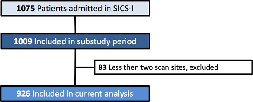
gible for inclusion. Patients were excluded if their ICU scribed by using the Cohen κ coefficient. Sensitivity,Cox et al. Critical Care (2020) 24:14 Page 3 of 7 Fig. 1 The six scan sites according to the BLUE-protocol [18] specificity, positive predictive value (PPV), negative pre- primary analyses are presented with 98.5%CIs and sec- dictive value (NPV), and diagnostic accuracy of lung ondary (subgroup) analyses with 95%CIs. ultrasound against auscultation to detect pulmonary edema were calculated. Analyses were performed using Results Stata version 15 (StataCorp, College Station, TX, USA). This SICS-I sub-study started on September 15, 2015, A subgroup analysis was performed to assess whether and continued until July 22, 2017, during which 1009 these results were robust in patients who were not patients were included. A total of 149 patients (15%) mechanically ventilated. We performed a sensitivity ana- were excluded because no bilateral or less than 2 scan lysis to assess the agreement and diagnostic accuracy of sites were scanned due to emphysema, drains, or wound LUS for pulmonary edema on chest X-ray, in patients dressings hampering the ultrasound windows, leaving where a chest X-ray was available shortly before or after 926 patients (85%) for the analysis (Fig. 2). Baseline study inclusion (i.e., on the same day). characteristics of all patients are shown in Table 1. The SICS-I was designed to address multiple hypoth- eses on six different outcomes, and therefore, the pul- Findings of lung ultrasound and auscultation monary edema outcome was adjusted for multiple The criteria for pulmonary edema diagnosed by LUS hypothesis testing. We refer to our SAP for more details, were met in 307 of 926 patients (33%). In 156 of these but in short, a p value of 0.015 indicated statistical sig- patients (51%), auscultation was normal. A total of 302 nificance and p values between 0.015 and 0.05 indicated of 926 patients (32%) had pulmonary edema diagnosed suggestive significance with an increased family-wise by pulmonary auscultation. From these patients, 151 pa- error rate [19]. For secondary or sensitivity analyses, a p tients (50%) had pulmonary edema on LUS. Of the 302 value below 0.05 indicated statistical significance due to patients with pulmonary edema on auscultation, 130 pa- the hypothesis-generating purpose. Accordingly, the tients had crepitations and 209 patients had rhonchi. Fig. 2 Flowchart. Less than two scan sites meaning if less than two out of six scan sites or no bilateral scan sites of LUS were available, the presence of pulmonary edema could not be assessed
Cox et al. Critical Care       (2020) 24:14                                                                                           Page 4 of 7

Table 1 Baseline characteristics of all included patients                            Sensitivity analysis
                                                                 N = 926             Diagnostic accuracy of auscultation improved if patients
Age, years (SD)                                                  62 (14)             were not mechanically ventilated (Table 3). The overall
Gender, male (%)                                                 598 (64)
                                                                                     accuracy for auscultation was 69% (95% CI 64–74) in
                                                                                     non-mechanically ventilated patients and 67% (98.5%CI
Height, cm (SD)                                                  176 (10)
                                                                                     64–70) in all patients (p < 0.001). The overall accuracy
Weight, kg (SD)                                                  83 (18)             for crepitations was 71% (95% CI 67–76) for rhonchi
Mechanical ventilation, n (%)                                    537 (57)            and 66% (95%CI 61–71) in non-ventilated patients. The
Vasoactive medication, n (%)                                     461 (49)            agreement between auscultation and LUS improved in
APACHE IV score, mean (SD)                                       76 (29)             non-mechanically ventilated patients (κ statistic 0.31).
Admission type
                                                                                       Radiologists’ reports assessing the chest X-ray were
                                                                                     analyzed in a subset of 315 patients as this was part of
  - Surgical, n (%)                                              292 (31)
                                                                                     the standard ICU management until November 21,
  - Medical, n (%)                                               645 (69)            2016. The baseline characteristics of these patients were
Outcomes                                                                             comparable to the overall population (Additional file 1:
  - Length of stay, days                                         3.3 (1.9–6.8)       Table S1). The median time lag between LUS and chest
  - 90-day mortality, n (%)                                      249 (27)            X-ray was 4 h (2–7 h). In 89 of these patients (28%), the
                                                                                     radiologist reported the diagnosis of edema; in 6 patients
                                                                                     (2%), it was unclear; and in 220 patients (70%), there was
  From 130 patients with crepitations, 86 patients (66%)                             no pulmonary edema on chest X-ray according to the
had pulmonary edema on LUS, and of the 209 patients                                  radiologist (Additional file 1: Table S2). The agreement
with rhonchi, 96 patients (46%) had pulmonary edema                                  and diagnostic accuracy of LUS for pulmonary edema as
on LUS. The agreement between auscultation and LUS                                   diagnosed on chest X-ray were limited (κ statistic 0.12;
was poor (κ statistic 0.25).                                                         Additional file 1: Table S3).

Diagnostic performance                                                               Discussion
Diagnostic performance measures of crepitations, rhon-                               In this prospective observational study, we found poor
chi, and auscultation for the detection of pulmonary                                 agreement between auscultation and LUS for the diag-
edema are displayed in Table 2. The sensitivity of crepi-                            nosis of pulmonary edema in acutely admitted critically
tations was 66% (98.5% CI 55–76), specificity was 71%                                ill patients.
(98.5% CI 67–75), positive predictive value was 28%                                     Several previous studies focused on the diagnostic ac-
(98.5% CI 22–34), and negative predictive value was 93%                              curacy of LUS compared to other imaging modalities,
(98.5% CI 90–95). The overall diagnostic accuracy of                                 such as chest X-ray and CT scan [4, 10, 20]. However,
crepitations was 72% (98.5% CI 69–74). The sensitivity                               few studies have compared the diagnostic accuracy of
of rhonchi was 47% (98.5% CI 39–56), specificity was                                 LUS with the stethoscope, one of the most frequently
69% (98.5% CI 65–74), positive predictive value was 31%                              used instruments at the bedside. Lichtenstein et al. pro-
(98.5% CI 25–38), and the negative predictive value was                              spectively compared the diagnostic performance of aus-
82% (98.5% CI 77–85). The overall diagnostic accuracy                                cultation, LUS, and chest X-ray for detecting alveolar
of rhonchi was 64% (98.5% CI 61–67).                                                 consolidation and alveolar-pulmonary edema with CT
  The sensitivity of abnormal auscultation overall was 52%                           scan in 32 patients with acute respiratory distress syn-
(98.5% CI 45–59), specificity was 74% (98.5% CI 70–79),                              drome and in 10 healthy volunteers [13]. The authors
positive predictive value was 49% (98.5% CI 42–56), and                              found that auscultation had a diagnostic accuracy of 55%
the negative predictive value was 76% (98.5% CI 72–80).                              for alveolar-pulmonary edema, which corresponds fairly
The overall diagnostic accuracy of auscultation was 67%                              to the 67% accuracy in our study [13]. In that study,
(98.5% CI 64–70).                                                                    LUS had a diagnostic accuracy of 97% for alveolar

Table 2 Test characteristics of specific findings compared to LUS in all patients
                      Abnormal,            Total,         Diagnostic performance in % (98.5% confidence intervals)
                      N                    N
                                                          Sensitivity            Specificity       PPV           NPV           Diagnostic accuracy
Crepitations          130                  917            66 (55–76)             71 (67–75)        28 (22–34)    93 (90–95)    72 (69–74)
Rhonchi               209                  913            47 (39–56)             69 (65–74)        31 (25–38)    82 (77–85)    64 (61–67)
Auscultation          302                  926            52 (45–59)             74 (70–79)        49 (42–56)    76 (72–80)    67 (64–70)
Abnormal auscultation was defined as the presence of crepitations and/or rhonchi at any of the sites
Cox et al. Critical Care (2020) 24:14 Page 5 of 7
Table 3 Test characteristics of specific findings compared to LUS in non-mechanically ventilated patients
Abnormal, Total, Diagnostic performance in % (95% confidence intervals)
N N
Sensitivity Specificity PPV NPV Diagnostic accuracy
Crepitations 73 387 36 (28–45) 90 (85–94) 66 (55–75) 73 (70–75) 71 (67–76)
Rhonchi 70 384 28 (21–36) 87 (82–91) 54 (44–64) 69 (66–71) 66 (61–71)
Auscultation 124 391 51 (43–60) 79 (73–84) 56 (49–63) 75 (72–79) 69 (64–74)
Abnormal auscultation was defined as the presence of crepitations and/or rhonchi at any of the sites
consolidation and 95% for alveolar-pulmonary edemas, Limitations
and chest X-ray had a diagnostic accuracy of 75% for al- Several limitations of this study must be acknowledged.
veolar consolidation and 72% for alveolar-pulmonary First, the clinical examination and ultrasonography were
edema [13]. In a sensitivity analysis, we observed that conducted as early as possible after ICU admission
the agreement and diagnostic accuracy of LUS for pul- which limits the applicability of use in patients with pro-
monary edema were limited when compared to chest X- longed admission. Further studies should explicate how
ray, which is in line with other studies [1]. auscultation and LUS compare in other departments
Another study by Torino et al. prospectively investigated and more specifically other pathologies such as a
the agreement between auscultation and LUS in non- pneumothorax. Second, we were not able to validate all
admitted patients before and after undergoing our LUS assessments by experts, also because there are
hemodialysis [11]. The authors similarly found a very poor no reference standards for the interpretation of LUS.
agreement (κ statistic 0.16, in this study κ statistic 0.25) Chest X-ray and CT are other diagnostic methods that
between the presence of crepitations on auscultation and are frequently used for the assessment of pulmonary
the presence of B lines on LUS in a total of 1106 measure- edema. However, previous studies have suggested that
ments in 79 patients [11]. Although their population LUS is superior to chest X-ray and comparable to chest
seems different to ours, patients receiving dialysis may also CT scan for diagnosing pulmonary edema [3, 8]. There-
suffer from pulmonary edema as a consequence of fluid fore, we decided not to use these modalities as a refer-
overload. Their results and conclusions are similar to ours, ence standard and only included a sensitivity analysis of
and therefore, these observations may be generalizable to chest X-ray. We limited LUS reporting to the number of
populations beyond the critically ill. B lines per field and did not use further qualitative com-
We found that the diagnostic accuracy of auscultation mentary. Third, the auscultation was not standardized.
improved if patients were not mechanically ventilated; During clinical examination, researchers performed both
no previous study has reported this finding. Acoustic auscultation and LUS; however, in contrast to LUS, we
disturbances caused by the ventilators might explain the did not describe in detail the location of auscultation. In
complicated appreciation of subtle auscultation findings. practice, these were similar to the LUS scan sites. There-
fore, we think the influence on our results is minimal.
Implications and generalizability Also, the researchers only specified whether they heard
Improved diagnostic accuracy for detecting pulmonary significant crepitation or rhonchi on auscultation. Other
edema could lead to improved treatment leading to in- abnormal breathing sounds were not recorded and
creased benefits and decreased harms for the patient. In we only documented their overall presence or absence;
critically ill patients, typically multiple pathophysio- we are unable to compare auscultation with LUS for
logical processes are co-occurring at the same time, each specific scan site. In addition, ideally, we ask the
which hampers the extrapolation of the test characteris- patient to cough to distinguish between rhonchi and/or
tics for diagnosing abnormalities in these patients, such crepitations. Unfortunately, the large majority of the pa-
as pulmonary edema. As some physicians still use aus- tients in the ICU are not cooperative with this request.
cultation to detect pulmonary edema, we think our study Fourth, even though the researchers who performed the
clarifies that auscultation may not be as reliable for de- measurements were not involved in patient care, they
tecting pulmonary edema as classically perceived, espe- were not blinded for patient information, such as admis-
cially in the ICU. Ultrasonography becomes increasingly sion diagnoses, other clinical variables and the results of
available, and our data add nuance to the discussion sur- auscultation when performing the CCUS. However, as
rounding how this technology might be properly inte- ultrasonography was always performed after ausculta-
grated into clinical practice in the care of the critically tion, we believe it is proper to discuss this potential
ill. These observations encourage further research of source of bias but do not believe that it substantially in-
LUS; the need for external validation remains to increase fluenced our results due to the objective nature of B line
the generalizability of this diagnostic modality. appearance. Fifth, since researchers were senior medicalCox et al. Critical Care (2020) 24:14 Page 6 of 7
students and junior residents, auscultation by more ex- Availability of data and materials
perienced medical doctors could potentially improve the The datasets used and/or analyzed during the current study are available
from the corresponding author on reasonable request.
diagnostic accuracy. Last, 83 (8%) patients were excluded
from the analyses due to the absence of LUS or ausculta- Ethics approval and consent to participate
tion data. However, the relatively small proportion of In unresponsive patients, informed consent was first obtained from the legal
representatives. Consent for the use of the study data was asked at a later
this excluded patient group makes it unlikely that ex- time if the patient recovered consciousness. If the patient died before
cluded patients would have altered the conclusions. Des- consent was obtained, the study data was used, and the legal
pite the potential biases and limitations, we showed that representatives were informed of the study. The study was approved by the
local institutional review board (METc M15.168207).
the agreement between auscultation and lung ultrasound
was poor. This is important as current data is scarce on Consent for publication
the diagnostic value of new non-invasive bed tools such Not applicable
as CCUS, especially in comparison with clinical examin-
Competing interests
ation in critically ill patients. The authors declare that they have no competing interests.
Author details
Conclusions 1
Department of Critical Care, University Medical Center Groningen, University
The agreement between auscultation and LUS for de- of Groningen, P.O. Box 30.001, 9700 RB Groningen, The Netherlands.
2
tecting pulmonary edema is poor. As some physicians Emergency, Cardiovascular, and Critical Care Research Group, Centre for
Health and Social Care Research, Kingston University and St George’s
still use auscultation to detect pulmonary edema, this University, London, UK. 3Department of Anesthesiology, University Medical
study clarifies that auscultation may not be as reliable Center Groningen, University of Groningen, Groningen, The Netherlands.
4
for detecting pulmonary edema as classically perceived, Department of Internal Medicine, University Medical Center Groningen,
University of Groningen, Groningen, The Netherlands. 5Department of
especially in the ICU. Anaesthesiology and Intensive Care, Royal Surrey County Hospital, Guildford,
UK. 6Department of Radiology, University Medical Center Groningen,
University of Groningen, Groningen, The Netherlands. 7Department of
Supplementary information Intensive Care, Maastricht University Medical Center+, Maastricht University,
Supplementary information accompanies this paper at https://doi.org/10. Maastricht, The Netherlands.
1186/s13054-019-2719-8.
Received: 15 October 2019 Accepted: 23 December 2019
Additional file 1: Table S1. Baseline characteristics of patients with and
without chest X-ray. Table S2. Pulmonary edema as diagnosed on chest-
X ray and LUS. * We have excluded 6 patients with a chest X-ray due to References
unclear images. Table S3. Diagnostic performance of LUS for pulmonary 1. Assaad S, Kratzert WB, Shelley B, Friedman MB, Perrino A. Assessment of
edema on chest X-ray. pulmonary edema: principles and practice. J Cardiothorac Vasc Anesth WB
Saunders. 2018;32:901–14.
2. Mojoli F, Bouhemad B, Mongodi S, Lichtenstein D. Lung ultrasound for
Abbrevations critically ill patients. Am J Respir Crit Care Med. Am Thorac Soc. 2019;199:
APACHE IV: Acute Physiology and Chronic Health Evaluation; BLUE- 701–14.
protocol: Bedside Lung Ultrasound in Emergency protocol; CCUS: Critical care 3. Shrestha GS, Weeratunga D, Baker K. Point-of-care lung ultrasound in
ultrasound; CT: Computerized tomography; ICU: Intensive care unit; critically ill patients. Rev Recent Clin Trials. 2018;13:15–26.
IQR: Interquartile range; LUS: Lung ultrasound; NPV: Negative predictive value; 4. Xirouchaki N, Magkanas E, Vaporidi K, Kondili E, Plataki M, Patrianakos A,
PPV: Positive predictive value; SAP: Statistical analysis plan; SD: Standard et al. Lung ultrasound in critically ill patients: comparison with bedside
deviation; SICS: Simple Intensive Care Studies; X-ray: Radiography chest radiography. Int Care Med. 2011;37:1488–93.
5. Miglioranza MH, Picano E, Badano LP, Sant’Anna R, Rover M, Zaffaroni F,
Acknowledgements et al. Pulmonary congestion evaluated by lung ultrasound predicts
We would like to thank all medical students and coordinators from the SICS decompensation in heart failure outpatients. Int J Cardiol. 2017;240:271–8.
Study Group for their devoted involvement with patient inclusions. 6. Saigal S, Joshi R, Sharma JP, Pandey V, Pakhare A. Lung ultrasound and
The following are the SICS Study Group members: project leaders—Geert blood gas-based classification of critically ill patients with dyspnea: a
Koster, MD; Frederik Keus, MD, PhD; Iwan CC van der Horst, MD, PhD. pathophysiologic approach. Indian J Crit Care Med. 2018;22:789–96.
Research coordinator—Willem Dieperink, PhD. Researchers who conducted 7. Amir R, Knio ZO, Mahmood F, Oren-Grinberg A, Leibowitz A, Bose R, et al.
patient inclusions: Roos Bleijendaal, MD; Yasmin F. Cawale, MD; Ramon P. Ultrasound as a screening tool for central venous catheter positioning and
Clement, MD; Devon Dijkhuizen, BSc; Ruben J Eck, MD; Bart Hiemstra, MD, exclusion of pneumothorax. Crit Care Med. 2017;45:1192–8.
PhD; Anja Haker, BSc; Casper D.H. Hilbink, MD; Thomas Kaufmann, MD; 8. Yang W, Wang Y, Qiu Z, Huang X, Lv M, Liu B, et al. Lung ultrasound is
Martiene Klasen; MD, Manon Klaver, MD; Laura J. Schokking, BSc; Victor W. accurate for the diagnosis of high-altitude pulmonary edema: a prospective
Sikkens, MD; Madelon Vos, MD; Justin Woerlee, MD; Renske Wiersema, BSc. study. Can Respir J. 2018;2018:5804942.
9. Cortellaro F, Ceriani E, Spinelli M, Campanella C, Bossi I, Coen D, et al. Lung
ultrasound for monitoring cardiogenic pulmonary edema. Intern Emerg
Authors’ contributions Med. 2017;12:1011–7.
EC, RW, and AB drafted the manuscript and conducted the analyses. IvdH 10. Martindale JL, Wakai A, Collins SP, Levy PD, Diercks D, Hiestand BC, et al.
and FK created the idea of the study. GK, TK, RE, and BH developed the Diagnosing acute heart failure in the emergency department: a systematic
protocol and implemented the study. AW, TCK, CV, and JT critically reviewed review and meta-analysis. Carpenter C, editor. Acad Emerg Med 2016;23:
the manuscript for important intellectual content. All authors critically 223–242.
reviewed the manuscript and agreed with the final version and findings. 11. Torino C, Gargani L, Sicari R, Letachowicz K, Ekart R, Fliser D, et al. The
agreement between auscultation and lung ultrasound in hemodialysis
Funding patients: the LUST Study. Clin J Am Soc Nephrol Am Soc Nephrol. 2016;11:
Not applicable 2005–11.Cox et al. Critical Care (2020) 24:14 Page 7 of 7
12. Tasci O, Hatipoglu ON, Cagli B, Ermis V. Sonography of the chest using
linear-array versus sector transducers: Correlation with auscultation, chest
radiography, and computed tomography. J Clin Ultrasound. 2016;44:383–9.
13. Lichtenstein D, Goldstein I, Mourgeon E, Cluzel P, Grenier P, Rouby J-J.
Comparative diagnostic performances of auscultation, chest radiography,
and lung ultrasonography in acute respiratory distress syndrome.
Anesthesiology. 2004;100:9–15.
14. Hiemstra B, Eck RJ, Koster G, Wetterslev J, Perner A, Pettilä V, et al. Clinical
examination, critical care ultrasonography and outcomes in the critically ill:
cohort profile of the Simple Intensive Care Studies-I. BMJ Open. 2017;7:
e017170.
15. Cohen JF, Korevaar DA, Altman DG, Bruns DE, Gatsonis CA, Hooft L, et al.
STARD 2015 guidelines for reporting diagnostic accuracy studies:
explanation and elaboration. BMJ Open. 2016;6:e012799.
16. Marini F, Frigieri FC, Guarducci D. Lung and pleural ultrasonography in
emergency and intensive care. Textb Echocardiogr Intensivists Emerg
Physicians. Cham: Springer International Publishing; 2019. p. 495–501.
17. Wiersema R, Castela Forte JN, Kaufmann T, de Haas RJ, Koster G, Hummel
YM, Koeze J, Franssen CFM, Vos ME, Hiemstra B, Keus F, van der Horst ICC.
Observational Study Protocol for Repeated Clinical Examination and Critical
Care Ultrasonography Within the Simple Intensive Care Studies. J Vis Exp.
2019;(143). PubMed PMID: 30735183. https://doi.org/10.3791/58802.
18. Lichtenstein DA. BLUE-protocol and FALLS-protocol: two applications of
lung ultrasound in the critically ill. Chest. 2015;147:1659–70.
19. Jakobsen JC, Wetterslev J, Winkel P, Lange T, Gluud C. Thresholds for
statistical and clinical significance in systematic reviews with meta-analytic
methods. BMC Med Res Methodol. 2014;14:120.
20. Martelius L, Heldt H, Lauerma K. B-lines on pediatric lung sonography. J
Ultrasound Med. 2016;35:153–7.
Publisher’s Note
Springer Nature remains neutral with regard to jurisdictional claims in
published maps and institutional affiliations.You can also read