Melanoma in Singapore: A 20-year review of disease and treatment outcomes
←
→
Page content transcription
If your browser does not render page correctly, please read the page content below
Ann Acad Med Singap 2021;50:456-66
ORIGINAL ARTICLE                                                                              https://doi.org/10.47102/annals-acadmedsg.2020535

Melanoma in Singapore: A 20-year review of disease and treatment outcomes
Pei Ming Yeo 1MRCP (UK), Ziying Vanessa Lim 1MRCP (UK), Wei Ding Virlynn Tan 1MSc, Xiahong Zhao 1PhD, Hui Yi Chia 1MRCP (UK),
Suat Hoon Tan 1FAMS (Dermatology), Melissa Ching Ching Teo 2FRCSEd, Melissa Wee Ping Tan 1FAMS (Dermatology)

                ABSTRACT
                Introduction: Melanomas in Asians have different clinicopathological characteristics and prognosis
                from melanomas in Caucasians. This study reviewed the epidemiology and treatment outcomes of
                cutaneous melanoma diagnosed at a tertiary referral dermatology centre in Singapore, which has a
                multiracial population. The study also determined whether Asians had comparable relapse-free and
                overall survival periods to Caucasians in Singapore.
                Method: This is a retrospective review of cutaneous melanoma cases in our centre between 1996
                and 2015.
                Results: Sixty-two cases of melanoma were diagnosed in 61 patients: 72.6% occurred in Chinese, 19.4%
                in Caucasians and 3.2% in Indians, with an over-representation of Caucasians. Superficial spreading
                melanoma, acral lentiginous melanoma and nodular melanoma comprised 37.1%, 35.5% and 22.6%
                of the cases, respectively. The median time interval to diagnosis was longer in Asians than Caucasians;
                median Breslow’s thickness in Asians were significantly thicker than in Caucasians (2.6mm versus
                0.9mm, P=0.018) and Asians tend to present at a later stage. The mortality rates for Asians and
                Caucasians were 52% and 0%, respectively.
                Conclusion: More physician and patient education on skin cancer awareness is needed in our
                Asian-predominant population for better outcomes.

                                                                                      Ann Acad Med Singap 2021;50:456-66

                Keywords: Asian, melanoma, nails, skin neoplasm, survival

INTRODUCTION                                                                Skin Centre, the only tertiary referral dermatology centre
Melanoma is rare in Asians.1 Asian studies of melanoma                      in Singapore, from January 1996 to December 2015.
have reported a larger proportion of patients diagnosed                     Epidemiologic, disease, treatment and outcome data
with advanced melanoma (stage III or IV), and overall                       were extracted from patients’ medical records from the
five-year survival rates ranging from 41.6% to 45.6%.2                      institute. Histopathology slides of all cases of melanoma
This is in contrast to the 2019 statistics from the                         were retrieved from the institution’s data bank and
American Cancer Society where the 5-year survival                           reviewed. For patients referred to a tertiary referral
rate for all surveillance, epidemiology, and end results                    cancer centre for further management, treatment records
(SEER) stages combined was 92% for melanoma in                              and outcome were retrieved from that institute. Clinical
the US. In addition, melanoma presents differently in                       or pathological staging was determined according to
Asian patients, with clear distinctions in the prevalent                    the 7th edition of the American Joint Committee
subtype, site of presentation, risk factors and tumour                      on Cancer.3
mutations. Our study aims to describe the clinical                            Prophylactic lymph node dissection (PLND) was
presentation, treatment and outcomes of cutaneous                           defined as lymph node removal without any clinical,
melanoma in Singapore over the last 20 years.                               radiological or histological evidence of lymph node
                                                                            involvement. Complete lymph node dissection (CLND)
METHODS                                                                     was defined as lymph node removal following sentinel
We conducted a retrospective single-centre review of                        lymph node positivity. In our institution, therapeutic
cutaneous invasive melanoma diagnosed at the National                       lymph node dissection (TLND) involves lymph node

1
 National Skin Centre, Singapore
2
 Department of Surgical Oncology, National Cancer Centre Singapore
Correspondence: Dr Ziying Vanessa Lim, National Skin Centre, 1 Mandalay Road, Singapore 308205.
Email: vlim@nsc.com.sg

                                          Ann Acad Med Singap Vol 50 No 6 June 2021 | annals.edu.sg
Melanoma in Singapore—Pei Ming Yeo et al. 457
level of 0.05. The Stata 15 software was used for all
CLINICAL IMPACT statistical analyses.4 This study was approved by the
institutional ethics review board.
What is New
• The incidence of melanoma in Singapore has
RESULTS
steadily increased in recent years. Epidemiological data
• Asians have a thicker Breslow’s thickness and Sixty-two cases of invasive cutaneous melanoma
tend to present later compared with Caucasians.
were diagnosed in 61 patients at our centre (1 patient
had 2 melanomas on different anatomical regions
Clinical Implications diagnosed at different time points). Cases of melanoma
• Melanomas in Asians confer a worse prognosis in situ (MIS) were excluded from the analysis.
than in Caucasians. Table 1 summarises the clinical characteristics of
• Primary care providers should examine their patients with cutaneous melanoma.
patients for early lesions in the soles, palms and
nails for unexplained pigmented lesions. There was a predominance of Chinese (72.6%),
followed by Caucasians (19.4%) and Indians (3.2%),
with an over-representation of Caucasians. Bangladeshi,
Filipino and Nepalese constitute the other ethnicities
removal in the presence of clinical and/or histological (4.8%). There were no Malays diagnosed with cutaneous
confirmed lymph node involvement by melanoma. melanoma although Malays constitute the second
Locoregional relapse was defined as clinically largest group in the racial composition of the population
recognisable lymph node metastases or histological in Singapore.
relapse in the regional lymph node, satellite or in-transit There were 39 male patients (62.9%), with a male
lesions. Distant relapse was defined as visceral relapse. to female ratio of 1.7 to 1, in keeping with an earlier
Melanoma BRAF and c-KIT mutational analysis by melanoma study in Singapore. 5 The most common
Sanger sequencing started to be performed in Singapore age of onset was in both the 6th and 7th decade and
from the year 2012. BRAF mutational analysis detects the median age at diagnosis was 64.0 years (range
the V600E in exon 15 of BRAF gene while c-KIT 9 and 27–99). Overall, there was a decreasing trend in age
11 mutational analysis detects gene mutations in exons at the time of initial diagnosis, contributed by the
9 and 11. If additional c-KIT rare mutations are younger age of presentation (65.0 years) in Asians
requested for, they are performed for gene mutations in the last 5 years (2011–2015) compared to a median
in exons 13 and 17. NRAS mutational analysis is not of 80.0 years in the earlier 5 years (2006–2010).
routinely performed. The median age of Caucasians was significantly lower
Median values were reported for non-normal at 47.0 years (range 36–72 years) compared with
continuous variables, and the Wilcoxon signed-rank Asians (67.0 years, range 27–99 years) during the
test and Kruskal-Wallis test were performed to test for entire study period (P=0.007).
differences between 2 groups and among more than After adjusting for population census, the incidence
2 groups, respectively. Counts and percentages were of cutaneous melanoma diagnosed at our centre
reported for categorical variables, and Fisher’s exact test between 2011 and 2015 was 0.13 patient per 100,000
was used to test for differences in proportions between patient-years, representing an approximately 3-fold
groups. Median survival time was analysed by the increase compared to the period from 1996 to 2010.
Kaplan-Meier method and compared using the Log-
rank test between groups. Univariate analysis of the Pathologic data
association between the prognostic factors and survival The most common histological subtype of cutaneous
was performed using the Cox proportional hazard melanoma diagnosed were superficial spreading
model. To avoid missing potential significant factors, melanoma (SSM) at 37.1% and acral lentiginous
variables with a P value458 Melanoma in Singapore—Pei Ming Yeo et al.
Table 1. Clinical characteristics of patients with cutaneous melanoma Table 2. Microtumoural characteristics of patients with cutaneous
melanoma
Clinical characteristics
Microtumoural characteristics no. (%)
Age, median (IQR), years 64.0 (27–99)
Histological subtype
Sex, no. (%) Superficial spreading melanoma 23 (37.1)
Male 39 (62.9) Acral lentiginous melanoma 22 (35.5)
Female 23 (37.1) Nodular melanoma 14 (22.6)
Others
Race, no. (%)
Lentigo maligna melanoma 1 (1.6)
Chinese 45 (72.6)
Melanoma arising from congenital 1 (1.6)
Caucasian 12 (19.4)
melanocytic nevus
Indians 2 (3.2)
Spitzoid melanoma with deep penetrating 1 (1.6)
Bangladeshi 1 (1.6)
nevus-like features
Filipino 1 (1.6)
Nepalese 1 (1.6) Breslow’s thickness, mm
4 16 (25.8)
Site, no. (%) Indeterminate 4 (6.5)
Limbs 15 (24.2)
Ulceration
Palmoplantar/ subungual 28 (45.2)
No 35 (56.5)
Trunk 14 (22.6)
Yes 27 (43.5)
Head and neck 4 (6.5)
Groin 1 (1.6) Mitotic rate
0/mm2 3 (4.8)
Staging, no. (%)
>1/mm2 28 (45.2)
I 19 (30.6)
Rare 4 (6.5)
II 25 (40.3)
Few 1 (1.6)
III 15 (24.2)
Moderate 1 (1.6)
IV 1 (1.6)
Multiple 1 (1.6)
Indeterminate 2 (3.2)
Seen throughout 1 (1.6)
Primary treatment (N=47) Unknown 23 (37.1)
Surgery, no. (%) 41 (87.2)
Presence of tumour-infiltrating lymphocytes
Wide excision only 15 (36.6)
Absent 52 (83.9)
Wide excision with SLNB in first surgery 16 (39.0)
Non-brisk 7 (11.3)
Wide excision with LND in first surgery 10 (24.4)
Brisk 3 (4.8)
Adjuvant therapy, no. (%) 2 (4.3)
Patient did not undergo surgery, no. (%) 6 (12.8) Presence of microsatellites
Yes 2 (3.2)
IQR: interquartile range; LND: lymph node dissection; SLNB: sentinel
No 60 (96.8)
lymph node biopsy
Regression
Yes 4 (6.5)
contrast to earlier Singapore studies in which ALM 5
No 58 (93.5)
and NM6 were the most common histological subtypes.
Lymphovascular/ perineural invasion
Among Asians in our study, the most common Yes 6 (9.7)
subtype of melanoma was ALM (44.0%) followed No 56 (90.3)
by SSM (32.0%) and NM (20.0%); and among
Caucasians, the most common subtype of melanoma
was SSM (58.3%) followed by NM (33.3%) (P=0.005).
Asians than Caucasians (median 2.6mm versus
There was no significant change in the proportion
0.9mm, P=0.02). Stratified by ethnicity, Caucasians
of melanoma subtypes in 2009–2015 compared to
showed a decrease in median Breslow’s thickness
1995–2008.
when comparing between the periods of 1995–2008
The most common site of presentation in Asians was and 2009–2015 (1.7mm versus 0.7mm, P=0.58) but
the sole of the foot (46.0%) versus the back (33.3%) this did not reach statistical significance. Asians did
and lower limb (33.3%) in Caucasians. There were no not show a significant change in median Breslow’s
cases of melanoma on the foot in Caucasians (Table 3). thickness (2.7mm versus 2.6mm, P=0.60) over time;
The median Breslow’s thickness was 2.5mm (range 38.6% of stage I and II melanoma were ulcerated at
0.2–15mm) with melanomas significantly thicker in diagnosis. Asians had significantly more ulcerated
Ann Acad Med Singap Vol 50 No 6 June 2021 | annals.edu.sgMelanoma in Singapore—Pei Ming Yeo et al. 459
Table 3. Comparison of clinico-pathological characteristics between Asians and Caucasians
Parameters Asians Caucasians P value
(n=50) (n=12)
Sex, no. (%) 0.508
Male 30 (60) 9 (75)
Female 20 (40) 3 (25)
Median age, years 67 47 0.007
Histological subtypes, no. (%) 0.005
ALM 22 (44) 0 (0)
SSM 16 (32) 7 (58)
NM 10 (20) 4 (33)
Lentigo maligna melanoma 0 (0) 1 (8)
Others 2 (4) 0 (0)
Anatomical site, no. (%) 0.012
Head and neck 3 (6) 1 (8)
Trunk, shoulder blade, groin 9 (18) 6 (50)
Upper limb excluding hand 5 (10) 1 (8)
Lower limb excluding foot 6 (12) 4 (33)
Hand, dorsum 1 (2) 0 (0)
Foot, dorsum 1 (2) 0 (0)
Foot, plantar 23 (46) 0 (0)
Foot, unspecified 2 (4) 0 (0)
Median Breslow’s thickness, mm 2.6 0.9 0.018
Ulceration, no. (%) 0.008
No 24 (48) 11 (92)
Yes 26 (52) 1 (8)
Mitotic ratea, no. (%) 0.653
1/mm 2
23 (79) 9 (90)
Staging, no. (%) 0.046
I 12 (24) 7 (58)
II 23 (46) 2 (17)
III/IV 14 (28) 2 (17)
Indeterminate 1 (2) 1 (8)
Median time to diagnosis, months 24 12 0.233
Primary treatment,a no. (%) 0.373
Surgery
Only wide excision 13 (32) 2 (33)
Wide excision with SLNB or LND 22 (54) 4 (67)
Ann Acad Med Singap Vol 50 No 6 June 2021 | annals.edu.sg460 Melanoma in Singapore—Pei Ming Yeo et al.
Table 3. Comparison of clinico-pathological characteristics between Asians and Caucasians (Cont’d)
Parameters Asians Caucasians P value
(n=50) (n=12)
Adjuvant therapy 1 (2) 1 (17)
Patient did not undergo surgery 6 (15) 0 (0)
Relapse, no. (%)
a
0.046
No 14 (34) 5 (83)
Yes 18 (44) 0 (0)
Not applicable 6 (15) 1 (17)
Unknown 3 (7) 0 (0)
Time to relapse (months) 8 NA NA
Dead,b no. (%) 0.022
No 17 (41) 6 (100)
Yes 23 (56) 0 (0)
Unknown 1 (2) 0 (0)
ALM: acral lentiginous melanoma; LND: lymph node dissection; NA: not applicable; NM: nodular melanoma; SLNB: sentinel lymph node biopsy;
SSM: superficial spreading melanoma
a
Excluded patients with unknown mitotic rate
b
Only 6 Caucasian patients and 41 Asian patients had treatment details
lesions compared to Caucasians (52% versus 8.3%, When stratified against ethnicity, the delay was longer
P=0.008). for Asians (24±50.3 months) than in Caucasians
Only 13 (21.0%) and 6 (9.7%) patients were tested (12±6.9 months) although this did not reach statistical
for BRAF and c-KIT mutation, respectively. Four significance (P=0.23). Time interval to diagnosis in
patients had BRAF and 1 had c-KIT mutation detected. the last 5 years (2011–2015) compared to the preceding
5-year interval was not significantly shorter (P=0.65).
Staging data
Nineteen (30.6%), 25 (40.3%), 15 (24.2%) and 1 (1.6%) Treatment and outcomes data
cases were diagnosed with stages I to IV disease, Follow-up data were not available for 15 cases and
respectively. Two cases (3.2%) did not undergo staging they were excluded from treatment outcome analysis.
procedure—1 patient opted to explore alternative Forty-one patients (87.2%) underwent wide excision
medicine while the other patient declined in view of (Table 1), while 2 (4.3%) had adjuvant chemotherapy
advanced age. Twenty eight percent of Asians were or radiotherapy.
diagnosed with advanced melanoma (stage III or IV) Twenty-one patients (44.7%), all with Breslow’s
at presentation, while 16.7% of Caucasians were thickness of 1mm or more, underwent sentinel lymph
diagnosed with advanced melanoma (P=0.046). node biopsy (SLNB), of which 6 (28.6%) yielded
Stratified by histological subtype of melanoma, only positive SLNB necessitating subsequent CLND. Five
27.3% and 13.0% of patients with ALM and SSM, patients (10.6%) had lymph node dissection (LND)
respectively had advanced melanoma at presentation, without prior SNLB—1 patient had fluorodeoxyglucose
while 50.0% of patients with nodular melanoma had avid nodes on positron emission tomography, 1 patient
advanced melanoma at presentation (P=0.04). had enlarged nodes on computed tomography, 1 patient
The median time interval to diagnosis (defined as had fine needle aspiration cytology proven disease,
the duration between the onset of a new pigmented and the reason for LND was not stated in 2 cases.
lesion, change in an existing mole or previous normal Three of the patients who had LND had lymph
surveillance and the diagnosis of melanoma) for the node metastases. Of the 21 cases who had neither
total study cohort was 24 months (range 1–240 months). SLNB nor LND, 3 had pT1a or pT1b disease, 9
Ann Acad Med Singap Vol 50 No 6 June 2021 | annals.edu.sgMelanoma in Singapore—Pei Ming Yeo et al. 461
declined or were considered unfit for surgery, 2 RFS time for ALM and non-ALM were 26 and 102
defaulted follow-ups, and 7 had unclear reasons that months respectively (P=0.75). The median OS and
were not documented. RFS for Asians are 55 and 23 months, respectively.
Follow-up duration was defined as the time from The 5-year survival rate stratified by American
the date of diagnosis to the date of the most recent Joint Committee on Cancer (AJCC) stage is 100%
review. Median follow-up duration was 22 months (95% CI 100–100), 45.7% (95% CI 27.1–77.1), 43.1%
(range 1–199 months). Eighteen (38.3%) cases had (95% CI 22.1–84.0) for stages I, II and III, respectively.
relapse, higher than that in earlier studies of 18.5%6 There was no significant improvement in overall
to 20.8%.5 Of the patients who experienced a relapse, survival in the last 5 years (2011–2015) compared
10 had stage III disease, 9 had stage II disease, to the preceding 15 years of our study (HR 0.73,
and 1 had stage I disease. The median duration 95% CI 0.29–1.83, P=0.498).
between the most recent treatment and date of Multivariable analysis demonstrated that the subtype
relapse was 8 months (range 1–31 months). Of the 18 of melanoma was not an independent prognostic
patients who relapsed, 50.0% had locoregional relapse, factor for OS and RFS, but an older age at diagnosis
27.8% had concurrent locoregional and distant relapse, (HR=1.07 and 1.06, P=0.006 and 0.02, respectively)
and 22.2% had distant relapse. Seven patients and thicker Breslow’s measurements (HR=1.21 and
(38.9%) declined further treatment, 7 patients (38.9%) 1.63, P=0.045 and 0.01, respectively) were independent
underwent repeat surgery, 2 (11.1%) had palliative adverse prognostic factors for OS and RFS, respectively
radiotherapy to distant metastases, and 1 (5.6%) had (Table 4).
palliative chemotherapy. One patient with c-KIT
Patients who relapsed were more likely to be Asian
mutation was treated with imatinib and is still alive at
(P=0.027), 60 years old or older (P=0.045), have
the time of writing.
deeper Breslow’s thickness (median 5.5mm versus
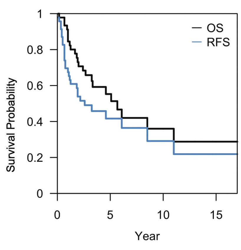
The median overall survival (OS) was 68 months, 1.8mm, P=0.0013), presence of tumour-infiltrating
the relapse-free survival (RFS) was 31 months and the lymphocytes (P=0.006) and positive sentinel lymph
5-year OS rate was 55.3% (95% CI 41.2–74.1) (Fig. 1). node involvement for melanomas thicker than 1mm
The median OS time for ALM and non-ALM were (P=0.04).
55 and 102 months, respectively (P=0.58). The median
Twenty-three patients in our cohort died, of which
the cause of death in 14 patients (60.9%) was
unknown. Significantly, all 8 patients (34.8%) who
died from melanoma were Asians. Of these, 46.2%
had ALM, 30.8% had SSM and 23.1% had NM. In the
study group, 88.4% had stage II or III disease at
diagnosis, and 34.6% either declined salvage treatment
or defaulted follow-up. The mortality rate for Asians
versus Caucasians was 52% and 0%, respectively.
DISCUSSION
(Cont’d)
In our study, the incidence rate of melanoma was
0.12 per 100,000 and 4.33 per 100,000 person-years
for Asians and Caucasians, respectively, between 2011
and 2015. This is in contrast to 0.2 to 0.65 per 100,000
person-years in other Asian studies,5-14 21.9 per 100,000
person-years in the US, and 1.3 to 35.8 per 100,000
person-years in Europe.15
Worrisome features observed in our study included
thicker Breslow’s thickness, later stage at diagnosis
and poorer overall prognosis for Asians. Despite a call
for greater education efforts on early melanoma
Fig. 1. Kaplan-Meier curves for overall survival (OS: black line)
and relapse free survival (RFS: blue line) of patients with melanoma in detection in Singapore since 2001, 6 the Breslow’s
our study. thickness has not decreased significantly in the last
Ann Acad Med Singap Vol 50 No 6 June 2021 | annals.edu.sg462 Melanoma in Singapore—Pei Ming Yeo et al.
Table 4. Multivariable analysis of prognostic factors for overall survival (OS) and relapse-free survival (RFS) for 47 patients diagnosed with primary
stage I–IV cutaneous melanoma 1996–2015
OS RFS
Hazard ratio P value Hazard ratio P value
(95% CI) (95% CI)
Age 1.07 (1.02, 1.13) 0.006 1.06 (1.01, 1.12) 0.02
Breslow’s thickness 1.21 (1, 1.46) 0.045 1.63 (1.12, 2.38) 0.01
AJCC Staging
I 1 1
II 8.56 (0.72, 101.91) 0.089 7.37 (0.68, 79.59) 0.10
III 9.10 (0.58, 142.76) 0.116 9.45 (0.53, 169.85) 0.13
IV/ Indeterminate 4.73 (0.11, 205.73) 0.420 3.94 (0.15, 100.08) 0.41
Sentinel lymph node biopsy performed
Negative 1 1
Positive 1.00 (0.16, 6.43) 0.997 0.16 (0.00, 17.80) 0.44
Not done/ Unknown 0.85 (0.12, 5.86) 0.869 0.40 (0.02, 9.61) 0.57
BRAF mutation done
No 1 1
Yes 1.79 (0.44, 7.25) 0.412 5.73 (0.61, 53.72) 0.13
Ulceration
No 1 1
Yes 0.31 (0.09, 1.05) 0.061 0.24 (0.02, 3.10) 0.28
Perineural invasion
No 1 1
Yes 3.09 (0.37, 25.62) 0.295 0.35 (0.01, 8.70) 0.53
Adjuvant treatment
No 1 –
Yes 0.27 (0.02, 3.47) 0.315 – –
Treatment modality
Surgery/ adjuvant chemotherapy/ adjuvant radiotherapy 1 –
None 1.92 (0.37, 9.87) 0.433 – –
Unknown 4.02 (0.38, 42.69) 0.249 –
None 1.92 (0.37, 9.87) 0.433 – –
Clark level
IV – 1
V – – 0.24 (0.02, 3.10) 0.28
Unknown – – 5.31 (0.73, 38.74) 0.10
AJCC: American Joint Committee on Cancer; CI: confidence interval
Ann Acad Med Singap Vol 50 No 6 June 2021 | annals.edu.sgMelanoma in Singapore—Pei Ming Yeo et al. 463 5 years. In fact, compared with previous melanoma T-lymphocyte-associated protein-4 (CTLA-4) inhibitors studies in Singapore, our study cohort has a higher and programmed death (PD-1) inhibitors have shown proportion of advanced disease at presentation, and a promising results mainly for patients with non-acral longer time interval to diagnosis (Table 5). Moreover, cutaneous melanoma in Caucasian populations.27-29 the incidence rate of melanoma between 2011 and 2015 However, 2 recent studies suggest that Asian patients had risen. We postulate that this could be due to the with advanced ALM are relatively unlikely to respond lack of skin cancer awareness among the Singapore to PD-1 blockade monotherapy 30 or toripalimab 31 population. In our study, 75% of Caucasians who had compared to Caucasians. In our study, no patients no previous skin cancer self-referred for a mole check received immune checkpoint, precluding us from and had the melanoma detected during the routine drawing meaningful conclusions about whether the skin check. In contrast, only 10% of Asians consulted introduction of these drugs has improved the survival a dermatologist for a total body skin examination or of melanoma in our population. This was because mole check. Education directed at both general PD-1 inhibitors were only approved to be used in practitioners (GPs) and the public should be tailored Singapore in 2016, which was after our study period. to our mainly Asian population17 with a predominance Another postulated reason may be due to the high of the ALM subtype of melanoma.18 The public should costs of checkpoint inhibitors. Among the various be taught how to self-monitor moles on the palms genetic markers, patients were only tested for BRAF and soles, and pigmented bands on nails. Opportunistic and c-KIT perhaps due to the low incidence of melanoma screening may be performed strategically NRAS mutation in melanomas occurring in the during annual health screening to include screening for Asian population.33 pigmented lesions on the body, soles and palms. One of the limitations of this study was its GPs may use the acronym CUBED19 which stands for retrospective nature and high dropout rates in the Coloured lesion, Uncertain diagnosis, Bleeding lesion Caucasian and non-resident population, precluding a on the foot or under the nail, Enlargement of a lesion, detailed assessment of treatment outcomes in these and Delay in healing to identify ALM. With regards to patients. This is not unusual as these patients may have nail unit melanoma, melanoma is suspected when the left the country and continued follow-up back in their first finger or toe is involved, two-thirds of the nail plate home countries. For Singapore patients who were lost is pigmented, black-grey pigmentation is present, to follow-up, we propose a closer working relationship irregularly-sized, coloured-band, and Hutchinson between oncologists and dermatologists to ensure signs are present.20 that these patients have dermatologic follow-up for Cost-effectiveness analysis for melanoma screening assessment for melanoma recurrence and regular skin had been conducted predominantly in Caucasian surveillance. populations, in which the incidence rate of melanoma There is a partial overlap of our current study population is significantly higher than in Singapore with a and an earlier melanoma study in Singapore.5 However predominantly Asian population.20-22 We did not find our duration of data collection and cohort size is any cost-effectiveness analysis studies for melanoma significantly larger than this previous study. We have screening in Asian countries. However, there are tabulated a comparison of epidemiology, clinical features, several reasons to support melanoma screening in staging and outcomes of patients in our study and 2 Singapore. Firstly, the incidence of melanoma in previous Singapore melanoma studies (Table 5). recent years is rising. Secondly, visual examination is a Our centre sees a significantly lower proportion of relatively simple and inexpensive screening modality. Malay patients and a significantly higher proportion Thirdly, melanoma is a potentially curable disease if of other ethnic groups, especially Caucasian patients identified early, and non-operable melanoma has a (P
464
Table 5. Comparison of patients in previous Singapore melanoma studies and current study
Study Tan et al.6 Lee et al.5 Current study
Study period 1989–1998 (10 years) 1998–2008 (11 years) 1996–2015 (20 years)
Study size 24 patients 48 patients 62 cases in 61 patientsa
Demographics Mean age, years 54 (range 15–83) 60 (range 29–95) 61 (range 27–99)
Male to female ratio 0.8:1 1.3:1 1.7:1
Resident versus expatriate Data on number of non-resident expatriate Consists of 38 (79.2%) resident ethnic Consists of 47 (75.8%) resident ethnic patients, and
population patients unavailable patients, and 10 (20.8%) non-resident 15 (24.2%) non-resident expatriate patients
expatriate patients
Clinical Most common site of The extremities (lower limbs more than On the palms and soles On the palms and soles
features presentation upper limbs) Asians: sole of foot (46.0%)
Caucasians: back (33.3%) and lower limb (33.3%)
Time interval to diagnosis 1 month to 240 months 1 to 120 months 1 month to 240 months
(mean 20 months) (mean 20 months) (mean 38 months)
(median 9 months) (data on median unavailable) (median 24 months)
Mean duration: Mean duration:
Asians: 22.8 months Asians: 24 months
Caucasians: 7.4 months Caucasians: 12 months
Histology Top 3 most common subtypes NM (41%), ALM (41%) and SSM (7%) ALM (50%), SSM (37.5%) and NM (12.5%) SSM (37.1%), ALM (35.5%) and NM (22.6%)
of melanoma in decreasing
order of frequency
Breslow’s thickness Median Breslow’s thickness was 3.1mm Mean Breslow’s thickness was 2.3mm Median Breslow’s thickness was 2.5mm
Melanoma in Singapore—Pei Ming Yeo et al.
(range 0.2–16mm) (data on range unavailable) (range 0.2–15mm)
Asians: 2.6mm
Caucasians: 0.9mm
Ann Acad Med Singap Vol 50 No 6 June 2021 | annals.edu.sg
Staging All patients presented with either stage I or 16 (36%), 23 (52%), 4 (9%), and 1 (2%) 19 (30.6%), 25 (40.3%), 15 (24.2%), and 1 (1.6%)
II disease patients were diagnosed with stages I to IV cases were diagnosed with stages I to IV disease,
disease, respectively respectively
Outcomes Number of patients who had 5 patients (20.8%) 10 patients (21%) 18 cases (38.3%)
relapse
Time to relapse Duration between initial diagnosis and date Duration between initial diagnosis and date of Duration between the most recent treatment and date
of relapse was between 12 and 84 months relapse was between 4 and 60 months of relapse was between 1 month and 31 months
Number of patients who died 3 patients (12.5%) Data on the number of patients who died from 8 patients (12.9%)
from melanoma melanoma is unavailable
ALM: acral lentiginous melanoma; NM: nodular melanoma; SSM: superficial spreading melanoma
a
One patient had 2 melanomas on different anatomical regions diagnosed at different time points
Superscript numbers: Refer to REFERENCESMelanoma in Singapore—Pei Ming Yeo et al. 465
tan easily, reducing the risk of many skin cancers 10. Chan KK, Chan RC, Ho RS, et al. Clinical Patterns of Melanoma
in Asians: 11-Year Experience in a Tertiary Referral Center. Ann
including melanoma. In addition, ethnic groups with
Plast Surg 2016;77:S6-S11.
more conservative clothing styles may have less sun
11. Chang JW. Cutaneous Melanoma: Taiwan experience and literature
exposed areas, resulting in less ultraviolet-induced review. Chang Gung Med J 2010;33:602-12.
types of melanoma. 12. Kim JE, Chung BY, Sim CY, et al. Clinicopathologic Features
and Prognostic Factors of Primary Cutaneous Melanoma: a
CONCLUSION Multicenter Study in Korea. J Korean Med Sci 2019;34:e126.
Given our study findings, we propose more thorough 13. Ishihara K, Saida T, Otsuka F, et al. Statistical profiles of malignant
melanoma and other skin cancers in Japan: 2007 update. Int J Clin
and targeted education for primary healthcare providers Oncol 2008;13:33-41.
and integration of efforts to teach patients self- 14. Panda S, Dash S, Besra K, et al. Clinicopathological study of
examination of pigmented lesions on the soles, palms malignant melanoma in a regional cancer center. Indian J Cancer
and nails alongside opportunistic screening efforts. 2018;55:292-6.
Prognosis is stage-dependent with good OS seen in 15. Melanoma Patient Network Europe. Melanoma – The Facts, December
patients with early disease and worse outcomes in 2019. Available at: http://www.melanomapatientnetworkeu.org/
melanoma.html. Last accessed on 15 April 2020.
patients with advanced melanoma. With our population,
16. Levit EK, Kagen MH, Scher RK, et al. The ABC rule for clinical
we should strive to improve survival of melanoma detection of subungual melanoma. J Am Acad Dermatol 2000;
with earlier detection. Further research is needed to 42:269-74.
identify modifiable risk factors for ALM to address 17. Kundu RV, Kamaria M, Ortiz S, et al. Effectiveness of a knowledge-
this increasing trend in the incidence of melanoma. based intervention for melanoma among those with ethnic skin.
Affordability of and healthcare financing models J Am Acad Dermatol 2010;62:777-84.
for genetic testing and targeted therapy or immune 18. Merkel EA, Gerami P. Malignant melanoma of sun-protected sites:
a review of clinical, histological and molecular features. Lab Invest
checkpoint inhibitors should also be re-evaluated as
2017;97:630-5.
they offer better disease control for patients with
19. Albreski D, Sloan SB. Melanoma of the feet: misdiagnosed and
metastatic or inoperable melanoma,34,35 yet their current misunderstood. Clin Dermatol 2009;27:556-63.
high costs at present may deter uptake of their use. 20. Benati E, Ribero S, Longo C, et al. Clinical and dermoscopic clues
to differentiate pigmented nail bands: an International Dermoscopy
Society study. J Eur Acad Dermatol Venereol 2017;31:732-6.
21. Losina E, Walensky RP, Geller A, et al. Visual Screening for
REFERENCES Malignant Melanoma: A Cost-effectiveness Analysis. Arch Dermatol
1. Ferlay J, Colombet M, Soerjomataram I, et al. Estimating the 2007;143:21-8.
global cancer incidence and mortality in 2018: GLOBOCAN 22. Gordon LG, Rowell D. Health system costs of skin cancer and
sources and methods. Int J Cancer 2019;144:1941-53. cost-effectiveness of skin cancer prevention and screening: a
2. Chang JW, Guo J, Hung CY, et al. Sunrise in melanoma systematic review. Eur J Cancer Prev 2015;24:141-9.
management: Time to focus on melanoma burden in Asia. Asia 23. Adamson AS, Jarmul JA, Pignone MP. Screening for Melanoma in
Pac J Clin Oncol 2017;13:423-7. Men: a Cost-Effectiveness Analysis. J Gen Intern Med 2020;
3. Edge SB, Compton CC. The American Joint Committee on 35:1175-81.
Cancer: the 7th edition of the AJCC cancer staging manual and 24. Curtin JA, Busam K, Pinkel D, et al. Somatic activation of KIT
the future of TNM. Ann Surg Oncol 2010;17:1471-74. in distinct subtypes of melanoma. J Clin Oncol 2006;24:4340-6.
4. StataCorp. 2017. Stata Statistical Software: Release 15. College 25. Hayward NK, Wilmott JS, Waddell N, et al. Whole-genome landscapes
Station, US: StataCorp LLC. of major melaoma subtypes. Nature 2017;545:175-80.
5. Lee HY, Chay WY, Tang MB, et al. Melanoma: differences 26. Liang WS, Hendricks W, Kiefer J, et al. Integrated genomic analyses
between Asian and Caucasian patients. Ann Acad Med Singap reveal frequent TERT aberrations in acral melanoma. Genome
2012;41:17-20. Res2017;27:524-32.
6. Tan E, Chua SH, Lim JT, et al. Malignant melanoma seen in a 27. Yeh I, Jorgenson E, Shen L, et al. Targeted Genomic Profiling of
tertiary dermatological centre, Singapore. Ann Acad Med Singap Acral Melanoma. J Natl Cancer Inst 2019;111:1068-77.
2001;30:414-8. 28. Ascierto PA, Long GV, Robert C, et al. Survival Outcomes in
7. Pailoor J, Mun KS, Leow M. Cutaneous malignant melanoma: Patients With Previously Untreated BRAF Wild-Type Advanced
clinical and histopathological review of cases in a Malaysian Melanoma Treated With Nivolumab Therapy: Three-Year Follow-up
tertiary referral center. Malays J Pathol 2012;34:97-101. of a Randomized Phase 3 Trial. JAMA Oncol 2019;5:187-94.
8. Ingkaninanda P, Visessiri Y, Rutnin S. Clinicopathological Features 29. Larkin J, Chiarion-Sileni V, Gonzalez R, et al. Five-Year Survival
and Prognostic Factors of Malignant Melanoma: A Retrospective with Combined Nivolumab and Ipilimumab in Advanced Melanoma.
Analysis of Thai Patients in Ramathibodi Hospital. J Med Assoc N Engl J Med 2019;381:1535-46.
Thai 2015;98:820-7. 30. Robert C, Ribas A, Schachter J, et al. Pembrolizumab versus
9. Chi Z, Li S, Sheng X, et al. Clinical presentation, histology, ipilimumab in advanced melanoma (KEYNOTE-006): post-hoc
and prognoses of malignant melanoma in ethnic Chinese: A study of 5-year results from an open-label, multicentre, randomised,
522 consecutive cases. BMC Cancer 2011;11:85. controlled, phase 3 study. The Lancet Oncology 2019; 20:1239-51.
Ann Acad Med Singap Vol 50 No 6 June 2021 | annals.edu.sg466 Melanoma in Singapore—Pei Ming Yeo et al.
31. Nakamura Y, Namikawa K, Yoshino K, et al. Anti-PD1 checkpoint 34. Sheen YS, Liao YH, Liau JY, et al. Prevalence of BRAF and
inhibitor therapy in acral melanoma: a multicenter study of 193 NRAS mutations in cutaneous melanoma patients in Taiwan.
Japanese patients. Ann Oncol 2020;3:1198-1206. J Formos Med Assoc 2016;115:121-7.
32. Tang B, Chi Z, Chen Y, et al. Safety, Efficacy and Biomarker 35. Guo J, Carvajal RD, Dummer R, et al. Efficacy and safety of
Analysis of Toripalimab in previously treated advanced melanoma: nilotinib in patients with KIT-mutated metastatic or inoperable
results of the POLARIS-01 multicenter phase II trial. Clin Cancer melanoma: final results from the global, single-arm, phase II TEAM
Res 2020;26:4250-9. trial. Ann Oncol 2017;28:1380-7.
33. Teh YL, Goh WL, Tan SH, et al. Treatment and outcomes of 36. Guo J, Si L, Kong Y, et al. Phase II, open-label, single-arm trial
melanoma in Asia: Results from the National Cancer Centre of imatinib mesylate in patients with metastatic melanoma harboring
Singapore. Asia Pac J Clin Oncol 2018;14:e95-e102. c-Kit mutation or amplification. J Clin Oncol 2011;29:2904-9.
Ann Acad Med Singap Vol 50 No 6 June 2021 | annals.edu.sgYou can also read