CERVICAL AND OCULAR VESTIBULAR EVOKED MYOGENIC POTENTIALS IN MIGRAINE PATIENTS
←
→
Page content transcription
If your browser does not render page correctly, please read the page content below
J Hear Sci, 2021; 11(2): 59–68
ISSN: 2083-389X,                                                                                                     DOI: 10.17430/JHS.2021.11.2.7
eISSN: 2084-3127                                                                                                               CC BY-NC-ND 3.0 PL

                               CERVICAL AND OCULAR VESTIBULAR EVOKED
                               MYOGENIC POTENTIALS IN MIGRAINE
Contributions:
A Study design/planning
                               PATIENTS
B Data collection/entry
C Data analysis/statistics     Abeir Osman Dabbous1,A,C-E, Nevin Mohieldin Shalaby2,A-B,D,
D Data interpretation
E Preparation of manuscript    Alaa El-Din Ahmed Abousetta3,A, Noha Ali Hosny1,F, Eman Adel Fadel3,B-C,F
F Literature analysis/search
G Funds collection             1
                                 Audio-Vestibular Unit, Department of Otolaryngology, Kasr-Al-Ainy
                                 Faculty of Medicine, Cairo University, Egypt
                               2
                                  Neurology Department, Kasr-Al-Ainy Faculty of Medicine, Cairo University, Egypt
                               3
                                   Audio-vestibular Unit, Department of Otolaryngology, Suez-Canal University, Egypt
                               Corresponding author: Abeir Osman Dabbous; Audio-Vestibular Unit, Department of
                               Otolaryngology, Kasr-Al-Ainy Faculty of Medicine, Cairo University, 12211, Cairo, Egypt;
                               email: abeirdabbous@kasralainy.edu.eg
Abstract

Introduction: In migraine, there is no anatomical correlate of vertigo and no structural abnormality is evident in conventional imaging. Cervical
vestibular-evoked myogenic potential (cVEMP) is an uncrossed inhibitory vestibulo-spinal reflex (VSR), while ocular VEMP (oVEMP) repre-
sents a crossed excitatory vestibulo-ocular reflex (VOR).
Objective: This study aims at functional evaluation of the findings of cVEMP and oVEMP in migraine patients.
Material and methods: This was a cross-sectional case-control study that included 20 migraine patients as the case group and 30 healthy adult
subjects as a control group. All participants were subjected to history taking, otological examination, basic audiological evaluation, bedside
examination of the dizzy patient, cVEMP, oVEMP, and posturography tests.
Results: 35% of migraine patients showed delayed cVEMP latency and 40% showed abnormal oVEMP in the form of statistically significant
delayed right oVEMP P1 (p = 0.050) and left oVEMP N1 latency (p = 0.038) compared with controls. cVEMP parameters were not corre-
lated to posturography results. The majority of migraine patients (70%) had normal equilibrium pattern and normal sensory analyses ratios
(65%). Only 30% had vestibular dysfunction.
Conclusions: VSR and VOR are affected in migraine patients. We recommend the use of cVEMP and oVEMP in migraine patients for func-
tional assessment of brainstem pathways.
Key words: vertigo • vestibulo-ocular reflex • vestibulo-spinal reflex

SZYJNE I OCZNE MIOGENNE PRZEDSIONKOWE POTENCJAŁY WYWOŁANE
U PACJENTÓW Z MIGRENĄ

Streszczenie

Wprowadzenie: W migrenie nie występują anatomiczne korelaty zawrotów głowy, a w konwencjonalnych badaniach obrazowych nie uwidaczniają
się nieprawidłowości strukturalne. Szyjny miogenny przedsionkowy potencjał wywołany (cVEMP) jest hamującym odruchem przedsionkowo-
rdzeniowym (VSR), realizowanym przez włókna nerwowe nieskrzyżowane, podczas gdy oczny miogenny przedsionkowy potencjał wywołany
(oVEMP) reprezentuje pobudzający odruch przedsionkowo-oczny (VOR), realizowany przez włókna nerwowe skrzyżowane.
Cel: Celem badania jest funkcjonalna ocena wyników badań cVEMP i oVEMP u pacjentów z migreną.
Materiał i metody: Przeprowadzono badanie przekrojowe z grupą kontrolną. W grupie badanej znalazło się 20 pacjentów z migreną, a w grupie
kontrolnej – 30 zdrowych dorosłych osób. Wszyscy uczestnicy przeszli następujące procedury: wywiad medyczny, badania otologiczne i audi-
ologiczne, badanie zawrotów głowy w pozycji leżącej, badania cVEMP i oVEMP, a także posturografię.
Wyniki: U 35% pacjentów z migreną, w porównaniu do grupy kontrolnej, stwierdzono wydłużoną latencję cVEMP, a 40% miało nieprawidłowe
oVEMP w postaci statystycznie istotnego opóźnienia latencji P1 oVEMP po stronie prawej (p = 0,050) i latencji N1 oVEMP po stronie lewej (p = 0,038).
Parametry cVEMP nie były skorelowane z wynikami badania posturograficznego. U większości pacjentów migrenowych (70%) stwierdzono
normalne wzorce równowagi i normalne wyniki analizy sensorycznej (65%). Tylko u 30% badanych występowały zaburzenia przedsionkowe.
Wnioski: Pacjenci z migreną mają zaburzone odruchy VSR i VOR. Zalecamy wykonywanie u pacjentów migrenowych badań cVEMP i oVEMP
w celu oceny funkcjonowania ścieżek pnia mózgu.
Słowa kluczowe: zawroty głowy • odruch przedsionkowo-oczny • odruch przedsionkowo-rdzeniowy

Introduction                                                                 specific features and associated symptoms; 2) Migraine
                                                                             with aura is primarily characterized by transient focal
Migraine is a common disabling primary headache disor-                       neurological symptoms that usually precede or some-
der. There are two major types: 1) Migraine without aura                     times accompany the headache. Some patients also expe-
is a clinical syndrome characterized by headache with                        rience a prodromal phase, occurring hours or days before
                                                                                                                                                 59
Original papers • 59–68

the headache, and/or a postdromal phase following head-            aura was diagnosed according to the diagnostic criteria
ache resolution. Prodromal and postdromal symptoms                 of the International Classification of Headache Disor-
include hyperactivity, hypoactivity, depression, cravings          ders (ICHD-3) [1,9]. All complained of dizziness, while
for particular foods, repetitive yawning, fatigue, and neck        12 (60%) had vertigo, and all fitted the new diagnostic cri-
stiffness and/or pain for years [1].                               teria of “vestibular migraine” according to the ICHD-3 [1].

Vestibular migraine (VM) is one of the most common                 The study group was compared to 30 healthy adult volun-
neurological disorders causing vertigo and dizziness (it           teer subjects as the control group. The study was approved
was previously termed migraine-associated vertigo/dizzi-           by the Research Ethical Committee and Otolaryngology
ness; migraine-related vestibulopathy; or migrainous ver-          department council of the Faculty of Medicine, Cairo Uni-
tigo) [2]. There are five major diagnostic criteria for VM,        versity. Informed consent was signed by all participants.
labelled A to E as follows. A) At least five episodes fulfilling   Tests were performed in the Audiology Unit outpatient
criteria C and D. B) A current or past history of Migraine         clinic during the period March 2016 to March 2020. Exclu-
without aura or Migraine with aura. C) Vestibular symp-            sion criteria were peripheral vestibular disorder, postural
toms of moderate or severe intensity, lasting between 5 min        hypotension, general diseases causing peripheral neuropa-
and 72 h. D) At least half the episodes are associated with        thy, patients with neurological disorders, or with peripheral
at least one of the following three migrainous features: 1.        extra-ocular muscle paresis or visual Scores defects hinder-
headache with at least two of the following four character-        ing the testing. Patients were tested during the interictal
istics: a) unilateral location, b) pulsating quality, c) moder-    period. Participants were subjected to the following 9 tests.
ate or severe intensity, d) aggravation by routine physical
activity, 2. photophobia and phonophobia, 3. visual aura.          1) History taking and assessment of the severity of dizzi-
E) Not better accounted for by another ICHD-3 diagno-              ness/vertigo using the Arabic translation [10] of the Dizzi-
sis or by another vestibular disorder [1].                         ness Handicap Inventory (DHI) [11]. The DHI is a 25-item
                                                                   self-assessment inventory that pertains to dizziness or
Although vertigo is reported by more than 60% of patients          unsteadiness problems. Questions are designed to address
with Migraine with brainstem aura, the International Clas-         the impacts of balance system disease on a person’s func-
sification of Headache Disorders, 3rd edition (ICHD-3)             tional (F) aspects (i.e., on their everyday life), emotional (E)
requires at least two brainstem symptoms in addition to            impacts (the effects on their emotional well-being), and
visual, sensory, or dysphasic aura symptoms for this diag-         physical (P) impacts (the effects on their stability). The DHI
nosis. Fewer than 10% of patients with Vestibular migraine         is therefore made up of an E-subscale score, an F-subscale
fulfill these criteria. Therefore, Vestibular migraine and         score, and a P-subscale score as well as a total score. Each
Migraine with brainstem aura are not synonymous, although          subscale was analyzed separately and by scoring the handi-
individual patients may meet the diagnostic criteria for           cap according to the total score. A total score of zero reflects
both disorders [1].                                                no handicap, while the maximum is 100. A total score of
                                                                   16–34 points reflects mild handicap, 36–52 points mod-
The cervical vestibular-evoked myogenic potential (cVEMP)          erate handicap, and ≥54 points severe handicap. 2) Gen-
assesses the descending vestibular pathway via the uncrossed       eral and neurological examination. 3) ENT examination.
ipsilateral sacculo-collic reflex, while the ocular VEMP           4) Basic audiological assessment, including pure tone audi-
(o-VEMP) test has been validated for evaluation of the             ometry, speech audiometry, tympanometry, and acoustic
ascending vestibular pathway via the crossed utriculo-ocu-         reflex threshold measurement. 5) Bedside examination of
lar reflex [3]. Central vestibular lesions may impair VEMP         the dizzy patient. 6) Video-nystagmography (Micromedical
responses along the descending and ascending tracts in             Corp, USA), including oculomotor and positional, as well
the brainstem [4–7]. Computerized dynamic posturogra-              as caloric testing. 7) Subjective visual vertical (SVV) test
phy (CDP) is a test of a person’s ability to maintain balance      (Difra, Belgium). 8) VEMP tests (Neuro-Audio, Neurosoft
by effectively using visual, vestibular, and somatosensory         Ltd, Russia). For cVEMP, the active electrode was placed
inputs separately as well as suppressing or compensating           on the middle of the sterno-cleido-mastoid (SCM) mus-
for inaccurate or challenging sensory information. CDP can         cle; the reference electrode on the upper sternum (supra-
complement the results of conventional vestibular tests in         sternal notch), and the ground electrode on the forehead.
specific situations, such as analyses of the vestibulo-spinal      Electrode impedance was kept below 5 kΩ. Subjects were
reflex (VSR) and sensory analyses of balance disorders [8].        instructed to sit upright and tense the muscle by turning
                                                                   their chin to the contralateral shoulder. Rectified EMG
In migraine, no structural abnormality shows on conven-            was monitored during recordings to ensure low noise.
tional imaging; that is, there is no anatomical correlate of       Stimuli were tone bursts of 500 Hz with rise and fall times
vertigo. There is then a need to assess functional abnor-          of 1 ms and plateau of 2 ms presented monoaurally at 5 Hz
mality in migraineurs using electrophysiological testing.          through insert phones at 100 dBnHL. At least 60 sweeps
This study aims to evaluate of the findings of cervical and        were obtained using a 30–2000 Hz filter. The time win-
ocular vestibular evoked myogenic potentials (cVEMP and            dow for analysis was 50 ms. For oVEMP, the positive elec-
oVEMP) in migraine patients and to relate VEMP param-              trode was placed on the orbital margin below the center
eters to the clinical presentation and other vestibular tests.     of the eye and the reference electrode placed 15–30 mm
                                                                   below the positive electrode, on the cheek, with the ground
Material and methods                                               electrode on the forehead. Subjects were instructed to sit
                                                                   upright, relax their facial muscles, and look up with their
The study group included 20 adult migraineurs having diz-          eyes without moving their head. At least 200 sweeps were
ziness with or without vertigo. Migraine with and without          made with a 1–1000 Hz filter. Other conditions were the
60                                                                                  Journal of Hearing Science · 2021 Vol. 11 · No. 2
Dabbous et al. – VEMPs in migraine
same as with cVEMP. VEMP responses were judged as There were statistically significant differences between the
either present or absent according to the presence or absence migraine group and the control group regarding EQ scores
of a biphasic response. Parameters measured were latency (under conditions C1, C2, and C6) and in the SOM ratio
in ms and amplitude in µV. For cVEMP, measurements (Table 1). Table 2 shows EQ deficits and affected sensory
were made of P13 latency, N23 latency, P13–N23 peak-to- analyses. Normal EQ was found in 13/20 (65%) and nor-
peak amplitude, and the inter-aural amplitude difference mal SA ratios in 14/20 (70%).
(IAAD) ratio or the amplitude asymmetry ratio, which is
the peak to peak amplitude difference between the 2 ears None of the migraine patients had any oculographic abnor-
divided by the total amplitude of both ears. For oVEMP, mality. There were no statistically significant differences
measures were N10 latency, P15 latency, N10–P15 peak- between migraine patients and normal controls regarding
to-peak amplitude, and IAAD. 9) A sensory organization the SVV test. cVEMP and oVEMP were present bilaterally
test (SOT) using computerized dynamic posturography in all subjects. There were no statistically significant dif-
(CDP). An equilibrium score (EQ) was calculated for each ferences between migraine patients and controls regard-
of the 6 SOT conditions, and a composite EQ was calcu- ing cVEMP, except for a statistically significant smaller
lated. Ratios were used to identify possible impairments IAAD%, but it was still within the normal range (Table 3).
of an individual’s sensory system. a) The somato-sensory For oVEMPs, there were statistically significant delayed Rt
(SOM) ratio (condition 2/condition 1); b) the visual (VIS) oVEMP P1 and Lt oVEMP N1 latency in the migraineurs
ratio (condition 4/condition 1); c) the vestibular (VEST) compared to their controls; there was also statistically
ratio (condition 5/condition 1), which assesses the abil- significant smaller Rt oVEMP rectified amplitude in the
ity to use input from each sensory system to control bal- migraine patients compared to their controls (Table 4). Fig-
ance; and d) the vision preference (PREF) ratio [(condi- ure 1 shows cVEMP and oVEMP traces in one of our nor-
tion 3 + 6) / (condition 2 + 5)], which assess the extent to mal controls, and for comparison, Figure 2 shows cVEMP
which a subject relies on visual input to control balance, and oVEMP traces in one of our migraine patients.
even when the visual information is incorrect.
According to our normative values for cVEMP,
Statistical analysis methods. Data collected was coded using IAAD% > 36.55% was considered abnormal, reflecting
Microsoft Excel 2010, and then imported into SPSS (Statis- amplitude asymmetry; similarly, P13 > 16.73 ms (Rt),
tical Package for Social Science) version 19.0 for analysis. 15.97 ms (Lt), N23 > 26.68 ms (Rt), and 25.74 ms (Lt)
According to the type of data, the following tests were per- were considered abnormal. For oVEMP, IAAD% > 36.10%,
formed to test differences for significance: Mann–Whitney N1 > 11.95 ms (Rt), 12.12 ms (Lt), P1 > 17.29 ms (Rt),
U-test (if the data were not normally distributed) and Chi- 19.50 ms (Lt) were considered abnormal. The final VEMP
square test with least significant difference. Pearson’s cor- result was considered normal if both the latency and ampli-
relation test was used to determine correlations between tude were normal and were considered abnormal if either
individual results. Differences were considered statistically or both were abnormal. Table 5 shows, for migraineurs, the
significant at p < 0.05. distribution of latency and amplitude asymmetry abnor-
malities, and the final cVEMP and oVEMP results. There
Results was no statistically significant difference regarding the
relation between the distribution of cVEMP and oVEMP
Cases comprised 16 (80%) females, and 4 (20%) males, abnormality results.
with a mean age of 35.1 ± 6.7 (range 22 to 44 years). Con-
trols comprised 17 (57%) females and 13 (43%) males, There were no statistically significant differences between
with a mean age of 32.8 ± 5.4 (range 24 to 44 years). cVEMP and oVEMP findings in migraineurs when com-
Groups were matched in terms of age and gender. The mean pared with those with/without nausea, vomiting, allodynia,
migraine duration was 6.0 ± 3.7 years (range 1–15) and phonophobia, or vertigo. Although migraine patients with
mean migraine attacks/month was 3.2 ± 1.4 (range 0–6). phonophobia had larger cVEMP amplitude than those with-
The mean migraine attack duration was 3.3 ± 1 h (range out phonophobia, all migraineurs with and without pho-
2–5). There were 14/20 (70%) of migraine patients who nophobia had symmetrical IAAD.
were under treatment. Twelve patients (60%) had phono-
phobia; 13 (65%) had photophobia, 3 (15%) had allodynia, Statistically, there was significantly more IAAD oVEMP
14 (70%) had nausea, 6 (30%) had vomiting, 16 (80%) had symmetry in those with photophobia compared to those
aura – visual in 12 (60%), sensory in 9 (45%), and motor without photophobia. All migraineurs with photophobia
in 3 (15%), diplopia in 4 (20%), blurring in 9 (45%), and had symmetrical IAAD while 71.4% of those without pho-
tinnitus in 4 (20%). All migraineurs complained of dizzi- tophobia had asymmetrical IAAD and this distribution
ness, while 12 (60%) had vertigo. was statistically significant. There was a statistically sig-
nificant larger Rt and Lt cVEMP P13–N23 amplitude and
Regarding the DHI, the mean F-score was 17.5 ± 2.7 (range Rt and Lt cVEMP rectified amplitude in those with aura
12–22), the mean P-score was 6.7 ± 2.18 (range 4–10), the compared to those without aura.
mean E-score was 15.4 ± 1.96 (range 12 to 18), and the
mean total score was 39.6 ± 5.72 (range 28–50). There were All migraineurs with vertigo had symmetrical oVEMP
5/20 (25%) who had mild handicap, while 15/20 (75%) had amplitude compared to 75% of those without vertigo,
moderate handicap. Regarding PTA: 19 (95%) and 18 (90%) and this distribution was not statistically significant. In
of migraine patients had normal hearing, while 1 (5%) migraineurs there was no statistically significant difference
and 2 (10%) had high frequency hearing loss (at 8 kHz) in the distribution of cVEMP and oVEMP abnormality
in the right and left ears respectively. in terms of the presence or absence of vertigo. Similarly,
Journal of Hearing Science · 2021 Vol. 11 · No. 2 61Original papers • 59–68
Table 1. Comparison between migraine patients and their controls according to equilibrium scores in the 6 SOT conditions
(C1–C6), composite scores, and sensory analysis ratios
Controls (n = 30) Migraine (n = 20)
Z** p
Equilibrium Mean SD Min Max Mean SD Min Max
scores
C1 95.24 2.4 90.3 98.6 93.05 4 79.33 97.5 –2.162 0.031*
C2 92.76 3.26 82.3 97.6 89.05 5.47 73 96 –2.834 0.005*
C3 90.21 4.47 81.3 98.6 87.8 5.48 78.33 95.6 –1.605 0.109
C4 85.69 5.26 77.3 98.4 82.17 8.15 54.3 89.6 –1.347 0.178
C5 70.41 7.38 57.6 92.4 64.65 14.94 38 90 –1.406 0.16
C6 69.71 8.79 55.3 90.1 58.12 18.71 17.33 94 –2.506 0.012*
Composite score 79.17 5.82 71 92 75.15 8.44 58 91 –1.836 0.066
SOM ratio 97.35 3.03 84.67 100 94.84 5.02 78.07 100 –2.125 0.034*
VIS ratio 89.68 5.34 80.3 100 88.4 8.73 56.56 96.17 –0.089 0.929
VEST ratio 73.6 7.51 60.4 97.8 68.7 15.37 40.42 94.24 –1.129 0.259
PREF ratio 96.48 4.46 83.2 100 91.97 8.87 65.16 100 –1.869 0.062
Z** of Mann–Whitney test
*p-value is statistically significant
there were no statistically significant differences between reflects vestibulo-ocular reflex (VOR) pathology, which
migraineurs with and without vertigo regarding posturog- agrees with Kim et al. [13]. Our result of oVEMP ampli-
raphy. There were no statistically significant differences tude asymmetry, accords with Inoue et al. [18], Makowiec
between migraineurs with and without normal compos- et al. [19], and Zaleski et al. [20]. Significant oVEMP N1–
ite score or VEST ratio score regarding cVEMP findings. P1 amplitude differences have been found between healthy
and migraine groups [19], but in other studies the differ-
As the age of migraineurs patients increased, the rectified Lt ence did not reach statistical significance [13,14].
oVEMP decreased (a weak negative correlation) (Table 6).
As F-score of the DHI increased, the cVEMP N23 latency In the current study, even though the mean cVEMP
on the Rt side was delayed (weak positive correlation). As latency of the whole group was comparable to the con-
P-score of the DHI increased, the Rt P13–N23 amplitude trols, 7 migraine cases (35%) had abnormal cVEMP due to
cVEMP decreased (fair negative correlation). As P-score of delayed latency, indicating that the vestibulo-spinal reflex
the DHI increased, the Rt and Lt rectified cVEMP ampli- (VSR) was affected. oVEMP abnormality in the form of
tude decreased (fair negative correlations). As the total and latency delay was found in 8/20 patients (40%), includ-
the E-score of the DHI increased, the Rt P13–N23 recti- ing 2 that also had amplitude asymmetry (IAAD%).
fied cVEMP amplitude decreased (fair negative correla-
tion) (Table 7). In comparison, Jung et al. [21] found abnormal cVEMP
measurements in 29% of VM patients. Inoue et al. [18] did
Discussion not find any significant difference between VM patients
and controls in terms of the prevalence of abnormal air-
In the current study, there was no statistically significant conducted cVEMPs or oVEMPs: 39% cVEMPs abnormality
difference in cVEMP latency between migraineurs and con- in VM versus 22% for controls, and 35% oVEMPs abnor-
trols. Although there was a statistically significant smaller mality in VM versus 8% for controls. At the same time,
IAAD% than the controls, it was within normal limits and Inoue and colleagues found no significant differences in
so all cases had symmetrical cVEMP. The amplitude results response amplitude or latencies for cVEMPs or oVEMPs
agree with Shalaby et al. [12], Kim et al. [13], and Khalil between VM patients and controls [18]. Khalil et al. [14]
et al. [14]. However, Kang et al. [15] found a prolonged found that 5 of 20 VM patients (25%) had normal cVEMP
latency in the cVEMP test in VM patients. In terms of responses and 15 (75%) had abnormal cVEMPs, while
amplitudes, Kang et al. [15], Baier et al. [16], and Salviza the majority (95%) of VM patients (19/20) had abnormal
et al. [17] all reported bilaterally reduced cVEMP ampli- oVEMP responses.
tudes in VM patients compared to controls. Makowiec et
al. [18] found that in patients with VM the most common In the current study, only 3/20 (15%) of migraine patients
cVEMP abnormality was either asymmetrical amplitude showed abnormality in both cVEMP and oVEMP, while
or complete absence of VEMP. 8/20 (40%) showed normal results in both, 5/20 (25%)
showed normal cVEMP but abnormal oVEMP, and
In our study, migraineurs showed statistically significant 4/20 (20%) showed normal oVEMP but abnormal cVEMP.
delayed oVEMP latencies compared to controls. This However, this abnormality distribution was not significant.
62 Journal of Hearing Science · 2021 Vol. 11 · No. 2Dabbous et al. – VEMPs in migraine
Table 2. Equilibrium deficit findings, affected sensory analyses ratios, and abnormalities in the migraine patients
Migraine
(n = 20)
Equilibrium deficit No. %
Normal (no deficits) 13 65
Visual preference/vestibular dysfunction pattern 1 5
Visual/vestibular dysfunction pattern 1 5
Severe dysfunction pattern 0 0
Vestibular dysfunction pattern 4 20
Visual preference pattern 1 5
Sensory analyses (affected sensory ratios) No. %
Normal SA ratios 14 70
Abnormalities No. %
VEST 4 20
VIS 0 0
PREF 0 0
VEST / SOM 1 5
VEST / VIS 1 5
Abnormalities No. %
SOM ratio 1 5
VIS ratio 1 5
VEST ratio 6 30
PREF ratio 0 0
Numbers are not mutually exclusive as a patient can have multiple abnormalities (i.e., numbers do not add up to the total numbers in
each group)
Table 3. Comparison of cVEMP parameters between migraine patients (n = 20) and normal controls (n = 30)
Migraine patients Controls
cVEMP p-value
(n = 20) (n = 30)
mean SD min max mean SD min max
P13 (in ms) 15.32 2.81 11.4 21.8 14.22 1.25 12 16.5 0.067
N23 (in ms) 22.22 2.55 18.4 26.5 22.18 2.25 16.5 27.5 0.957
Rt P13–N23 amplitude in µV 46.55 17.36 19 91 44.41 19.36 10.7 89.7 0.691
rectified P13–N23 amplitude 0.89 0.43 0.3 1.9 1.05 0.57 0.2 2.7 0.282
in µV
P13 (in ms) 14.96 2.42 11 19.6 14.07 0.95 12.3 16.1 0.077
N23 (in ms) 21.73 2.71 17.5 26.5 21.92 1.91 17.3 26.2 0.763
Lt P13–N23 amplitude in µV 53.62 25.79 22.1 111.1 43.97 22.03 8.1 92.1 0.163
Rectified P13–N23 amplitude 1.07 0.58 0.4 2.3 1.09 0.64 0.2 3.2 0.888
in µV
IAAD% 9.39 6.51 0.2 22.8 16.53 10.01 0.2 30.4 0.007*
Rectified IAAD% 15.42 12.44 0 41 16.22 10.52 0 35 0.806
* p-value is statistically significant
Journal of Hearing Science · 2021 Vol. 11 · No. 2 63Original papers • 59–68
Table 4. Comparison of oVEMP parameters between migraine patients (n = 20) and normal controls (n = 30)
Migraine patients Controls
oVEMP p-value
(n = 20) (n = 30)
mean SD min max mean SD min max
N1 (in ms) 11.1 1.45 9.4 16.4 10.61 0.67 9.4 12 0.119
P1 (in ms) 16.21 1.49 14.2 19.9 15.53 0.88 12.5 17.5 0.050*
Rt
N1–P1 amplitude in µV 4.68 3.56 1.4 17.4 4.05 3.61 0.6 17.2 0.546
Rectified N1–P1 amplitude (in µV) 0.24 0.2 0.1 0.8 0.48 0.36 0.1 1.2 0.008*
N1 (in ms) 11.34 1.53 9 16.3 10.64 0.74 9 12.4 0.038*
P1 (in ms) 16.52 3.08 12.3 28.4 15.3 2.1 5.7 17.7 0.103
Lt
N1–P1 amplitude in µV 4.44 4.02 1.2 20.1 4.4 3.42 0.5 13.4 0.971
Rectified N1–P1 amplitude in µV 0.28 0.3 0.1 1.4 0.41 0.25 0.1 0.9 0.114
IAAD% 17.67 10.85 0 37.5 17.59 9.25 0.2 30.4 0.980
Rectified IAAD% 8.91 15.93 0 50 16.79 12.92 0 42.8 0.060
*p-value is statistically significant
Figure 1. Example traces of cVEMP (A) and oVEMP (B) in one of our normal controls (female, 36 years old)
64 Journal of Hearing Science · 2021 Vol. 11 · No. 2Dabbous et al. – VEMPs in migraine
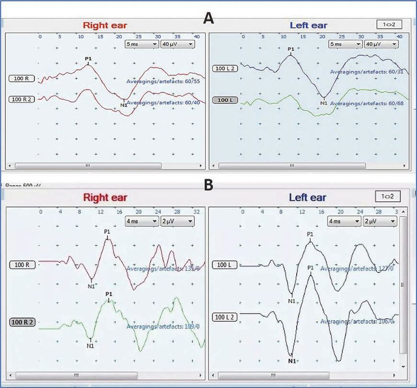
Figure 2. Example traces of cVEMP (A) and oVEMP (B) in one of our migraine patients (female, 38 years old)
Table 5. Relation between of cVEMP and oVEMP results (including latency, asymmetry, or loss) in migraine patients. There
was no statistically significant difference regarding the relation between the distribution cVEMP and oVEMP abnormality
results
cVEMP result
Migraine Total
Abnormal Normal
Abnormal 3 (15%) 5 (25%) ‡8 (40%)
oVEMP result
Normal 4 (20%) 8 (40%) 12 (60%)
Total †7 (35%) 13 (65%) 20 (100%)
χ2 = 0.037, p = 1.000
†All migraine cases with abnormal cVEMP (n = 7) had delayed latency, even though they all had symmetrical amplitude. All cVEMP
abnormalities in migraine patients were in the form of latency delay. ‡Migraine cases with abnormal oVEMP (n = 8) had delayed
latency including 2 with amplitude asymmetry
In comparison, Makowiec et al. [19] concluded that patients Our cVEMP and oVEMP findings were not correlated
with VM are more likely than subjects with vestibular to any clinical symptom, except that cVEMP amplitude
disorders other than migraine to exhibit normal cVEMP was statistically significant larger in both ears of migraine
responses in the presence of unilaterally abnormal oVEMP patients with aura compared to those without aura. How-
responses. Such a VEMP pattern may be a biomarker of VM ever, Sürmeli et al. [22] found that the cVEMP P13–
and further supports a possible pathophysiologic relation- N23 amplitudes in migraine patients with aura were sig-
ship between the utriculo-ocular reflex and VM. nificantly smaller than in patients without aura. In their
Journal of Hearing Science · 2021 Vol. 11 · No. 2 65Original papers • 59–68
Table 6. Correlation between cVEMP and oVEMP findings and age of migraine patients (n =20)
cVEMP
Rt Lt
P13–N23 P13-N23 Asymmetry Rectified
P13 N23 Rectified P13 N23 Rectified IAAD%
amplitude amplitude
Age of r –0.106 0.102 –0.245 –0.318 –0.053 0.078 –0.212 –0.16 0.378 0.138
migraine
patients p 0.658 0.67 0.298 0.172 0.826 0.745 0.37 0.5 0.875 0.563
oVEMP
Rt Lt Asymmetry Rectified
IAAD%
N1–P1 N1–P1
N1 P1 Rectified N1 P1 Rectified
amplitude amplitude
Age of r 0.051 0.055 0.347 –0.388 0.020 –0.127 0.244 –0.454 –0.251 –0.049
migraine
patients p 0.831 0.819 0.134 0.091 0.932 0.594 0.300 0.044* 0.286 0.836
*p-value is statistically significant
Table 7. Correlation between cVEMP and oVEMP findings and Dizziness Handicap Inventory in migraine patients (n = 20)
cVEMP
Dizziness Rt Lt
Handicap rectified
Inventory P13–N23 rectified P13–N23 rectified asymmetry
P13 N23 P13 N23 IAAD%
amplitude amplitude amplitude amplitude
r 0.268 0.462 –0.22 –0.189 0.083 0.312 –0.151 –0.067 0.091 0.037
F-score
p 0.253 0.040* 0.352 0.426 0.729 0.181 0.526 0.78 0.703 0.878
r 0.005 0.297 –0.599 –0.645 –0.048 0.207 –0.505 –0.416 –0.276 0.137
P-score
p 0.983 0.204 0.005* 0.002*
0.842 0.381 0.023* 0.068 0.239 0.565
r 0.138 0.131 –0.289 –0.521 0.121 0.197 –0.337 –0.356 –0.397 0.074
E-score
p 0.562 0.581 0.217 0.018* 0.612 0.405 0.147 0.124 0.083 0.755
r 0.174 0.373 –0.43 –0.512 0.062 0.292 –0.378 –0.311 –0.198 0.095
total score
p 0.462 0.105 0.059 0.021* 0.796 0.212 0.100 0.181 0.402 0.691
oVEMP
Dizziness Rt Lt
Handicap rectified
Inventory N1–P1 rectified N1–P1 rectified asymmetry
N1 P1 N1 P1 IAAD%
amplitude amplitude amplitude amplitude
r –0.02 –0.089 0.041 0.113 0.225 0.182 –0.097 –0.039 0.401 –0.035
F-score
p 0.934 0.709 0.864 0.635 0.34 0.442 0.686 0.869 0.08 0.885
r –0.375 –0.043 –0.297 –0.011 –0.324 –0.291 –0.198 –0.203 0.035 –0.283
P-score
p 0.103 0.857 0.204 0.964 0.163 0.213 0.404 0.391 0.884 0.227
r 0.255 0.296 –0.142 –0.051 0.19 0.084 –0.111 –0.021 –0.201 –0.306
E-score
p 0.279 0.205 0.55 0.831 0.423 0.723 0.642 0.928 0.395 0.189
r –0.065 0.043 –0.143 0.031 0.046 0.003 –0.158 –0.103 0.131 –0.229
total score
p 0.786 0.856 0.549 0.896 0.846 0.99 0.505 0.665 0.581 0.332
*p-value is statistically significant; E = emotional; F= functional; P= physical
study, the patients had vertigo as the aura. In compari- In contrast to our study, Lipton et al. [23] and Young et al.
son, Shalaby et al. [12] found that VEMP abnormality [24] found that allodynia is very common, affecting roughly
was found in 67% of those having aura, 62% of those hav- 60% of migraine patients. A potential mechanism for how
ing vertigo, 100% of those having dizziness, and 69% of migraine might be associated with dizziness or vertigo is
those having symptoms of brainstem dysfunction. There sensory exaggeration: in migraine, all senses can be more
was no statistically significant difference between the fre- acute, so allodynia is possible. This may make patients
quency of these symptoms in migraineurs with and with- with migraine more likely to experience motion sickness
out VEMP abnormality. and amplify the effects of small amounts of vestibular
66 Journal of Hearing Science · 2021 Vol. 11 · No. 2Dabbous et al. – VEMPs in migraine
disturbance that other people might not notice [25]. Our found that amplitude of the oVEMP response decreased
results were not in agreement with these researchers, prob- significantly with age, suggestive of an age-related decline
ably because of the smaller percentage of patients having in utricular function. But Khalil et al. [14] found that there
allodynia, even though there were those who had sensory was no significant correlation between age of patients and
exaggeration to sound and light (phonophobia and pho- either cVEMP or oVEMP responses.
tophobia were as high as in previous reports).
Assessed by DHI, the majority of the migraine group,
Our study agrees with previous studies in that there were 15/20 patients (75%), had a moderate degree of handicap.
comparable cVEMP and oVEMP parameters in migraineurs In contrast to our study, Yip and Strupp [32] did not find
either with or without vertigo [12,26,27]. any significant correlation between DHI and cVEMP or
oVEMP amplitude, or of amplitude asymmetry. They did
Hong et al. [28] assumed that VM patients have difficulty not find any significant correlation between DHI and pos-
in incorporating somatosensory information. They found tural sway on posturography. They found that patients with
an abnormal vestibular ratio in SOT in 45% of patients central vestibular disorders, excluding migraine, had higher
with VM, abnormal visual ratio in 58%, and abnormal DHI than those with peripheral or functional disorders.
SOM ratio in 19%. Jung et al. [20] found that 16%, 49%, However, their study did not include migraine as a central
58%, and 21% showed abnormal SOM, VIS, VEST, and VIS disorder as it has central and peripheral effects. However,
PREF ratios, respectively. They thought that an abnormal the connection between DHI scores and cVEMP ampli-
VEST ratio on posturography and an abnormal VEMP tude were fairly well correlated.
response were frequent findings in VM patients who had
had recurrent attacks for more than 6 months and they Conclusions
regarded these indicators as pointing to a poor progno-
sis. Çelebisoy et al. [29] reported peripheral vestibular cVEMP and oVEMP were present bilaterally in all the
dysfunction in VM during the symptom-free period to migraine patients. In 35% of patients there was a cVEMP
be more common than a central deficit. However, in the abnormality in the form of delayed latency, reflecting
present study the majority of migraineurs had normal effects of migraine on the vestibulo-spinal reflex. oVEMP
equilibrium; after exclusion of peripheral vestibulopathy, was abnormal in 40% of patients in the form of latency
vestibular dysfunction was found in 30%. In agreement delay, and there was another 10% with amplitude asym-
with the current findings, Çelebisoy et al. [29] also found metry as well, reflecting vestibulo-ocular reflex pathology.
comparable posturography results between migraineurs On posturography, migraineurs showed a somatosensory
with and without vertigo. defect but only 30% showed vestibular dysfunction and
the majority had a normal equilibrium pattern and nor-
In agreement with our study, Mallinson et al. [30] also mal sensory analyses ratios.
found that cVEMP and oVEMP abnormalities do not cor-
relate with CDP findings. CDP measures dynamic sway However, cVEMP, oVEMP, and posturography findings
and VEMPs are related to activities involving head move- did not correlate with clinical symptoms. As age of the
ment and are not a measure of body sway [30]. However, migraineurs increased, the oVEMP amplitude decreased.
Khalil et al. [14] found that there was a significant corre- Duration of migraine did not correlate with VEMP
lation between abnormal Fukuda, modified clinical test of parameters.
sensory integration of balance, tandem gait tests, and both
cVEMPs and oVEMPs abnormalities. Our conclusion is that in migraineurs we recommend the
use of cVEMP for the assessment of VSR and the use of
In the present study, duration of migraine was not cor- oVEMP in assessing VOR. We also recommend the use of
related to any of the studied VEMP parameters, agree- posturography as complementary testing for full assess-
ing with Khalil et al. [14]. Like us, Agrawal et al. [31] also ment of the VSR in migraine patients.
References
1. Headache Classification Committee of the International Head- 6. Welgampola MS and Colebatch JG. Characteristics and clinical
ache Society (IHS). The International Classification of Headache applications of vestibular-evoked myogenic potentials. Neurolo-
Disorders, 3rd edition. Cephalalgia, 2018; 38(1): 1–211. gy, 2005; 24: 1682–8.
2. Lempert T, von Brevern M. Vestibular Migraine. Neurol Clin, 7. Todd NPM, Rosengren SM, Aw ST, Colebatch JG. Ocular vestib-
2019; 37(4): 695–706. ular evoked myogenic potentials (OVEMPs) produced by air- and
3. Iwasaki S, Smulders Y, Burgess A, McGarvie L, MacDougall H, bone-conducted sound. Clin Neurophysiol, 2007; 118: 381–90.
Halmagyi G. Ocular vestibular evoked myogenic potentials to 8. Pang MY, Lam FM, Wong GH, Au IH, Chow DL. Balance per-
bone conducted vibration of the midline forehead at Fz in healthy formance in headshake computerized dynamic posturography:
subjects. Clin Neurophysiol, 2008; 119(9): 2135–47. aging effects and test–retest reliability. Phys Ther, 2011; 91(2):
4. Curthoys IS. A critical review of the neurophysiological evidence 246–53.
underlying clinical vestibular testing using sound, vibration and 9. Headache Classification Committee of the International Head-
galvanic stimuli. Clin Neurophysiol 2010; (121): 132–44. ache Society (IHS): The International Classification of Headache
5. Rosengren SM, Welgampola MS, Colebatch JG. Vestibular evoked Disorders, 3rd edition (beta version). Cephalalgia, 2013 Jul; 33(9):
myogenic potentials: past, present and future. Clin Neurophysi- 629–808.
ol, 2010; 121: 636–51. 10. Alsanosi A. Adaptation of the dizziness handicap inventory for
use in the Arab population. Neurosciences, 2012; 17(2): 139–44.
Journal of Hearing Science · 2021 Vol. 11 · No. 2 67Original papers • 59–68
11. Jacobson G, Newman CW. The development of the Dizziness 22. Sürmeli M, Sürmeli R, Deveci I, Önder S, Yalçın AD, Oysu Ç.
Handicap Inventory. Arch Otolaryngol Head Neck Surg, 1990; Correlation between cVEMP and ABR for the evaluation of ves-
116: 424–7. tibular migraine. J Int Adv Otol, 2016; 12: 326–31.
12. Shalaby NM, Ramzy GM, Nada MAF, Hussein AF, El-Fayomy 23. Lipton, RB, Bigal ME, Ashina S, Burstein R, Silberstein S, Reed
NM, El-Minawi MS, Dabbous AO, El-Dessouky T. Assessment ML, Serrano D, Stewart WF. Cutaneous allodynia in the migraine
of the vestibulo-spinal reflex in migraine patients. Egypt J Neu- population. Ann Neurol, 2008; 63(2): 148–58.
rol Psychiat Neurosurg, 2010; 47(1): 67–74. 24. Young WB. Allodynia as a complication of migraine: background
13. Kim CH, Jang MU, Choi HC, Sohn JH. Subclinical vestibular and management. Curr Treat Options Neurol, 2009; 11(1): 3–9.
dysfunction in migraine patients: a preliminary study of ocular 25. Hain T. How Migraine Causes Dizziness. Available at , accessed June 2020.
14. Khalil LH, Hazzaa NM, Nour AA. Vestibular migraine: a correla- 26. Roceanu A, Allena M, de Pasqua V et al. Abnormalities of the
tion study between clinical findings and vestibular evoked myo- vestibulo-collic reflex are similar in migraineurs with and with-
genic potentials (VEMPs). Egypt J Ear Nose Throat Allied Sci, out vertigo. Cephalalgia, 2008; 28: 988–90.
2016; 17: 11–16. 27. Özdemir O, Akpınar CK, Küçüköner O, Mehel DM, Bedir A,
15. Kang WS, Lee SH, Yang CJ, Ahn JH, Chung JW, Park HJ. Vestib- Akgül G, Can E, Özgür A. Vestibular evoked myogenic potential
ular function tests for vestibular migraine: clinical implication of (VEMP) results in migraine and migrainous vertigo. Acta Oto-
video head impulse and caloric tests. Front Neurol, 2016; 7: 166. laryngol, 2020; 140(2): 140–43.
16. Baier B, Stieber N, Dieterich M. Vestibular-evoked myogenic po- 28. Hong HR, Shim DB, Kim TS, Shim BS, Ahn JH, Chung JW, Yoon
tentials in vestibular migraine. J Neurol, 2009; 256(9): 1447–54 TH, Park HJ. Results of caloric and sensory organization test-
17. Salviza M, Yucec T, Acarb H, Taylana I, Yuceanta GA, Karata- ing of dynamic posturography in migrainous vertigo: compari-
sa A. Diagnostic value of vestibular-evoked myogenic potentials son with Meniere’s disease and vestibular neuritis. Acta Otolar-
in Ménière’s disease and vestibular migraine. J Vestib Res, 2015; yngol, 2013; 133: 1236–41.
25: 261–6. 29. Çelebisoy N, Gökçay F, Şirin H, Biçak N. Migrainous vertigo:
18. Inoue A, Egami N, Fujimoto C, Kinoshita M, Yamasoba T, Iwa- clinical, oculographic and posturographic findings. Cephalal-
saki S. Vestibular evoked myogenic potentials in vestibular mi- gia, 2008; 28: 72–7.
graine: do they help differentiating from Menière’s disease? Ann 30. Mallinson AI, Kuijpers ACM, Van Zwieten G, Kakal J, Mullings
Otol Rhinol Laryngol, 2016; 125(11): 931–7. W, Longridge NS. Computerized dynamic posturography does
19. Makowiec KF, Piker EG, Jacobson GP, Ramadan NM, Roberts not detect measured CVEMP and OVEMP abnormalities. Gait
RA. Ocular and cervical vestibular evoked myogenic potentials Posture, 2019; 67: 248–50.
in patients with vestibular migraine. Otol Neurotol, 2018; 39: 31. Agrawal Y, Zuniga G, Davalos-Bichara M, Schubert MC, Walston
561–7. JD, Hughes J, Carey JP. Decline in semicircular canal and otolith
20. Zaleski A, Bogle J, Starling A, Zapala DA, Davis L, Wester M, Ce- function with age. Otol Neurotol, 2013; 33(5): 832–9.
vette M. Vestibular evoked myogenic potentials in patients with 32. Yip CW, Strupp M. The Dizziness Handicap Inventory does not
vestibular migraine. Otol Neurotol, 2015; 36: 295–302. correlate with vestibular function tests: a prospective study. J Neu-
21. Jung JH, Yoo MH, Song CI, Lee JR, Park HJ. Prognostic signifi- rol, 2018; 265(5): 1210–18.
cance of vestibulospinal abnormalities in patients with vestibu-
lar migraine. Otol Neurotol, 2015; 36(2): 282–8.
68 Journal of Hearing Science · 2021 Vol. 11 · No. 2You can also read