Clinical Outcomes of Inside-Out Meniscal Repair According to Anatomic Zone of the Meniscal Tear
←
→
Page content transcription
If your browser does not render page correctly, please read the page content below
Original Research

Clinical Outcomes of Inside-Out
Meniscal Repair According to
Anatomic Zone of the Meniscal Tear
Mark E. Cinque,*† MD, Nicholas N. DePhillipo,‡ MS, ATC, OTC, Gilbert Moatshe,*§|| MD,
Jorge Chahla,{ MD, PhD, Mitchell I. Kennedy,* BS, Grant J. Dornan,* MSc,
and Robert F. LaPrade,#** MD, PhD
Investigation performed at The Steadman Philippon Research Institute, Vail, Colorado, USA

Background: There is significant discrepancy in the reported vascularity within the meniscus, and a progressively diminishing
blood supply may indicate a differential healing capacity of tears that is dependent on the affected meniscal zone.
Purpose: To examine the outcomes after inside-out meniscal repair in all 3 meniscal vascularity zones.
Study Design: Cohort study; Level of evidence, 3.
Methods: Patients were included if they underwent inside-out meniscal repair by a single surgeon between 2010 and 2014 and had
a minimum 2-year follow-up. Patients were divided into 3 groups based on the meniscal tear location (red-red, red-white, and
white-white zones) as determined during an intraoperative assessment. Patient-reported outcome scores were obtained at final
follow-up.
Results: A total of 173 patients (mean age, 33.6 ± 14.3 years) were included, with a mean follow-up of 2.9 ± 0.9 years. All patients
demonstrated significant improvements with inside-out meniscal repair from preoperatively to postoperatively, regardless of the
meniscal tear location. Patients who underwent meniscal repair in the red-red and red-white zones had significantly increased
postoperative Tegner, Lysholm, and Western Ontario and McMaster Universities Osteoarthritis Index (WOMAC) scores compared
with patients who underwent meniscal repair in the white-white zone (P < .05). Patients who underwent acute repair (6 weeks)
demonstrated significantly higher improvements on the Tegner activity scale (acute: 5.8 ± 2.2; chronic: 4.6 ± 2.2; P ¼ .001) and
Lysholm score (acute: 85.6 ± 13.3; chronic: 80.8 ± 13.5; P ¼ .025) compared with patients treated beyond 6 weeks from injury,
regardless of the meniscal tear zone. Patients with grade IV femoral condyle chondral lesions at the time of surgery had significantly
inferior outcomes compared with patients with grade I through III chondral lesions, regardless of the meniscal tear zone. Three
patients (1.7%) subsequently underwent revision inside-out repair, and 3 (1.7%) underwent partial meniscectomy.
Conclusion: Patients who underwent inside-out meniscal repair demonstrated significant improvements on subjective outcome
measures at a minimum 2-year follow-up, regardless of the meniscal tear zone. Inside-out meniscal repair is recommended for
potentially reparable meniscal tears in all 3 vascular zones; however, improved outcomes can be achieved when performed
acutely, in the absence of full-thickness femoral condyle chondral injuries, and in the red-red and red-white zones.
Keywords: inside-out meniscal repair; meniscal repair outcomes; meniscal repair

Meniscal tears are the most common knee abnormality,                                    longevity of the joint.26 Recent studies have demonstrated
with a mean annual incidence of 66 per 100,000 people.16                                excellent clinical outcomes after the repair of vertical and
The importance of the meniscus cannot be understated                                    more complex meniscal tears.10,24
because approximately 40% to 60% of the tibiofemoral load                                  The reported blood supply for the medial meniscus involves
is transmitted onto the menisci,31 potentially increasing up                            20% to 30% of the periphery, while the vascular supply for the
to 90% of the load in deep knee flexion.4,19,23,33 It is widely                         lateral meniscus is 10% to 25% of its periphery.1 The central
reported that osteoarthritis progresses more rapidly in the                             70% to 75% of both menisci has been reported to receive nutri-
absence of a functional meniscus.14 Therefore, it is impor-                             tion via diffusion only.1 Consequently, there is significant
tant to promptly identify and repair meniscal tears when                                variability in the reported vascularity within the meniscus,
possible because these could significantly affect the                                   with the peripheral tissue (“red-red” and “red-white” zones)
                                                                                        being more vascular than the central zone (“white-white”
The Orthopaedic Journal of Sports Medicine, 7(7), 2325967119860806
                                                                                        zone).3,27,28,30 This progressively diminishing blood supply
DOI: 10.1177/2325967119860806                                                           thereby indicates a differential healing capacity of tears,
ª The Author(s) 2019                                                                    dependent on the affected meniscal tear zone.

This open-access article is published and distributed under the Creative Commons Attribution - NonCommercial - No Derivatives License (http://creativecommons.org/
licenses/by-nc-nd/4.0/), which permits the noncommercial use, distribution, and reproduction of the article in any medium, provided the original author and source are
credited. You may not alter, transform, or build upon this article without the permission of the Author(s). For article reuse guidelines, please visit SAGE’s website at
http://www.sagepub.com/journals-permissions.

                                                                                   1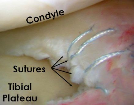
2   Cinque et al                                                                                        The Orthopaedic Journal of Sports Medicine

   Animal models have demonstrated that meniscal tears
heal in a manner similar to other connective tissue.2,5,20
Theoretically, these tears require vascularization to pro-
vide the biological factors necessary for tissue repair.
However, animal studies have also demonstrated that
meniscal tissue may be able to heal without significant
vascular contributions. In this regard, prior studies have
demonstrated that meniscal repair outcomes were compa-
rable in patients with and without a concomitant ligamen-
tous injury, 6,10,11 which further demonstrates that
patients undergoing meniscal repair can achieve good out-
comes (with a concomitant ligamentous injury) and with-
out (no concomitant ligamentous injury) biological
enhancement. Therefore, tear extension into the avascu-
lar zone is not a contraindication for repair, particularly in                     Figure 1. Illustration of a lateral meniscus demonstrating the
the young and active patient, because of the clinical ben-                         classic 3 zones according to the reported vascularity. R-R,
efits of meniscal preservation.11                                                  red-red; R-W, red-white; W-W, white-white.
   The majority of the current literature has examined the
outcomes of meniscal repair in the red-red and/or red-white                           Patients were divided into 3 groups based on the menis-
zones. The purpose of this study was to examine the out-                           cal tear location: red-red, red-white, or white-white zones
comes after inside-out meniscal repair in all 3 meniscal                           (Figure 1). Acute injuries were defined as occurring 6
vascularity zones. Our hypothesis was that there would                             weeks from injury to surgery; chronic injuries were defined
be no difference in outcomes between the different meniscal                        as being >6 weeks from injury to surgery. Patients with
zones repaired.                                                                    degenerative meniscal tears were excluded. The senior
                                                                                   author determined and documented the tear type and loca-
                                                                                   tion at the time of arthroscopic surgery.
METHODS
                                                                                   Surgical Techniques
Study Design
                                                                                   Inside-out meniscal repair with vertical mattress sutures
This study was approved by the Vail Valley Medical Center                          was performed in all cases. Meniscal repair was performed
Institutional Review Board. All data were queried from a                           using a posterolateral or posteromedial approach according
prospectively collected data registry. Patients were                               to a previously reported technique, depending on whether
included if they underwent inside-out meniscal repair by                           the tear was on the lateral or medial meniscus.4 A self-
the senior author (R.F.L.) between 2010 and 2014 and had a                         delivery device fitted with a cannula (Ivy Sports Medicine)
minimum 2-year follow-up. Additionally, patients were                              was used to pass double-loaded nonabsorbable No. 2-0
included if they underwent anterior cruciate ligament                              sutures into the meniscus. To pass the sutures, the knee
reconstruction (ACLR) during the same surgical procedure                           was positioned in 20 of flexion, and the meniscal needle
because previous literature has demonstrated outcomes of                           was advanced approximately 1 cm through the superior or
ligament reconstruction with and without meniscal repair                           inferior aspect of the meniscus; then, the knee was flexed to
to be comparable. Patients were also excluded from this                            70 to 90 while the needle was further advanced to help the
study if they underwent prior ipsilateral or contralateral                         assistant retrieve the needle through the previously made
knee surgery, partial meniscectomy, and meniscal root or                           incision. The same process was repeated adjacent to the
radial tear repair. Patients were also excluded if they pre-                       previous suture with the second needle penetrating
sented with an associated fracture or underwent concomi-                           the joint capsule, such that the sutures were placed on both
tant multiligament knee reconstruction, posterior cruciate                         the superior and the inferior borders of the meniscus
ligament reconstruction (PCLR), cartilage resurfacing pro-                         between 3 to 4 mm apart to create a vertical mattress pat-
cedures, or osteotomy.                                                             tern. If another tear configuration was encountered,

   **Address correspondence to Robert F. LaPrade, MD, PhD, Twin Cities Orthopedics, Edina, MN, USA (email: laprademdphd@gmail.com).
   *Steadman Philippon Research Institute, Vail, Colorado, USA.
    †
       Department of Orthopaedic Surgery, Stanford University Medical Center, Stanford, California, USA.
    ‡
       The Steadman Clinic, Vail, Colorado, USA.
    §
       Oslo University Hospital, University of Oslo, Oslo, Norway.
    ||
       Oslo Sports Trauma Research Center, Norwegian School of Sport Sciences, Oslo, Norway.
    {
       Rush University Medical Center, Chicago, Illinois, USA.
    #
       Twin Cities Orthopedics, Edina, Minnesota, USA.
   One or more of the authors has declared the following potential conflict of interest or source of funding: R.F.L. is a consultant for and receives royalties
from Arthrex, Ossur, and Smith & Nephew. AOSSM checks author disclosures against the Open Payments Database (OPD). AOSSM has not conducted an
independent investigation on the OPD and disclaims any liability or responsibility relating thereto.
   Ethical approval for this study was obtained from the Vail Valley Medical Center Institutional Review Board (protocol 2002-03).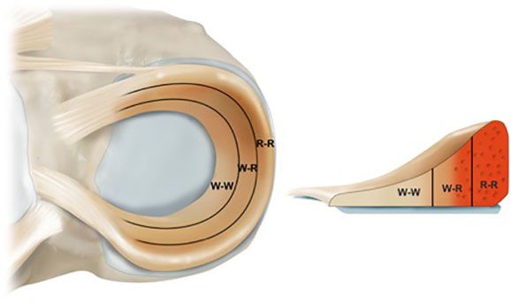
The Orthopaedic Journal of Sports Medicine                                                Meniscal Repair Outcomes by Zone         3

                                                                  Rehabilitation
                                                                  For all patients, physical therapy began within 24 hours after
                                                                  surgery to initiate early range of motion (ROM), muscle reacti-
                                                                  vation, and edema control. For patients who had undergone
                                                                  isolated meniscal repair, all were nonweightbearing for 6 weeks
                                                                  in a knee immobilizer after surgery. ROM outside the immobi-
                                                                  lizer was restricted from 0 to 90 for the first 2 weeks, followed
                                                                  by a progressive increase in knee ROM. Stationary cycling
                                                                  began at week 6, with generalized knee strengthening focused
                                                                  on endurance and progressing to strength and hypertrophy.
                                                                  The time to return to full activities after isolated meniscal
                                                                  repair was from 5 to 6 months postoperatively. For rehabilita-
                                                                  tion after meniscal repair with concomitant ACLR, patients
Figure 2. Arthroscopic image of a right knee demonstrating        were weightbearing as tolerated with the use of crutches for
inside-out meniscal repair in the red-white zone of the           2 weeks. ROM was not restricted, and patients were allowed
meniscus.                                                         to progress in knee ROM as tolerated. Patients returned to
                                                                  full activities around 7 to 9 months after combined meniscal
                                                                  repair and ACLR after passing a functional sports test.12
horizontal mattress sutures were utilized to maintain per-
pendicularity of the tear-suture complex. With the knee
flexed to 90 , all sutures were tied, being cautious to not      Statistical Analysis
overtighten the tissue or entrap nearby soft tissue
structures.                                                       A power analysis was performed a priori. Assuming an
   For medial meniscal repair, a vertical incision was made       alpha of 0.05 and an independent-samples t test, 23
posterior to the medial collateral ligament, and dissection was   patients per group was sufficient to detect an effect size
performed to enter the interval between the medial head of the    of d ¼ 0.85 with 80% statistical power. Data were tested
gastrocnemius and the direct arm of the semimembranosus,          for normal distribution using the Kolmogorov-Smirnov Z
where a retractor was placed. For lateral meniscal repair, an     test. For preoperative versus postoperative comparisons of
incision was made along the inferior aspect of the iliotibial     dependent variables, the paired-samples t test was utilized
band, and the interval between the lateral gastrocnemius ten-     for normally distributed data, and the Wilcoxon signed-
don and the posterolateral capsule, proximal to the biceps        rank test was utilized for nonnormally distributed data.
femoris, was entered, where a retractor was placed (Figure        The pre- and postoperative SF-12 PCS scores for patients
2). All sutures were tied directly over the joint capsule.8 The   in each meniscal tear zone were analyzed with analysis of
ACLR technique used in this study has been previously             variance. The pre- and postoperative Lysholm, Tegner, and
reported and validated.6,7 Grade IV chondral lesions were         WOMAC scores for patients in each meniscal tear zone
treated with chondroplasty if the edges were unstable.            were analyzed using the Kruskal-Wallis test. Comparisons
                                                                  of categorical data, including age, sex, BMI, and tear pat-
                                                                  tern, were performed using the chi-square test and Fisher
Patient-Reported Outcomes, Patient Satisfaction,                  exact test. All P values were 2-tailed, and P values of
4 Cinque et al The Orthopaedic Journal of Sports Medicine
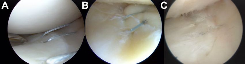
Figure 3. Arthroscopic view of inside-out meniscal repair with vertical mattress sutures performed on the 3 different vascularity
zones: (A) white-white, (B) red-white, and (C) red-red.
while 87 patients (50.3%) underwent combined meniscal TABLE 1
repair and ACLR. There was no significant difference in Patient Demographics and Characteristics
the frequency of concurrent ACLR between the 3 zones of by Meniscal Tear Locationa
meniscal injury (P ¼ .189). The majority of patients (n ¼
108; 62.4%) had an acute injury (6 weeks), while 65 White-
patients (37.6%) had a chronic injury (>6 weeks). There White Red-White Red-Red P
(n ¼ 33) (n ¼ 80) (n ¼ 60) Value
was no correlation between acuity of the tear and its loca-
tion. There were 25 patients (14.5%) with a grade IV chon- Age, mean ± SD, y 37.1 ± 16.4 33.4 ± 13.0 32.0 ± 14.6 .254
dral lesion present on the distal femur (medial or lateral Sex .078
femoral condyle) at the time of meniscal repair. Male 18 (55) 52 (65) 42 (70)
There were no significant differences among the 3 study Female 15 (45) 28 (35) 18 (30)
groups regarding patient sex (P ¼ .078) or BMI (P ¼ .546). Body mass index, 25.4 ± 5.4 24.5 ± 3.8 24.7 ± 3.8 .546
Patient demographics and clinical characteristics are pre- mean ± SD, kg/m2
sented in Table 1. The mean number of sutures used for Tear pattern .039b
repair was 7.7 (range, 2-18) in the red-red zone, 8.9 (range, Vertical 23 (70) 59 (74) 47 (78)
Bucket-handle 2 (6) 17 (21) 13 (22)
2-22) in the red-white zone, and 3.8 (range, 2-8) in the
Horizontal 8 (24) 4 (5) 0 (0)
white-white zone. No. of sutures used, 5.9 (2-10) 8.9 (2-22) 7.7 (2-18) .066
mean (range)
Analysis by Zone and Morphology of Meniscal Tear Chronicity .004b
Acute 14 (42) 48 (60) 46 (77)
Overall, there was a significant difference in the frequency Chronic 19 (58) 32 (40) 14 (23)
of each meniscal tear type (vertical, horizontal, and bucket- Grade IV chondral 9 (27) 13 (16) 3 (5) .011b
handle) in the different zones (P ¼ .039). Specifically, we lesion
observed a significantly greater number of horizontal tears Meniscal tear side .372
Medial 17 (52) 53 (66) 38 (63)
in the white-white zone compared with the red-white and
Lateral 10 (30) 17 (21) 15 (25)
red-red zones (both P < .001). However, there were no sig-
Bicompartmental 6 (18) 10 (13) 7 (12)
nificant differences between the 3 tear zones with respect to
the frequency of bucket-handle or vertical tears (P > .05). A a
Data are shown as n (%) unless otherwise indicated.
b
detailed post hoc analysis of patient-specific outcomes for Statistically significant difference in the rate of each demo-
each meniscal tear zone is provided in Table 2. graphic analyzed (P < .05).
All patients demonstrated significant improvements in
their outcome scores from preoperatively to postoperatively,
regardless of the zone of meniscal injury (P < .001) (Table 3). meniscal injury (Table 4). In both the red-red and red-white
Subjective outcomes after inside-out repair of meniscal tears zones, there was no significant difference in outcomes
in the red-red and red-white zones were significantly between patients who underwent isolated meniscal repair
improved compared with the outcomes of patients with tears and those who underwent concurrent meniscal repair and
in the white-white zone (P < .05). The preoperative to post- ACLR (both P > .05). Given the small sample size of repairs
operative improvement in subjective outcome scores for all 3 in the white-white zone, there was insufficient power to
meniscal tear zones met the minimal clinically important subanalyze these patients.
difference (MCID) for the SF-12 PCS (4.5), 34 WOMAC
(11.5),34 and Lysholm score (10.1).34 Chronicity of Meniscal Repair Analysis
Concurrent ACLR Analysis Across all zones of meniscal injury, there was a signif-
icantly greater number of meniscal repairs performed
A subgroup analysis was performed to evaluate the effect of in the acute phase than the chronic phase (P ¼ .004).
ACLR (n ¼ 87; 50.3%) on postoperative outcomes by zone of There was a significantly greater number of red-redThe Orthopaedic Journal of Sports Medicine Meniscal Repair Outcomes by Zone 5
TABLE 2 TABLE 3
Post Hoc Analysis Comparing Outcomes Preoperative and Postoperative Outcome Scores
by Meniscal Tear Locationa by Meniscal Tear Locationa
P Value Preoperative Postoperative P Value
SF-12 PCS SF-12 PCS
Red-white/white-white .049b White-white 35.1 ± 10.8 46.9 ± 9.66 Cinque et al The Orthopaedic Journal of Sports Medicine
TABLE 4 attempted for all potentially reparable meniscal tears,
Postoperative Outcome Scores by regardless of the zone of the tear or the morphology of the
Meniscal Repair Location, Stratified by the tear. Of note also is that resecting less meniscal tissue with
Presence or Absence of Concurrent ACLRa a tear in the white-white zone has less of a biomechanical
impact on the development of osteoarthritis compared with
With ACLR Without ACLR
tears in the red-red or red-white zone.
(n ¼ 87 (n ¼ 86
The peripheral meniscal vascularity has been reported to
[35 Red-Red, [25 Red-Red,
40 Red-White, 40 Red-White, diminish and become more peripheral with age.29 There-
12 White-White]) 21 White-White]) P Value fore, the healing potential of the meniscus depends largely
on the location of the lesion and the age of the patient.1,18
SF-12 PCS Because of their higher vascularity, peripheral meniscal
Red-red 53.3 (34-62) 50.0 (30-58) .381 tears (red-red zone and peripheral part of red-white zone)
Red-white 52.7 (30-62) 51.5 (35-59) .580
have been reported to have the greatest potential for
White-white 42.3 (30-57) 52.7 (45-58) N/A
WOMAC total
healing,18 and they are typically treated with repair. In
Red-red 6.7 (0-45) 7.8 (0-26) .713 contrast, tears in the white-white zone have often been
Red-white 7.7 (0-47) 7.6 (0-46) .984 treated with debridement and partial meniscectomy, given
White-white 21.8 (6-40) 25.8 (6-54) N/A the lower likelihood of successful healing of repair in avas-
Lysholm cular tissue.13 However, recent studies have demonstrated
Red-red 87.6 (59-100) 88.6 (71-100) .750 that the repair of meniscal tears in the avascular white-
Red-white 84.9 (48-100) 82.7 (60-100) .524 white zone yielded satisfactory outcomes either in the
White-white 70.0 (47-95) 70.0 (58-85) N/A setting of concomitant ACLR21 or a marrow venting pro-
Tegner
cedure.11,22 The addition of marrow venting and tunnel
Red-red 5.9 (2-10) 6.0 (2-9) .483
Red-white 5.6 (2-10) 5.1 (2-9) .355 reaming may increase intra-articular growth factors that
White-white 4.3 (4-5) 4.0 (3-10) N/A enhance meniscal healing.
Satisfaction In the present study, there were low meniscal repair fail-
Red-red 7.3 (5-10) 7.6 (4-10) .573 ure rates, with only 6 patients (3.5%) undergoing a second
Red-white 7.8 (4-10) 7.7 (4-10) .862 surgical procedure for failed meniscal repair. Noyes et al28
White-white 8.0 (7-9) 8.0 (7-9) N/A reported that 62% (18/29) of inside-out meniscal repairs in
a the red-white zone had normal or nearly normal character-
Data are shown as mean (range). ACLR, anterior cruciate lig-
ament reconstruction; N/A, not applicable; SF-12 PCS, 12-Item istics for pain, swelling, jumping, and the Cincinnati score,
Short Form Health Survey Physical Component Summary; while 38% (11/29) were documented as failures at a mean
WOMAC, Western Ontario and McMaster Universities Osteoar- follow-up of 16.8 years. Taken with the findings of the
thritis Index. present study, clinicians can expect improved outcomes
with low revision rates after inside-out meniscal repair for
a range of tear morphologies10,24 and zones.
2 years after inside-out meniscal repair, regardless of the The results of the present study suggest that patient-
vascular zone of the meniscal tear. However, patients who specific factors, including chronicity of the mensical
underwent meniscal repair in the red-red and red-white injury and the presence of grade IV chondral lesions,
zones displayed significantly improved outcomes compared may be of more clinical importance than the meniscal
with patients who underwent meniscal repair in the white- injury location. In a systematic review by Barber-
white zone. Furthermore, patients who underwent acute Westin and Noyes,3 a total of 637 meniscal repairs were
meniscal repair had significantly improved outcome scores evaluated. The range in the healing rates was high; how-
compared with patients who underwent chronic meniscal ever, age, chronicity of injury, involved tibiofemoral com-
repair, regardless of the tear location. Finally, the presence partment, sex, and ACLR were not demonstrated to
of grade IV femoral condyle cartilage lesions at the time of adversely affect the results.
surgery predicted worse outcomes after repair. This study has some limitations. Patients were only
The preoperative to postoperative improvement in sub- available for 2-year clinical follow-up, absent of examina-
jective outcome scores for all 3 meniscal tear zones met the tions by second-look arthroscopic surgery or magnetic res-
MCID for the SF-12 PCS (4.5), WOMAC (11.5), and onance imaging, and no multivariate analysis was
Lysholm score (10.1).34 These findings are supported by performed to identify potential confounding variables.
previous studies that reported significant improvements Patients did not return for follow-up magnetic resonance
in outcome scores, greater than or equal to their respective imaging, which previous studies have shown to be useful
MCID, regardless of tear morphology.10,24 Two recent sys- in the evaluation of meniscal repair healing.9,15,17,32 The
tematic reviews found meniscal repair to yield superior lower number of meniscal repairs in the white-white zone
long-term results with low failure rates (10%).25,26 More- weakens the direct comparison with patients who under-
over, long-term outcomes become more important when went meniscal repair in the red-white and red-red zones.
considering that meniscal tears are often seen in young However, this mimics the clinical scenario, in which most
patients. The current literature, coupled with the findings reparable meniscal tears are in the red-red and red-white
of this study, indicates that inside-out repair should be zones. Furthermore, 50.3% of patients in this studyThe Orthopaedic Journal of Sports Medicine Meniscal Repair Outcomes by Zone 7
TABLE 5
Patients With Failed Inside-Out Meniscal Repaira
Grade IV Chondral Time From Surgery
Patient Meniscal Tear Location Chronicity Lesion Present to Failure, mo Reason for Failure Subsequent Procedure
1 Red-white Chronic No 12 Unknown PMM
2 Red-white Chronic No 29 Reinjury PMM
3 Red-white Acute No 6 Reinjury PMM
4 Red-red Acute No 22 Reinjury MM repair
5 Red-red Chronic No 5 Unknown MM repair
6 Red-red Chronic No 60 Reinjury MM repair
a
All retears were noted in the medial meniscus. Acute: 6 weeks; chronic: 6 weeks. MM, medial meniscus; PMM, partial medial
meniscectomy.
underwent concomitant ACLR; this may influence the gen- comparable results to inside-out repair of vertical meniscus tears.
eralizability of meniscal repair outcomes. Am J Sports Med. 2017;45(10):2253-2259.
11. Dean CS, Chahla J, Matheny LM, Mitchell JJ, LaPrade RF. Outcomes
after biologically augmented isolated meniscal repair with marrow
venting are comparable with those after meniscal repair with concom-
CONCLUSION itant anterior cruciate ligament reconstruction. Am J Sports Med.
2017;45(6):1341-1348.
Patients who underwent inside-out meniscal repair demon- 12. Garrison JC, Shanley E, Thigpen C, Geary R, Osler M, Delgiorno J.
strated significant improvements in subjective outcome The reliability of the Vail Sport Test™ as a measure of physical per-
scores and a low revision rate at a minimum 2-year formance following anterior cruciate ligament reconstruction. Int J
follow-up, regardless of the tear zone. Although worse out- Sports Phys Ther. 2012;7(1):20-30.
13. Grant JA, Wilde J, Miller BS, Bedi A. Comparison of inside-out and all-
comes were observed in the white-white zone relative to the
inside techniques for the repair of isolated meniscal tears: a system-
others, inside-out meniscal repair is recommended for
atic review. Am J Sports Med. 2012;40(2):459-468.
potentially reparable meniscal tears in all 3 vascular zones; 14. Greis PE, Bardana DD, Holmstrom MC, Burks RT. Meniscal injury, I:
however, improved outcomes can be achieved when repair basic science and evaluation. J Am Acad Orthop Surg. 2002;10(3):
is performed acutely and in the absence of full-thickness 168-176.
femoral condyle chondral injuries. 15. Hantes ME, Zachos VC, Zibis AH, et al. Evaluation of meniscal repair
with serial magnetic resonance imaging: a comparative study
between conventional MRI and indirect MR arthrography. Eur J
Radiol. 2004;50(3):231-237.
REFERENCES 16. Hede A, Jensen DB, Blyme P, Sonne-Holm S. Epidemiology of menis-
cal lesions in the knee: 1,215 open operations in Copenhagen 1982-
1. Arnoczky SP, Warren RF. Microvasculature of the human meniscus. 84. Acta Orthop Scand. 1990;61(5):435-437.
Am J Sports Med. 1982;10(2):90-95. 17. Hoffelner T, Resch H, Forstner R, Michael M, Minnich B, Tauber M.
2. Arnoczky SP, Warren RF. The microvasculature of the meniscus and
Arthroscopic all-inside meniscal repair: does the meniscus heal? A
its response to injury: an experimental study in the dog. Am J Sports
clinical and radiological follow-up examination to verify meniscal heal-
Med. 1983;11(3):131-141.
ing using a 3-T MRI. Skeletal Radiol. 2011;40(2):181-187.
3. Barber-Westin SD, Noyes FR. Clinical healing rates of meniscus
18. Johnson D, Weiss B. Meniscal repair using the inside-out suture tech-
repairs of tears in the central-third (red-white) zone. Arthroscopy.
nique. Sports Med Arthrosc. 2012;20(2):68-76.
2014;30(1):134-146.
19. Jones S, Caplan N, St Clair Gibson A, Kader N, Kader D. Interactions
4. Brophy RH, Wright RW, David TS, et al. Association between previous
between severity and location of chondral lesions and meniscal tears
meniscal surgery and the incidence of chondral lesions at revision
found at arthroscopy. Knee Surg Sports Traumatol Arthrosc. 2011;
anterior cruciate ligament reconstruction. Am J Sports Med. 2012;
40(4):808-814. 19(10):1699-1703.
5. Cassidy RE, Shaffer AJ. Repair of peripheral meniscus tears: a pre- 20. King D. The healing of semilunar cartilages. J Bone Joint Surg Am.
liminary report. Am J Sports Med. 1981;9(4):209-214. 1936;18(2):333-342.
6. Chahla J, Dean CS, Matheny LM, Mitchell JJ, Cinque ME, LaPrade 21. LaPrade CM, Dornan GJ, Granan LP, LaPrade RF, Engebretsen L.
RF. Outcomes of inside-out meniscal repair in the setting of multi- Outcomes after anterior cruciate ligament reconstruction using the
ligament reconstruction in the knee. Am J Sports Med. 2017;45(9): Norwegian knee ligament registry of 4691 patients: how does menis-
2098-2104. cal repair or resection affect short-term outcomes? Am J Sports Med.
7. Chahla J, Nitri M, Civitarese D, Dean CS, Moulton SG, LaPrade RF. 2015;43(7):1591-1597.
Anatomic double-bundle posterior cruciate ligament reconstruction. 22. Longo UG, Campi S, Romeo G, Spiezia F, Maffulli N, Denaro V.
Arthrosc Tech. 2016;5(1):e149-e156. Biological strategies to enhance healing of the avascular area of the
8. Chahla J, Serra Cruz R, Cram TR, Dean CS, LaPrade RF. Inside-out meniscus. Stem Cells Int. 2012;2012:528359.
meniscal repair: medial and lateral approach. Arthrosc Tech. 2016; 23. Lutz C, Sonnery-Cottet B, Niglis L, Freychet B, Clavert P, Imbert P.
5(1):e163-e168. Behavior of the anterolateral structures of the knee during internal
9. Choi NH, Kim TH, Victoroff BN. Comparison of arthroscopic medial rotation. Orthop Traumatol Surg Res. 2015;101(5):523-528.
meniscal suture repair techniques: inside-out versus all-inside repair. 24. Moatshe G, Cinque ME, Godin JA, Vap AR, Chahla J, LaPrade RF.
Am J Sports Med. 2009;37(11):2144-2150. Comparable outcomes after bucket-handle meniscal repair and ver-
10. Cinque ME, Geeslin AG, Chahla J, Dornan GJ, LaPrade RF. Two- tical meniscal repair can be achieved at a minimum 2 years’ follow-up.
tunnel transtibial repair of radial meniscus tears produces Am J Sports Med. 2017;45(13):3104-3110.8 Cinque et al The Orthopaedic Journal of Sports Medicine
25. Moulton SG, Geeslin AG, LaPrade RF. A systematic review of the 30. Rubman MH, Noyes FR, Barber-Westin SD. Arthroscopic repair of
outcomes of posterolateral corner knee injuries, part 2: surgical treat- meniscal tears that extend into the avascular zone: a review of 198
ment of chronic injuries. Am J Sports Med. 2016;44(6):1616-1623. single and complex tears. Am J Sports Med. 1998;26(1):87-95.
26. Nepple JJ, Dunn WR, Wright RW. Meniscal repair outcomes at greater 31. Shrive NG, O’Connor JJ, Goodfellow JW. Load-bearing in the knee
than five years: a systematic literature review and meta-analysis. joint. Clin Orthop Relat Res. 1978;131:279-287.
J Bone Joint Surg Am. 2012;94(24):2222-2227. 32. Tachibana Y, Sakaguchi K, Goto T, Oda H, Yamazaki K, Iida S.
27. Noyes FR, Barber-Westin SD. Arthroscopic repair of meniscal tears Repair integrity evaluated by second-look arthroscopy after
extending into the avascular zone in patients younger than twenty arthroscopic meniscal repair with the FasT-Fix during anterior
years of age. Am J Sports Med. 2002;30(4):589-600. cruciate ligament reconstruction. Am J Sports Med. 2010;38(5):
28. Noyes FR, Chen RC, Barber-Westin SD, Potter HG. Greater than 965-971.
10-year results of red-white longitudinal meniscal repairs in patients 33. Walker PS, Erkman MJ. The role of the menisci in force transmission
20 years of age or younger. Am J Sports Med. 2011;39(5):1008-1017. across the knee. Clin Orthop Relat Res. 1975;109:184-192.
29. Petersen W, Tillmann B. Age-related blood and lymph supply of the knee 34. Wright RW. Knee injury outcomes measures. J Am Acad Orthop Surg.
menisci: a cadaver study. Acta Orthop Scand. 1995;66(4):308-312. 2009;17(1):31-39.You can also read