Clinical Evaluations of Intraoperative Injection of Platelet-Rich Plasma in Arthroscopic Single-Row Rotator Cuff Repair at 2- Year Follow-Up ...
←
→
Page content transcription
If your browser does not render page correctly, please read the page content below
Hindawi
BioMed Research International
Volume 2021, Article ID 6675097, 9 pages
https://doi.org/10.1155/2021/6675097

Research Article
Clinical Evaluations of Intraoperative Injection of Platelet-Rich
Plasma in Arthroscopic Single-Row Rotator Cuff Repair at 2-
Year Follow-Up

          Ming Li ,1 Kan Wang,2 Haojun Zhang,1 Chaohua Fang,1 Hua Liu,1 and Yunfeng Zhang1
          1
              Department of Joint Surgery, Ningbo No. 6 Hospital, Ningbo, Zhejiang 315000, China
          2
              Emergency Orthopedics, Ningbo No. 6 Hospital, Ningbo, Zhejiang 315000, China

          Correspondence should be addressed to Ming Li; lmjoint2019@163.com

          Received 25 October 2020; Revised 14 February 2021; Accepted 10 March 2021; Published 17 March 2021

          Academic Editor: Francesco Inchingolo

          Copyright © 2021 Ming Li et al. This is an open access article distributed under the Creative Commons Attribution License, which
          permits unrestricted use, distribution, and reproduction in any medium, provided the original work is properly cited.

          Background. The clinical evidence is conflicted on whether platelet-rich plasma (PRP) therapies have a positive effect on tendon
          healing and improved functional outcomes. Purpose. To evaluate the potentials of intraoperative injection PRP on the speed and
          quality of healing in patients undergoing arthroscopic repair for small to medium rotator cuff tears. Methods. A total of 86
          patients scheduled for arthroscopic single-row repair of small to medium rotator cuff tears were assigned to undergo either PRP
          injection (PRP group) or conventional repair (control group). The PRP group (N = 43) consisted of patients who received an
          intraoperative injection of liquid PRP. The control group (N = 43) consisted of patients who did not receive that treatment. The
          visual analogue scale (VAS) for pain before treatment and at 1, 14 days, 3, 6, and 24 months after surgery were recorded. The
          clinical outcomes were assessed by the University of California, Los Angeles (UCLA) and Constant scores before treatment and
          at 3, 6, and 24 months after surgery and magnetic resonance imaging or ultrasound examination at 24 months. Patient
          satisfaction and retear rate were also assessed. Results. No statistical differences in baseline characteristics such as age, gender,
          dominant arm, and tear size were observed between the two groups (P > 0:05). For the PRP group, the mean operation time was
          40.22 minutes, and for the control group, the mean operation time was 36.3 minutes. There was a statistically significant
          difference (P = 0:036). After surgery, all VAS measurements significantly decreased over time until final follow-up in both
          groups. No significant difference between the 2 groups was found for any VAS pain measurement at any time point except for
          the VAS at 1 day postoperatively, which was significantly lower in the PRP group (2:39 ± 1:03) than that in the control group
          (3:21 ± 1:85) (P = 0:014). Analysis of the PRP and control groups demonstrated a statistically significant improvement in UCLA
          and Constant scores from baseline to the 3-, 6-, and 24-month follow-up assessments (P < 0:05). However, no significant
          intergroup differences were observed in the clinical scores between the three follow-up time points (P > 0:05). At the 24-month
          follow-up, patient satisfaction rates reached 95.65% and 93.48% for the PRP and control groups, respectively. The retear rate of
          the PRP group (2/43, 4.65%) was lower than that of the control group (6/43, 13.95%). Conclusions. Although the pain at 1 day
          after surgery and the retear rate in the PRP group were significantly lower than those in the control group, the liquid PRP
          injection did not promote better clinical outcomes at the 2-year follow-up.

1. Introduction                                                          The patient’s age, activity level, and tear size are all important
                                                                         factors that determine the treatment plan. When conserva-
Rotator cuff tears are the most commonly encountered                      tive treatment fails, surgical repair provides a reliable treat-
shoulder disorder affecting millions of people across all parts           ment alternative [1]. Single-row and double-row fixation
of the globe. They can be degenerative or traumatic. The pur-            techniques under shoulder arthroscopy have been verified
pose of rotator cuff tear treatment is to relieve pain and                in the repair of rotator cuff tears. Due to multiple surgical
restore function. There are many treatments for rotator cuff              techniques to improve bone-to-tendon healing, the results
tears, and the best treatment is different for different patients.         after rotator cuff repair are usually good. However, rerupture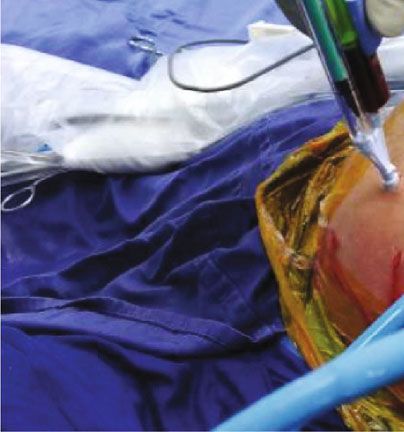
2 BioMed Research International
of the rotator cuff is still a significant postoperative issue and tional arthroscopic rotator cuff repair (control group). All
can be as high as 27% [2]. It is important to explore methods patients with rotator cuff tears in the associated study period
of biological augmentation to reduce the postsurgical rerup- were screened for inclusion. The inclusion criteria were as
ture rate and improve long-term shoulder function after follows: (i) age between 18 and 80 years old, (ii) symptoms
rotator cuff repair. In the past few years, the biomechanical or signs of rotator cuff tears, and (iii) MRI findings
repair of rotator cuff tears has made significant progress, (Figure 1(a)) of minor to medium rotator cuff tears (antero-
which has promoted research on bioassisted rotator cuff posterior size 0 mm and 30 mm). The exclusion criteria
repair. Biological methods are aimed at optimizing tissue included the following: (i) a history of shoulder surgery, a
healing to improve clinical outcomes. chronic dislocation or pyogenic infection, or rotator cuff
During the inflammation and repair phase of tendon arthropathy with glenohumeral osteoarthritis; (ii) a large or
healing, platelets accumulate at the tissue injury site and massive tear (anteroposterior size > 30 mm) during surgery;
release a large number of growth factors (GFs), which pro- (iii) pregnant or lactating women; (iv) rheumatoid arthritis;
mote cell migration and differentiation at the injury site. (v) gout; (vi) blood diseases; (vii) severe cardiovascular dis-
Platelet-rich plasma (PRP) is a fraction of the plasma con- eases; (viii) infections; (ix) immunodepression; (x) patients
taining platelets and GF concentrations above baseline that receiving anticoagulant therapy; and (xi) patients with
can be produced by centrifugal separation of whole blood haemoglobin values < 11 g/dl and platelet values < 150, 000/
[3, 4]. Basic scientific studies have shown the potential benefit mm3 . Patients who did not complete 24 months of follow-
of PRP for tendon healing. In vitro studies have shown that up were also excluded from the study.
the GFs in PRP, including transforming growth factor beta
(TGF-b), fibroblast growth factor (FGF), platelet-derived
2.2. PRP Preparation. For each preparation, a 50 ml blood
growth factor (PDGF), vascular endothelial growth factor
sample was collected from the median elbow vein using a
(VEGF), connective tissue growth factors, and epidermal
50-G needle, such that the ratio of blood to anticoagulant
growth factor (EGF), can influence healing and reduce
reached 9 : 1. PRP was prepared using a separation set (Wei-
inflammation [5].
gao New Polymer Materials Co., Ltd.) and a standard collec-
PRP has been used to successfully treat chronic elbow
tion programme during surgery as previously described [13].
tendinosis and refractory wounds [6–8], and a number of
A total of 4.5 ml of PRP was obtained, of which 3.5 ml was
basic studies have demonstrated the favourable effective-
immediately transferred to a sterile syringe for injection
ness of PRP in rotator cuff repair [9]. However, compara-
(Figure 1(b)), and the remainder was sent to the laboratory
tive clinical studies have reported conflicting results. The
for platelet concentration analysis. The PRP platelet and leu-
results of one systematic review indicated that the use of
kocyte counts were 802:26 ± 171:56 × 109 /l and 33:2 ± 9:56
PRP in rotator cuff repair can improve healing rates, pain
levels, and functional outcomes [10]. A randomized con- × 1012 /l, respectively, which were 6:35 ± 1:08 and 6:21 ±
trolled trial used PRP as an augment to rotator cuff repair 0:97 times greater than those in the peripheral blood, respec-
versus a conventional repair in patients undergoing arthro- tively. All procedures were performed in 30 minutes in the
scopic repair for medium to large rotator cuff tears. They same operating room.
reported that PRP significantly improved the quality, as
evidenced by a decreased retear rate, but not the speed of 2.3. Surgical Procedures. All surgical procedures were per-
healing [11]. However, another meta-analysis described formed by the same surgeon (ML) with patients in the lateral
unfavourable results [12]. decubitus position under general anaesthesia. Systematic
The objective of this study was to evaluate the potentials glenohumeral joint and subacromial exploration was per-
of intraoperative injection PRP on the speed and quality of formed, the rotator cuff tear was carefully evaluated
healing in patients undergoing arthroscopic single-row (Figure 1(c)), and the anteroposterior size and the presence
repair for small to medium rotator cuff tears. The speed of of subacromial impingement were documented. Tenotomy
healing was measured by clinical scores, and the quality of of the biceps tendon was performed in cases with severe ten-
healing was evaluated by retear rate. Our hypothesis was that dinitis, partial tears, subluxations, and complete dislocations.
PRP injection would accelerate the speed of healing and Debridement of bursal tissue and acromioplasty was mini-
improve the quality of healing in this population. mally performed. Rotator cuff repair was performed to cover
the original footprint using a single-row technique whenever
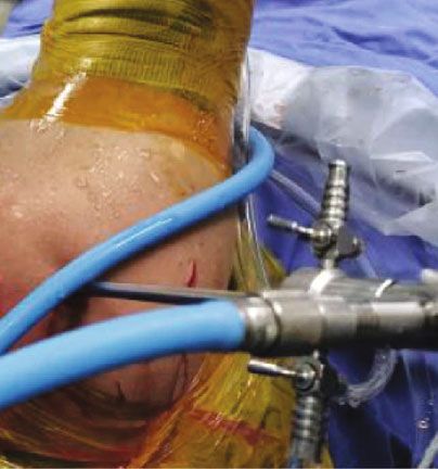
2. Materials and Methods possible (Figure 1(d)). Absorbable anchors (Twinfix; Smith &
Nephew, USA) of 5.5 mm in diameter were used to repair the
2.1. Patients. The present study was a retrospective com- rotator cuff tears.
parative study using conventional treatment as the control. At the end of the arthroscopic procedure of the PRP
This study was carried out in accordance with the ethical group, the portals were sutured except for the posterior por-
standards recognized by the Declaration of Helsinki rules tal, which was left for observation. The posterior portal was
and the principles of Good Clinical Practice guidelines. sutured after the positioning needle was placed at the
Additionally, the study was approved by the Hospital Ethics tendon-to-bone interface through the lateral portal. Then,
Committee prior to study commencement (No. 2015006). the fluid remaining in the subacromial space was aspirated,
Enrolled patients were allocated to undergo either arthro- and 3.5 ml of PRP was injected at the position through the
scopic rotator cuff repair with PRP (PRP group) or conven- needle (Figure 1(e)). The injection site was covered with a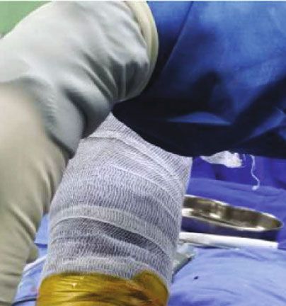
BioMed Research International                                      3

                                (a)                          (b)

                                (c)                          (d)

                                      Figure 1: Continued.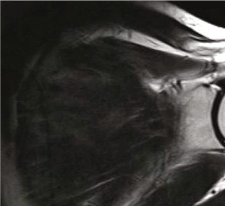
4                                                                                                            BioMed Research International

                                           (e)                                                         (f)

Figure 1: (a) MRI showed a torn rotator cuff. (b) PRP was transferred to a sterile syringe. (c) Arthroscopic exploration of a torn rotator cuff.
(d) Single-row rotator cuff repair. (e) Intra-articular injection of PRP. (f) MRI showed an intact rotator cuff.

sterile dressing, and the assistant was asked to press the por-          using a two-sided hypothesis test at an alpha level of 0.05
tals for two minutes to prevent PRP leakage.                             and a power of 80%, and a total of 42 participants is needed
                                                                         in each group. Taking into account the possibility of 20% vio-
2.4. Clinical Evaluation. Outcome assessments were per-                  lators or dropouts, we will include at least 52 patients in each
formed by a clinician blinded to the treatment. Each patient             group.
was evaluated at a preoperative clinical evaluation, as well                 The data are expressed as the mean ± SD unless other-
as at 3, 6, and 24 months postoperatively. Additionally, pain            wise indicated. Two-way ANOVA was performed to assess
was assessed using the visual analogue scale (VAS) at 1 and              the differences between groups at different follow-up times.
14 days postoperatively. The functional assessment included              Friedman’s test followed by the Wilcoxon signed rank test
the University of California, Los Angeles (UCLA) and the                 with Bonferroni correction was used to evaluate the data at
Constant shoulder scale and pain as measured by the VAS.                 different time points within a single group. Statistical analy-
Patients with a tear size of less than 10 mm were classified              ses were performed using SPSS version 23.0 (IBM Corp.),
as the small tear group, and patients with a tear size between           and P < 0:05 was considered statistically significant.
10 and 30 mm were classified as the medium tear group, so
patients in the PRP or control groups were divided into two              3. Results
subgroups. The clinical outcomes of the patients in the two
subgroups were compared.                                                 Between October 2017 and September 2018, 123 patients
    To evaluate the structural integrity, ultrasound or mag-             with rotator cuff tears received arthroscopic repair in our
netic resonance imaging (MRI) (Achieva 3.0-T; Philips                    department; due to incomplete treatment data, 37 of these
Medical Systems) with a dedicated shoulder coil was per-                 patients were excluded from the present study. In total, 86
formed at a minimum of 24 months after surgery. Criteria                 patients (86 shoulders) met the study criteria (43 patients
for retear were lack of continuity of the tendon in 1 slice of           and 43 shoulders in each group) and completed 24 months
the coronal plane. We only differentiated between retear                  of follow-up examinations.
and intact tendons (Figure 1(f)). All images were inter-
preted by a single radiologist with extensive experience in              3.1. Baseline Data. No differences in baseline characteristics
the interpretation of shoulder ultrasound or MRI. The radi-              of age, gender, dominant arm, tear size, percentage of acro-
ologist was blinded to the treatment group and was not                   mioplasty, and follow-up were observed between the two
involved in the clinical evaluation.                                     groups (Table 1). Tenotomy of the biceps tendon was
                                                                         performed in 5 cases in the PRP group and in 6 cases in the
2.5. Statistical Analysis. Sample size calculation is performed          control group due to severe tendinitis, partial tears, subluxa-
with the VAS score as the outcome measure. Based on the                  tions, and complete dislocations. The number of anchors
previous study, the smallest change score for the VAS score              ranged from 1 to 2, with a mean of 1:78 ± 0:42 in the PRP
to be considered clinically relevant is 2 points (on a 0-10              group and 1:74 ± 0:45 in the control group, with no signifi-
scale) between the PRP group and the control group. Power                cant difference (P = 0:37). For the PRP group, the mean oper-
calculation is performed based on the VAS score difference                ation time was 40.22 minutes, and for the control group, the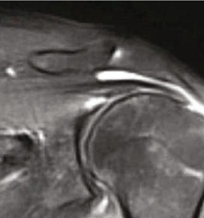
BioMed Research International                                                                                                                                                         5

                                          Table 1: Basal characteristics of patients in the two groups.

Characteristic                                                PRP group                                                           Control group                              P value
No. of patients                                                    43                                                                      43
Age                                                           57:35 ± 6:85                                                            55:70 ± 7:30                             0.21
Percentage of males, n (%)                                    22 (47.83%)                                                             20 (43.48%)
Percentage of right shoulder, n (%)                           30 (65.22%)                                                             28 (60.87%)
Duration (mo)                                                 7:61 ± 6:27                                                             6:78 ± 4:19                              0.30
Size (anteroposterior) (mm)                                   21:30 ± 8:29                                                            23:57 ± 7:45                             0.17
Percentage of acromioplasty, n (%)                            40 (86.96%)                                                             41 (89.13%)
Biceps procedure (tenotomy), n (%)                             5 (11.63%)                                                              6 (13.95%)
Number of anchor, n                                           1:74 ± 0:45                                                             1:78 ± 0:42                              0.37
Operation time (min)                                          40:22 ± 6:65                                                            36:30 ± 7:72                             0.03
Follow-up (mo)                                                24:87 ± 1:22                                                            24:70 ± 1:06                             0.25
MRI follow-up (mo)                                            24:63 ± 1:01                                                            24:14 ± 0:87                             0.30
PRP: platelet-rich plasma; MRI: magnetic resonance imaging.

                                                                                                           8
mean operation time was 36.30 minutes. There was a statisti-
cally significant difference (P = 0:036).                                           Visual analog scale      6

3.2. Clinical Evaluations. No significant difference was found                                               4

in VAS scores between the PRP and control groups at base-                                                  2
line (P = 0:15). After surgery, all VAS measurements signifi-
cantly decreased over time until final follow-up in both                                                    0
groups (Figure 2). No significant difference between the 2                                                              Preop       1-day    2-weeks 3-months 6-months 2-years
groups was found for any VAS pain measurement at any time                                                                 PRP
point except for the VAS at 1 day postoperatively, which was                                                              Control
significantly lower in the PRP group (2:39 ± 1:03) than that
in the control group (3:21 ± 1:85) (P = 0:014).                              Figure 2: Pain level assessment: visual analogue scale score. PRP:
    No significant difference was found in the UCLA and                        platelet-rich plasma.
Constant scores between the PRP and control groups at
baseline (P > 0:05). Preliminary analysis of the PRP and
control groups demonstrated a statistically significant                                                               40
                                                                                                                                                                   NS
improvement in UCLA and Constant scores from baseline                                                                                                   NS
to the 3-, 6-, and 24-month follow-up assessments (P <                                                               30                      NS
0:05). However, no significant intergroup differences were
                                                                                                        UCLA score

observed in the clinical scores between the three follow-up
                                                                                                                     20
time points (P > 0:05). For example, in the PRP group,                                                                        NS
the UCLA score increased from 10:52 ± 4:99 at the baseline
evaluation to 24 ± 3:50 at 3 months, 29:3 ± 2:73 at 6                                                                10
months, and 32:13 ± 1:79 at 24 months. In the control
group, the UCLA score increased from 11:83 ± 4:33 at the                                                              0
baseline evaluation to 23:52 ± 3:28 at 3 months, 28 ± 2:97
                                                                                                                              Preop

                                                                                                                                             3-months

                                                                                                                                                        6-months

                                                                                                                                                                   2-years

at 6 months, and 32:08 ± 2:02 at 24 months (Figure 3).
Similar results were documented for the Constant scores
(Figure 4). There were 19 patients in the small tear sub-
                                                                                                                              PRP
group: 10 patients in the PRP group and 9 patients in the
                                                                                                                              Control
control group. There were 67 patients in the medium tear
subgroup: 33 patients in the PRP group and 34 patients                       Figure 3: UCLA score in the PRP and control groups. NS (not
in the control group. There was no significant difference                      significant) indicated a P > 0:05 for the pairwise comparison
in functional scores between the subgroups at baseline                       between the two groups. UCLA: University of California, Los
and at the 3-, 6-, 12-, and 24-month follow-up postopera-                    Angeles.
tively (Table 2).
    Complications such as infection, haematoma, or other
major adverse events were not observed in either group. At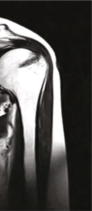
6                                                                                                     BioMed Research International

                        100                                   NS        was no significant difference in the overall gain of outcome
                                                   NS                   scores or retears, but they noticed that when PRP was applied
                         80             NS                              to the tendon-bone interface and PRP was applied to the top
       Constant score

                                                                        of the repaired tendon, the shoulder Constant score increased
                         60                                             significantly [12]. Hurley et al. [10] performed a systematic
                              NS
                                                                        review of 1147 patients in the literature to ascertain whether
                         40
                                                                        PRP improved patient outcomes in arthroscopic rotator cuff
                         20                                             repair. PRP resulted in significantly decreased rates of incom-
                                                                        plete tendon healing for all tears combined, incomplete ten-
                          0                                             don healing in small to medium tears, and incomplete
                              Preop

                                        3-months

                                                   6-months

                                                              2-years
                                                                        tendon healing in medium to large tears compared to the
                                                                        control. They concluded that the use of PRP in rotator cuff
                                                                        repair results in improved healing rates, pain levels, and func-
                              PRP
                                                                        tional outcomes [10]. However, studies have also reported
                                                                        the negative aspects of rotator cuff repair. Malavolta et al.
                              Control
                                                                        [16] published a prospective randomized study of 54 patients
Figure 4: Constant score in the PRP and control groups. NS (not         who underwent arthroscopic single-row repair of small to
significant) indicated a P > 0:05 for the pairwise comparison            medium supraspinatus tears. The clinical evaluations were
between the two groups.                                                 conducted using the UCLA and Constant scales and the
                                                                        VAS for pain at 6, 12, 24, and 60 months after surgery and
                                                                        MRI at 12 and 60 months. Statistical analysis revealed that
the 24-month follow-up, patient satisfaction rates reached              PRP did not promote better clinical or structural results at
95.65% and 93.48% for the PRP and control groups, respec-               the 60-month follow-up [16]. Our study did not demonstrate
tively, indicating no significant difference between the two              any difference between groups at any of the evaluation times
treatment options. At 24 months, 16 patients underwent                  in relation to the clinical scales, similar to the findings
MRI, and 70 patients were examined by ultrasound. The con-              described by Malavolta et al. One meta-analysis including
trol group exhibited 6 partial-thickness retears, while the             seven randomized controlled studies compared rotator cuff
PRP group had 2 partial-thickness retears. The retear rate              repair with and without PRP and suggested that PRP use at
of the PRP group (2/43, 4.65%) was lower than that of the               the time of arthroscopic rotator cuff repair does not univer-
control group (6/43, 13.95%).                                           sally improve retear rates or affect clinical outcome scores
                                                                        [17]. In addition, Moraes et al. [18] collected 19 studies,
4. Discussion                                                           and a total of 1088 participants used PRP, including not only
                                                                        the rotator cuff but also 5 other tendinopathies. They found
The most important findings of the study are that intraoper-             no significant improvement in functional outcomes and
ative injection PRP in patients undergoing arthroscopic                 insufficient evidence to support the use of PRP in clinical
single-row repair for small to medium rotator cuff tears did             practice [18]. Although the existing research results were
not accelerate the speed of healing but improved the quality            conflicting, the potential of PRP to promote rotator cuff
of healing. Previous studies have demonstrated the positive             repair is worthy of further study. We found that some com-
effects of PRP on rotator cuff repair. A randomized con-                  mon limitations in the above studies may affect the results
trolled trial is aimed at assessing the efficacy of PRP augmen-           and conclusions, such as the lack of standardization in the
tation on the speed and quality of healing in patients                  operative technique, inconsistent use of double-row repair
undergoing arthroscopic repair for medium to large rotator              and single-row repair among studies, and a combination of
cuff tears. Compared with repairs without PRP augmenta-                  all tear sizes.
tion, the PRP preparation and application methods signifi-                    PRP can be used as a liquid, gel, or matrix scaffold. The
cantly improved the quality of healing [11]. Randelli et al.            conventional and most commonly used method is the addi-
[14] reported a prospective randomized controlled trial in              tion of calcium and thrombin to obtain PRP gel or matrix
which 26 patients received an intraoperative application of             scaffolds [19–22]. Although the gel may produce a longer-
PRP in combination with an autologous thrombin compo-                   lasting release effect, the fixation of the gel is not a simple
nent. The results of the study showed that autologous PRP               procedure. The liquid form of PRP can also be activated by
reduced pain in the first postoperative months. These results            endogenous methods, such as by contacting type I collagen
are different from the results of our study. We found that the           in the rotator cuff tendon to act as an activator. The advan-
pain scores of patients in the PRP group were significantly              tage of liquid PRP injection is that the liquid form can be
lower than those in the control group at 1 day after surgery,           applied directly to the tendon-bone interface after the suba-
which may be related to the potential role of PRP. Studies              cromial fluid is evacuated. Another reason we chose liquid
have concluded that the PRP effect is likely to last the first            PRP instead of gel PRP in our study is that the injection is
24 postoperative hours but no longer than 48 hours [15].                relatively simple and time-saving.
Warth et al. [12] conducted a systematic review of all level I               PRP can be applied intraoperatively or postoperatively.
and level II studies, comparing the clinical and structural             Although it is not clear which method has the best effect on
results of rotator cuff repair with or without PRP. There                tendon-bone healing, most studies chose intraoperative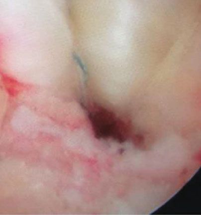
BioMed Research International                                                                                                                     7

Table 2: The Clinical outcomes in the small and medium tear subgroups before surgery and at 3-, 6-, 12-, and 24-month follow-up postop.

                                                   Preop                         3 months                          6 months          24 months
VAS of PRP group
   Small tear subgroup                           4:8 ± 1:78                      1:2 ± 0:44                        0:6 ± 0:54        0:2 ± 0:44
   Medium tear subgroup                         4:17 ± 1:72                     1:47 ± 0:51                    0:72 ± 0:45           0:44 ± 0:51
 P value                                            0.51                            0.34                             0.57               0.34
VAS of control group
   Small tear subgroup                           4:4 ± 2:3                        1 ± 0:71                         0:4 ± 0:54        0:2 ± 0:44
   Medium tear subgroup                         6:53 ± 1:58                     1:28 ± 0:46                    0:55 ± 0:51           0:39 ± 0:50
  P value                                           0.54                            0.29                             0.55               0.46
ULCA of PRP group
   Small tear subgroup                          13:2 ± 7:79                     24:8 ± 4:81                         30 ± 4           32:6 ± 2:30
   Medium tear subgroup                         9:77 ± 3:91                    23:78 ± 3:19                    29:11 ± 2:39           32 ± 3:24
  P value                                           0.18                            0.57                             0.53               0.52
ULCA of control group
   Small tear subgroup                           11 ± 7:58                      25:4 ± 3:21                    28:8 ± 2:92           32:6 ± 2:30
   Medium tear subgroup                        12:05 ± 3:24                      23 ± 3:19                     27:78 ± 2:92         31:94 ± 1:98
  P value                                           0.64                            0.15                             0.51               0.53
Constant of PRP group
   Small tear subgroup                         42:8 ± 24:96                    62:6 ± 10:89                        81 ± 2:23         93:6 ± 3:51
   Medium tear subgroup                        58:89 ± 16:28                   62:22 ± 8:01                    78:22 ± 5:56         91:44 ± 2:77
  P value                                           0.16                            0.93                             0.29               0.16
Constant of control group
   Small tear subgroup                         33:2 ± 19:43                      70 ± 9:06                     79:2 ± 7:12           92:8 ± 2:58
   Medium tear subgroup                        38:06 ± 10:61                   62:17 ± 8:59                    83:43 ± 0:96         90:88 ± 2:24
   P value                                          0.46                           0.089                             0.95               0.12
VAS: visual analog scale score for pain; PRP: platelet-rich plasma; UCLA: University of California, Los Angeles.

injection. Wang et al. [23] studied the effect of PRP injection                   et al. [26] found that retear rates (56.2% vs. 38.1%) were sig-
after rotator cuff repair and found that it did not improve                       nificantly higher in the platelet-rich fibrin matrix group than
tendon-bone healing or functional recovery. Moraes et al.                        the controls. The retear rate of the PRP group in our study
[18] found that PRP injection under arthroscopy did not                          (2/43, 4.65%) was lower than that of the control group
affect the retear rate or affect the functional outcome. How-                      (6/43, 13.95%). It seems that our results showed better retear
ever, when it is applied to the tendon-bone interface,                           rates, which may be related to the fact that the patients we
double-row repair, and small and medium-sized rotator cuff                        enrolled all had small and medium tears, and other studies
tears, there is a tendency to reduce the retear rate [20]. In                    also included large and massive tears.
addition, in a study by Randelli et al. [14], compared with                          There was no indication that the use of PRP in our study
the control group, the early functional results of intraopera-                   was associated with the occurrence of more complications
tive PRP treatment of rotator cuff repair were significantly                       within 24 months compared with the non-PRP group. This
improved.                                                                        is consistent with previous data. Clinical reports recording
    Our study showed that the clinical outcomes of patients                      the occurrence of adverse events have shown that the PRP
were not significantly different between the small tear group                      group does not have an increased incidence of adverse events
and the medium tear group at the 3-, 6-, 12-, and 24-month                       compared with the control group [27, 28].
follow-up postoperatively. However, some data support                                There are a number of limitations in the present study,
PRP use in some patients. Two meta-analyses focusing on                          including the nonrandomized double-blind design, the small
the potential of PRP application showed improvement in                           sample size, and a lack of direct evidence of rotator cuff heal-
tendon healing of small and medium-sized tears but not large                     ing (such as arthroscopic findings). The results of our study
tears [24, 25]. The retear rate of patients who undergo treat-                   indicated that intra-articular injection of liquid PRP for small
ment for small and medium rotator cuffs may be reduced. In                        to medium rotator cuff repair did not accelerate the speed of
a meta-analysis of 5 studies of 300 patients, Cai et al. [24]                    healing but improved the quality of healing compared with
found significant differences in repair failures of small to                       repairs without PRP application. In addition, the method of
medium rotator cuffs when PRP was not used. Bergeson                              PRP injection neither increased the operation time nor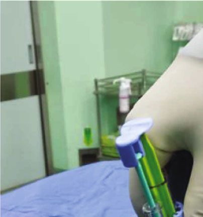
8                                                                                                           BioMed Research International

increased the occurrence of adverse events such as infection.                   parative study,” Journal of Orthopaedics, vol. 13, no. 1,
Previous literature has shown that PRP presents a wide                          pp. 10–14, 2016.
variation in the different preparation protocols and dosages,              [8]   D. Rainys, G. Samulėnas, M. Kievišas, E. Samulėnienė,
activation methods, white blood cell concentrations, and                        L. Pilipaitytė, and R. Rimdeika, “Platelet biology and the ratio-
concentrations of platelets and GFs. These factors may bias                     nale of PRP therapy in chronic wounds,” European Journal of
research results. Further studies may be needed to investigate                  Plastic Surgery, vol. 40, no. 2, pp. 87–96, 2017.
the effects of different characteristics of PRP on the speed and            [9]   N. Baksh, C. P. Hannon, C. D. Murawski, N. A. Smyth, and
quality of rotator cuff repair healing.                                          J. G. Kennedy, “Platelet-rich plasma in tendon models: a
                                                                                systematic review of basic science literature,” Arthroscopy,
                                                                                vol. 29, no. 3, pp. 596–607, 2013.
Data Availability                                                        [10]   E. T. Hurley, D. L. Fat, C. J. Moran, and H. Mullett, “The effi-
                                                                                cacy of platelet-rich plasma and platelet-rich fibrin in arthro-
The datasets used and/or analysed during the present study                      scopic rotator cuff repair: a meta-analysis of randomized
are available from the corresponding author on reasonable                       controlled trials,” The American Journal of Sports Medicine,
request.                                                                        vol. 47, no. 3, pp. 753–761, 2019.
                                                                         [11]   C. H. Jo, J. S. Shin, W. H. Shin, S. Y. Lee, K. S. Yoon, and
Conflicts of Interest                                                           S. Shin, “Platelet-rich plasma for arthroscopic repair of
                                                                                medium to large rotator cuff tears: a randomized controlled
The authors declare that they have no competing interests.                      trial,” The American Journal of Sports Medicine, vol. 43,
                                                                                no. 9, pp. 2102–2110, 2015.
                                                                         [12]   R. J. Warth, G. J. Dornan, E. W. James, M. P. Horan, and P. J.
Acknowledgments                                                                 Millett, “Clinical and structural outcomes after arthroscopic
                                                                                repair of full-thickness rotator cuff tears with and without
The present study was supported by grants from the Ningbo                       platelet-rich product supplementation: a meta-analysis and
Medical Science and Technology Project (grant no. 2020Y50)                      meta-regression,” Arthroscopy, vol. 31, no. 2, pp. 306–320,
and Zhejiang Provincial Key Laboratory of Pathophysiology                       2015.
(grant no. 201911).                                                      [13]   M. Li, C. Q. Zhang, T. Yuan, S. B. Chen, and R. J. Lv, “Evalu-
                                                                                ation of platelet rich plasma preparation kit (in Chinese),”
References                                                                      Chin J Repair Reconstr Surg, vol. 1, pp. 112–116, 2011.
                                                                         [14]   P. Randelli, P. Arrigoni, V. Ragone, A. Aliprandi, and
 [1] C. C. Schmidt, C. D. Jarrett, and B. T. Brown, “Management of              P. Cabitza, “Platelet rich plasma in arthroscopic rotator cuff
     rotator cuff tears,” The Journal of Hand Surgery, vol. 40, no. 2,           repair: a prospective RCT study, 2-year follow-up,” Journal
     pp. 399–408, 2015.                                                         of Shoulder and Elbow Surgery, vol. 20, no. 4, pp. 518–528,
 [2] M. D. McElvany, E. McGoldrick, A. O. Gee, M. B. Neradilek,                 2011.
     and F. A. Matsen, “Rotator cuff repair: published evidence on        [15]   C. Fontana, A. Di Donato, G. Di Giacomo et al., “Postoperative
     factors associated with repair integrity and clinical outcome,”            analgesia for arthroscopic shoulder surgery: a prospective ran-
     The American Journal of Sports Medicine, vol. 43, no. 2,                   domized controlled study of intraarticular, subacromial injec-
     pp. 491–500, 2014.                                                         tion, interscalenic brachial plexus block and intraarticular plus
 [3] H. Masuki, T. Okudera, T. Watanebe et al., “Growth factor and              subacromial injection efficacy,” European Journal of Anaesthe-
     pro-inflammatory cytokine contents in platelet-rich plasma                  siology, vol. 26, no. 8, pp. 689–693, 2009.
     (PRP), plasma rich in growth factors (PRGF), advanced               [16]   E. A. Malavolta, M. E. C. Gracitelli, J. H. Assunção, A. A. F.
     platelet-rich fibrin (A-PRF), and concentrated growth factors               Neto, M. Bordalo-Rodrigues, and O. P. de Camargo, “Clinical
     (CGF),” Int J Implant Dent., vol. 2, no. 1, p. 19, 2016.                   and structural evaluations of rotator cuff repair with and with-
 [4] F. Mussano, T. Genova, L. Munaron, S. Petrillo, F. Erovigni,               out added platelet-rich plasma at 5-year follow-up: a prospec-
     and S. Carossa, “Cytokine, chemokine, and growth factor pro-               tive randomized study,” The American Journal of Sports
     file of platelet-rich plasma,” Platelets, vol. 27, no. 5, pp. 467–          Medicine, vol. 46, no. 13, pp. 3134–3141, 2018.
     471, 2016.                                                          [17]   B. M. Saltzman, A. Jain, K. A. Campbell et al., “Does the use of
 [5] K. Yamaguchi, K. Ditsios, W. D. Middleton, C. F. Hildebolt,                platelet-rich plasma at the time of surgery improve clinical
     L. M. Galatz, and S. A. Teefey, “The demographic and mor-                  outcomes in arthroscopic rotator cuff repair when compared
     phological features of rotator cuff disease. A comparison of                with control cohorts? A systematic review of meta-analyses,”
     asymptomatic and symptomatic shoulders,” The Journal of                    Arthroscopy, vol. 32, no. 5, pp. 906–918, 2016.
     Bone and Joint Surgery. American Volume, vol. 88, no. 8,            [18]   V. Y. Moraes, M. Lenza, M. J. Tamaoki, F. Faloppa, and J. C.
     pp. 1699–1704, 2006.                                                       Belloti, “Platelet-rich therapies for musculoskeletal soft tissue
 [6] M. Alessio-Mazzola, I. Repetto, B. Biti, R. Trentini,                      injuries,” Cochrane Database of Systematic Reviews, vol. 4,
     M. Formica, and L. Felli, “Autologous US-guided PRP injec-                 2013.
     tion versus US-guided focal extracorporeal shock wave therapy       [19]   W. Wu, F. Chen, Y. Liu, Q. Ma, and T. Mao, “Autologous
     for chronic lateral epicondylitis: a minimum of 2-year follow-             injectable tissue-engineered cartilage by using platelet-rich
     up retrospective comparative study,” Journal of Orthopaedic                plasma: experimental study in a rabbit model,” Journal of Oral
     Surgery, vol. 26, no. 1, p. 230949901774998, 2018.                         and Maxillofacial Surgery, vol. 65, no. 10, pp. 1951–1957, 2007.
 [7] M. Karaduman, M. C. Okkaoglu, H. Sesen, A. Taskesen,                [20]   M. Saito, K. A. Takahashi, Y. Arai et al., “Intraarticular admin-
     M. Ozdemir, and M. Altay, “Platelet-rich plasma versus open                istration of platelet-rich plasma with biodegradable gelatin
     surgical release in chronic tennis elbow: a retrospective com-             hydrogel microspheres prevents osteoarthritis progression in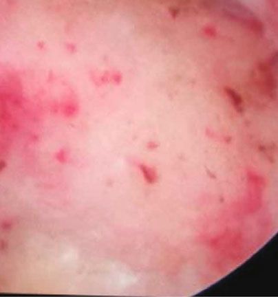
BioMed Research International                                               9

       the rabbit knee,” Clinical and Experimental Rheumatology,
       vol. 27, no. 2, pp. 201–207, 2009.
[21]   Y. Sun, Y. Feng, C. Q. Zhang, S. B. Chen, and X. G. Cheng,
       “The regenerative effect of platelet-rich plasma on healing in
       large osteochondral defects,” International Orthopaedics,
       vol. 34, no. 4, pp. 589–597, 2010.
[22]   M. Sánchez, E. Anitua, J. Azofra, J. J. Aguirre, and I. Andia,
       “Intra-articular injection of an autologous preparation rich in
       growth factors for the treatment of knee OA: a retrospective
       cohort study,” Clinical and Experimental Rheumatology,
       vol. 26, no. 5, pp. 910–913, 2008.
[23]   A. Wang, P. McCann, J. Colliver et al., “Do postoperative
       platelet-rich plasma injections accelerate early tendon healing
       and functional recovery after arthroscopic supraspinatus
       repair? A randomized controlled trial,” The American Journal
       of Sports Medicine, vol. 43, no. 6, pp. 1430–1437, 2015.
[24]   Y. Z. Cai, C. Zhang, and X. J. Lin, “Efficacy of platelet-rich
       plasma in arthroscopic repair of full-thickness rotator cuff
       tears: a meta-analysis,” Journal of Shoulder and Elbow Surgery,
       vol. 24, no. 12, pp. 1852–1859, 2015.
[25]   J. Chahal, G. S. Van Thiel, N. Mall et al., “The role of platelet-
       rich plasma in arthroscopic rotator cuff repair: a systematic
       review with quantitative synthesis,” Arthroscopy, vol. 28,
       no. 11, pp. 1718–1727, 2012.
[26]   A. G. Bergeson, R. Z. Tashjian, P. E. Greis, J. Crim, G. J. Stod-
       dard, and R. T. Burks, “Effects of platelet-rich fibrin matrix on
       repair integrity of at-risk rotator cuff tears,” The American
       Journal of Sports Medicine, vol. 40, no. 2, pp. 286–293, 2012.
[27]   C. H. Jo, J. E. Kim, K. S. Yoon, and S. Shin, “Platelet-rich
       plasma stimulates cell proliferation and enhances matrix gene
       expression and synthesis in tenocytes from human rotator cuff
       tendons with degenerative tears,” The American Journal of
       Sports Medicine, vol. 40, no. 5, pp. 1035–1045, 2012.
[28]   C. Charousset, A. Zaoui, L. Bellaiche, and B. Bouyer, “Are mul-
       tiple platelet-rich plasma injections useful for treatment of
       chronic patellar tendinopathy in athletes? A prospective
       study,” The American Journal of Sports Medicine, vol. 42,
       no. 4, pp. 906–911, 2014.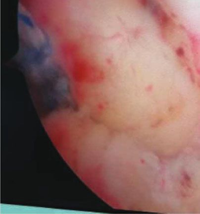
You can also read