Heterogeneity of non-cystic-fibrosis bronchiectasis in multiethnic Singapore: A prospective cohort study at a tertiary pulmonology centre ...
←
→
Page content transcription
If your browser does not render page correctly, please read the page content below
Ann Acad Med Singap 2021;50:556-65
ORIGINAL ARTICLE                                                                                 https://doi.org/10.47102/annals-acadmedsg.202178

Heterogeneity of non-cystic-fibrosis bronchiectasis in multiethnic Singapore:
A prospective cohort study at a tertiary pulmonology centre
Si Ling Young 1MRCP, Youxin Puan 1MRCP, Si Yuan Chew 1MRCP, Haja Mohideen Salahudeen Mohamed 2FRCR,
Pei Yee Tiew 1MRCP, Gan Liang Tan 3MRCP, Mariko Siyue Koh 1MRCP, Ken Cheah Hooi Lee 1MRCP

                 ABSTRACT
                 Introduction: Non-cystic fibrosis bronchiectasis (NCFB) is a highly heterogenous disease. We describe
                 the clinical characteristics of NCFB patients and evaluate the performance of Bronchiectasis Severity Index
                 (BSI) in predicting mortality.
                 Methods: Patients attending the bronchiectasis clinic between August 2015 and April 2020 with
                 radiologically proven bronchiectasis on computed tomography were recruited. Clinical characteristics,
                 spirometry, radiology, microbiology and clinical course over a median period of 2.4 years is presented.
                 Results: A total of 168 patients were enrolled in this prospective cohort study. They were predominantly
                 women (67.8%), Chinese (87.5%) and never-smokers (76.9%). Median age of diagnosis was 64 years
                 (interquartile range 56–71) and the most common aetiology was “idiopathic” bronchiectasis (44.6%).
                 Thirty-nine percent had normal spirometries. Compared to female patients, there were more smokers
                 among the male patients (53.8% versus 8.5%, P9) compared to
                 those with mild or moderate disease (BSI9) compared to
                 mild disease (BSI 0–4) was 14.8 (confidence interval 1.929–114.235, P=0.01).
                 Conclusion: The NCFB cohort in Singapore has unique characteristics with sex differences. Over
                 half the patients had a history of haemoptysis. The BSI score is a useful predictor of mortality in
                 our population.

                                                                                        Ann Acad Med Singap 2021;50:556-65

                 Keywords: Bronchiectasis, exacerbations, gender, haemoptysis, mortality, Reiff score, sex

INTRODUCTION                                                                 growing trend is reported in the US.3 There is less
Bronchiectasis is a chronic lung disease of significant                      epidemiologic data on non-cystic fibrosis bronchiectasis
morbidity and mortality. The pathological hallmarks                          (NCFB) in Asian countries. A cross-sectional
of the disease are abnormal dilatation of airways                            survey from China reported a 1.2% prevalence of
resulting from recurrent inflammation, airway                                bronchiectasis among those aged 40 years and older.6
obstruction and mucous plugging.1 The past 2 decades                         More recently, Choi et al. reported a prevalence of
have seen a significant increase in its prevalence,                          464 patients per 100,000 person-years with NCFB in
exceeding the threshold of 5 per 10,000 persons for the                      South Korea, with a mean age of 63.8±13.1 years.7
definition of an “orphan disease”.2-7 In the UK, a rising                    These observations suggest that unlike cystic fibrosis
incidence and prevalence was reported across nearly                          that predominantly affects Caucasians, NCFB occurs
all age groups between 2004 and 2013, most notably                           commonly in both Caucasians and Asians, especially
among women above 70 years of age. 2 A similar                               in the older age groups.

1
  Department of Respiratory and Critical Care Medicine, Singapore General Hospital, Singapore
2
  Department of Diagnostic Radiology, Singapore General Hospital, Singapore
3
  Department of Respiratory and Critical Care Medicine, Sengkang General Hospital, Singapore
Correspondence: Dr Si Ling Young, Department of Respiratory and Critical Care Medicine, Singapore General Hospital, Outram Road, Singapore 169608.
Email: siling.young@mohh.com.sg

                                           Ann Acad Med Singap Vol 50 No 7 July 2021 | annals.edu.sg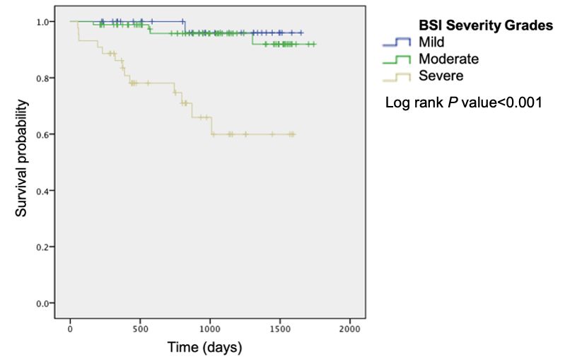
Non-CF bronchiectasis in Singapore—Si Ling Young et al.                                  557

                                                                       (CT), and attending the bronchiectasis clinic in
                  CLINICAL IMPACT                                      Singapore General Hospital, a tertiary hospital in
                                                                       Singapore, were recruited into this prospective cohort
                      What is New                                      study from 2017. The patients underwent a systematic
   •   This is one of the first studies describing the                 evaluation of potential underlying aetiologies with a
   characteristics of non-cystic fibrosis bronchiectasis               thorough assessment of disease symptoms, past history
   (NCFB) patients in Singapore, highlighting key                      of sino-pulmonary infections including tuberculosis,
   features such as a high incidence of haemoptysis                    ear infections, gastro-oesophageal reflux, subfertility,
   among these patients.                                               autoimmune disease and inflammatory bowel disease.
   •  The Bronchiectasis Severity Index (BSI) is a                     Serum immunoglobulins and full blood count were
   useful prognostic marker in our NCFB population.                    performed for all patients, in accordance with the
                                                                       British Thoracic Society and European Respiratory
                 Clinical Implications                                 Society guidelines.19,20 Other investigations such as
   •  This study highlights the heterogeneity of NCFB                  autoimmune markers, alpha-1-antitrypsin level, and
   and importance of further research to identify                      genetic testing for cystic fibrosis were performed if
   phenotypes that may help guide future management.                   relevant clinical features were present. The aetiology
   •   The BSI can aid clinicians in their communication               of bronchiectasis was determined on the basis of the
   with NCFB patients regarding the prognosis of                       aforementioned investigations by the treating physician
   their disease.
                                                                       via a clinical-radiological approach. Spirometry results,
                                                                       respiratory microbiology and exacerbation history
                                                                       from time of diagnosis were also collected. Interpretation
  Geographic variation in the aetiology and                            of spirometry was in accordance with the 2005
microbiology of NCFB has been described, such as                       American Thoracic Society interpretative strategies
the higher prevalence of idiopathic and post-infectious                for lung function tests.21 Bacterial colonisation was
NCFB patients reported in European and Asian                           defined by the growth of the same bacteria on 2 or
countries, 8 compared to the US where NCFB was                         more occasions at least 3 months apart on either sputum
frequently associated with immune dysregulation.9 For                  or broncheo-alveolar lavage specimens.
microbiology, the rates of Pseudomonas aeruginosa                         The CT images were independently reviewed by an
and Hemophilus influenzae colonisation vary across the                 experienced thoracic radiologist, and the morphological
US, Europe and Asia Pacific region. Non-tuberculous                    characteristics and severity of bronchiectasis were
mycobacterium (NTM) colonisation was found in 63%                      determined. The modified Reiff score was used to
of NCFB patients in the US bronchiectasis research                     assess the number of lobes involved and degree
registry, 10 but much lower rates were reported in                     of dilatation.22,23 The left lingula was considered a
Chinese studies. 9 Other organisms like Klebsiella                     separate lobe. The extent of bronchiectasis within each
pneumoniae were significantly prevalent in NCFB                        lobe was also graded with a score of 1, 2 or 3, according
patients in Thailand and South Korea.11,12                             to the proportion of airways involved: 50%, respectively. A radiology severity score
its diversity in clinical presentation, radiologic                     was obtained by a summation of scores for all the lobes.
involvement, spirometry patterns and prognosis as                         Exacerbation was defined as a deterioration in 3
reported by the various global registries on patients                  or more of the following symptoms for at least 48
with NCFB.10,13-17 Such heterogeneity has led to a keen                hours—cough, sputum volume or consistency, sputum
interest to identify phenotypes and endotypes with the                 purulence, breathlessness or exercise tolerance, fatigue
aim of individualising treatment to improve outcomes.18                and haemoptysis—associated with a requirement for
To date, information about the NCFB population in                      treatment with antibiotics, which is modified from
Singapore remains scarce. In this study, we describe                   the original definition by Hill et al. 24 A history of
the characteristics of NCFB patients in Singapore and                  haemoptysis was defined as the patient having
evaluated the performance of Bronchiectasis Severity                   reported any amount of haemoptysis before in their
Index (BSI) in predicting mortality.                                   lifetime that is attributed to bronchiectasis. Clinically
                                                                       significant haemoptysis referred to haemoptysis
METHODS                                                                requiring bronchoscopy, intubation, bronchial artery
Consecutive subjects (aged ≥21 years) with diagnosis                   embolisation or surgery. The BSI score was first derived
of bronchiectasis based on computed tomography                         and validated by Chalmers et al. and provided an easily

                                      Ann Acad Med Singap Vol 50 No 7 July 2021 | annals.edu.sg
558 Non-CF bronchiectasis in Singapore—Si Ling Young et al.
Table 1. Clinical characteristics of patients with NCFB in Singapore and mortality subgroup analysis
Characteristic Baseline characteristics Mortality group Survival group P value
(N=168) (n=18) (n=150)
Age at diagnosis, median (IQR), years 64 (56–71) 56 (54–60) 64 (57–71) 0.693
BMI, median (IQR) 19.3 (17.3–21.8) 18.5 (18.1–21.5) 19.3 (17.3–21.7) 0.003
Female, no. (%) 114 (67.8) 11 (61.1) 103 (68.7) 0.517
Smoking status, no. (%)
Never 123 (76.9) 8 (53.3) 113 (78.5) 0.030
Active 4 (2.5) 0 (0.0) 4 (2.8) 0.513
Ever 33 (20.6) 7 (46.7) 26 (18.1) 0.009
Asthma, no. (%) 15 (8.9) 0 (0.0) 15 (10.0) 0.160
COPD, no. (%) 9 (5.3) 3 (16.7) 6 (4.0) 0.024
Aetiology, no. (%)
Idiopathic 75 (44.6) 5 (27.8) 70 (46.7) 0.128
Post-TB 42 (25.0) 6 (33.3) 32 (21.3) 0.250
Post-infectious 38 (22.6) 5 (27.8) 37 (24.7) 0.773
FEV1 % predicted (baseline), median (IQR) 79 (63–95) 70 (51–84) 80 (65–95) 0.138
Spirometry pattern, no. (%)
Normal 52 (39.0) 3 (25.0) 49 (40.5) 0.294
Restrictive 29 (21.8) 5 (41.7) 24 (19.8) 0.081
Obstructive 15 (11.2) 1 (8.3) 14 (11.6) 0.735
Non-specific 13 (9.7) 1 (8.3) 12 (9.9) 0.860
Microbiology, no. (%)
Pseudomonas aeruginosa 35 (22.3) 6 (33.3) 29 (20.9) 0.232
Pseudomonas aeruginosa colonisation 24 (15.2) 5 (29.4) 19 (14.4) 0.113
Klebsiella pneumoniae 16 (10.2) 1 (5.6) 15 (10.8) 0.490
Hemophilus influenzae 6 (3.8) 1 (5.6) 5 (3.6) 0.683
Staphylococcus aureus 10 (6.4) 3 (16.7) 7 (5.0) 0.057
NTM 74 (46.3) 12 (66.7) 62 (43.7) 0.065
Radiology, no. (%)
Upper lobes 103 (61.3) 14 (77.8) 89 (59.3) 0.129
Middle lobes 146 (86.9) 17 (94.4) 129 (86.0) 0.316
Lower lobes 129 (76.7) 14 (77.8) 115 (76.7) 0.916
Lobes involved, median (IQR) 3 (2–4) 4 (3–4) 3 (2–4) 0.004
Radiology severity score, median (IQR) 6 (4–9) 7 (4–9) 6 (5–6) 0.007
Modified Reiff score, median (IQR) 4 (3–5) 4 (3–4) 4 (3–5) 0.015
Radiology pattern, no. (%)
Cylindrical 132 (78.5) 12 (66.7) 120 (80.0) 0.193
Cystic 22 (13.0) 5 (27.8) 17 (11.3) 0.051
Varicose 14 (8.3) 1 (5.6) 13 (8.7) 0.652
Exacerbations in the past year, no. (%) 45 (25.7) 10 (55.6) 35 (23.3) 0.004
Haemoptysis ever, no. (%) 92 (54.7) 8 (44.4) 84 (56.0) 0.352
Significant haemoptysis, no. (%) 36 (21.4) 3 (16.7) 33 (22.0) 0.602
BSI score, median (IQR) 6 (5–8) 8 (6–9) 6 (5–8)Non-CF bronchiectasis in Singapore—Si Ling Young et al. 559
accessible clinical score to aid in prognostication of Sixteen (10.2%) subjects had Klebsiella pneumoniae
patients with NCFB, which can influence clinical and 6 (3.8%) had Hemophilus influenzae. Seventy-
decision making and management. 23 This was a four subjects (46.3%) had sputum positive for NTM.
composite score of clinical variables used to classify The most common NTM isolated was Mycobacterium
bronchiectasis severity, and prospectively validated abscessus (41.9%), followed by M. fortuitum (25.7%),
to predict 1- and 4-year morbidity and mortality. M. avium complex (20.3%), and M. kansasii (8.1%).
The BSI scores, as well as chronic obstructive Eighteen (24.3%) subjects were initiated on NTM
pulmonary disease (COPD) assessment test scores were treatment, of whom 14 completed the course of
obtained.23,25 Mortality outcome was defined in this treatment. Two patients did not complete treatment
study as the point of death from any cause. In due to intolerable side effects, and 2 were still
calculating the clinical scores, missing data for undergoing treatment at the time of this writing. The
variables were assumed to be normal. All data were median treatment duration was 12 months (10–18).
entered into a secure digital platform (Research
Electronic Data Capture). Radiology
Statistical analyses were done using SPSS Statistics The median number of lobes involved was 3 (2–4). The
software version 23.0 (IBM Corp, Armonk, US). median modified Reiff score was 4 (3–5), and median
Chi-square test and Mann-Whitney U test were applied radiology severity score was 6 (4–9). Majority had
in the comparison of categorical and continuous middle lobe involvement (86.9%). The most common
data, respectively. Data were expressed as median radiological pattern observed was a cylindrical pattern
(interquartile range) for non-normally distributed (78.5%), followed by cystic pattern (13.0%).
continuous variables. Kaplan-Meier survival curves
were plotted to determine the relationship between BSI Clinical history
severity grades and mortality, and hazard ratios were Fifty-seven subjects (33.9%) had no history of
obtained using Cox proportional hazard regression exacerbations. Eight subjects (4.8%) had 3 or more
models. Statistical significance was defined as a exacerbations per year in their lifetime. Seventy-two
P value less than or equal to 0.05. subjects (60.0%) had a COPD assessment test score
of 10 and above. Eighteen subjects (10.7%) had
RESULTS demised during the period of follow-up. Ninety-two
A total of 168 subjects were recruited. The clinical (54.7%) subjects had a history of haemoptysis, of
characteristics are presented in Table 1. There was which 36 (21.4%) were clinically significant.
preponderance of women (67.8%) and Chinese
ethnicity (87.5%). The median age at diagnosis was Sex differences
64 years (56–71). Most subjects were never-smokers There were more smokers (51.8% vs 7.9%, P560 Non-CF bronchiectasis in Singapore—Si Ling Young et al. Table 2. Comparison of female and male NCFB patients in Singapore Characteristics Female (n=113) Male (n=54) P value Age, median (IQR), years 62 (56–69) 69 (60–74) 0.115 BMI, median (IQR) 19.3 (17.4–21.2) 19.1 (17.0–26.2) 0.658 Smoking status, no. (%) Active 0 (0) 4 (7.4) 0.004 Previous 9 (7.9) 24 (44.4)
Non-CF bronchiectasis in Singapore—Si Ling Young et al.                                               561

Table 3. Comparison of NCFB patients with and without a history of haemoptysis

Characteristics                                                 Haemoptysis (n=92)                No haemoptysis (n=76)               P value

Age, median (IQR), years                                             65 (56–71)                          63 (56–73)                    0.554
BMI, median (IQR)                                                 19.0 (16.9–21.1)                    20.2 (17.9–23.4)                 0.247
Female gender, no. (%)                                                64 (69.6)                           50 (65.8)                    0.602

Smoking status, no. (%)
 Active                                                                3 (3.4)                            1 (1.4)                      0.438
 Previous                                                             21 (23.6)                          12 (17.1)                     0.319
 Never                                                                64 (71.9)                          57 (81.4)                     0.162

Aetiology, no. (%)
 Idiopathic                                                           41 (44.6)                          34 (44.7)                     0.982
 Post-infectious                                                      21 (22.8)                          21 (27.6)                     0.474
 Post-TB                                                              25 (27.2)                          13 (17.1)                     0.121

Asthma, no. (%)                                                         4 (4.3)                          11 (14.5)                     0.022
COPD, no. (%)                                                           6 (6.5)                           3 (3.9)                      0.461

Spirometry, no. (%)
 Normal                                                              35 (48.6)                           17 (27.9)                     0.015
 Restrictive                                                         11 (15.3)                           18 (29.5)                     0.048
 Obstructive                                                           7 (9.7)                            8 (13.1)                     0.538
 Non-specific                                                          7 (9.7)                             6 (9.8)                     0.982
FEV1 % predicted (baseline)                                         84 (67–100)                         72 (63–87)                     0.004

Sputum cultures, no. (%)
 Pseudomonas aeruginosa                                               22 (24.7)                          13 (19.1)                     0.403
 Klebsiella pneumoniae                                                 8 (9.0)                            8 (11.8)                     0.569
 Hemophilus influenzae                                                 3 (3.4)                            3 (5.9)                      0.736
 Staphylococcus aureus                                                 8 (9.0)                            2 (2.9)                      0.124
 NTM                                                                  44 (48.9)                          30 (42.9)                     0.448

Radiology pattern, no. (%)
  Cylindrical                                                         73 (79.3)                          59 (77.6)                     0.787
  Cystic                                                               8 (8.7)                           14 (18.4)                     0.063
  Varicose                                                            11 (12.0)                           3 (3.9)                      0.062
Involvement, no. (%)
  Upper lobes                                                         53 (51.5)                          50 (48.5)                     0.305
  Middle lobes                                                        81 (55.5)                          65 (44.5)                     0.488
  Lower lobes                                                         71 (55.0)                          58 (45.0)                     0.544
Lobes involved, median (IQR)                                           3 (2–4)                            4 (2–5)                      0.080
Radiology severity score, median (IQR)                                 6 (4–9)                           7 (5–10)                      0.326
Modified Reiff score, median (IQR)                                     4 (2–5)                            4 (3–6)                      0.073

Use of antiplatelets or anticoagulation, no. (%)                      11 (12.1)                          12 (15.8)                     0.489

BSI score, median (IQR)                                                7 (5–9)                            6 (4–8)                      0.398
CAT score, median (IQR)                                               12 (5–19)                          15 (8–20)                     0.433

BMI: body mass index; BSI: Bronchiectasis Severity Index; CAT: COPD assessment test; COPD: chronic obstructive pulmonary disease; IQR: interquartile
range; NCFB: non-cystic fibrosis bronchiectasis; NTM: non-tuberculous mycobacteria; TB: tuberculosis
P values in bold are significant

Table 4. Hazard ratios of Bronchiectasis Severity Index (BSI) severity grades

BSI                                                       Hazard ratios                  95% confidence interval                    P value

Grade 1: Mild                                               Reference                            Reference                         Reference
Grade 2: Moderate                                             1.682                            0.188–15.052                          0.642
Grade 3: Severe                                              14.844                            1.929–114.235                          0.01

                                              Ann Acad Med Singap Vol 50 No 7 July 2021 | annals.edu.sg
562 Non-CF bronchiectasis in Singapore—Si Ling Young et al.
Most patients had normal spirometry patterns, and
fewer than 2 exacerbations per year. There was a high
prevalence of NTM, P. aeruginosa and K. pneumonia
infection. Over half the patients had a history of
haemoptysis, and approximately one-fifth of the
patients had clinically significant haemoptysis. There is
a higher proportion of NCFB among Chinese (87.5%)
compared to other races and this is out of proportion
to Singapore’s ethnic distribution (74.3% Chinese,
13.5% Malays and 9.0% Indians, according to
Singapore Department of Statistics’ 2020 figures).
The reason for this is unknown but postulated to be
due to differences in disease aetiology and sputum
microbiology.26 We are unable to confirm these findings
Fig. 1. Kaplan-Meier survival curves according to Bronchiectasis Severity due to the small number of non-Chinese patients in
Index (BSI) severity grades. our cohort.
Female preponderance of NCFB has been widely
described in various global registries including the UK,
US and Australia.10,13,18 Reasons postulated for this sex
distribution include the smaller conducting airways
in females, as well as the effects of oestrogen and
progesterone on mucociliary clearance.27 However, the
sex ratio is reversed in TB-endemic countries like
China, India and Pakistan.6,14,16 In these countries, TB
is a significant cause of NCFB and is more common
among men.28 NCFB patients in India and Pakistan are
also younger—83.1% of Pakistani patients are younger
than 60, and the mean age of diagnosis in India was 56
(41–66)—compared to the European cohort with mean
age of 67 (57–74).14,16 Factors such as the incidence
of childhood pulmonary infections (including TB) and
poor access to healthcare may contribute to the earlier
onset, higher burden and increased severity of the
disease.29,30 Southeast Asia accounts for over 40% of
the global TB incidence.31 Despite a high incidence
of TB in Singapore (47 per 100,000 population), the
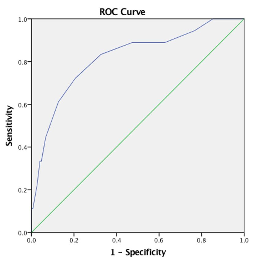
Fig. 2. Receiver operating characteristic curve for mortality according to overall mean age of diagnosis of NCFB remains
Bronchiectasis Severity Index score. comparable to the UK and US.10,18 In the current study,
ROC: receiver operating characteristic
40% of the NCFB patients with known aetiology were
due to previous TB infection. Idiopathic and post-TB
demonstrated a lower survival at the median overall bronchiectasis were equally common as aetiology
follow-up period of 2.4 years for subjects with severe of NCFB among male patients. On the other hand,
BSI grade as compared to those with mild or moderate the proportion of female patients with idiopathic
grades (PNon-CF bronchiectasis in Singapore—Si Ling Young et al. 563
in smoking habits is similarly reflected in the larger clinically significant. This is significantly higher than
proportion of COPD (11.1% vs 2.6%, P=0.023), and the prevalence of 20.9% and 23% that were reported
obstructive spirometry (20.8% vs 5.9%, P=0.009) seen in studies from Pakistan and the US, respectively.10,16
in the male NCFB patients. The frequency of positive In bronchiectasis, chronic airway inflammation causes
microbiology for NTM was significantly higher among hypertrophy and tortuosity of the vessels accompanying
females (55.0% vs 27.5%, P=0.001). the airways.39,40 Haemoptysis occurs due to the rupture
The spirometry findings of the present study contradict of these vessels, usually in the setting of an acute
previous reports that NCFB patients often have an infection or exacerbation. Despite our understanding
obstructive lung pattern.33 Proposed mechanisms for of the pathophysiology, it remains uncertain why
the airway obstruction include collapse of the major haemoptysis occurs more frequently in some patients
airways during expiration, bronchial wall thickening, but not others. We did not find an association between
presence of endobronchial secretions and obliterative haemoptysis and the presence of hypertrophied
bronchitis.34 However, a high proportion of smokers in bronchial arteries on computed tomography imaging
some older studies might have contributed to the high in our population, which may suggest the presence of
frequency of airway obstruction observed. Other studies o t h e r f a c t o r s a ff e c t i n g t h e d e v e l o p m e n t o f
assessing lung function in NCFB patients have reported haemoptysis. Other factors associated with
that obstructive and restrictive spirometry were haemoptysis in NCFB include the use of inhaled
both associated with increased disease severity and anticholinergics and short-acting beta agonists, 41
hospitalisation rate.33 The high proportion of patients presence of cystic pattern of on CT42 and post-TB
with normal spirometry and fairly preserved FEV1 aetiology.37 However, these findings were not replicated
may signal better clinical outcomes in our NCFB in our study.
population. Lower BMI, smoking status, history of exacerbations,
Nearly half the NCFB patients (46.3%) in our study modified Reiff scores and radiological severity were
had positive NTM culture, which is higher than the associated with mortality in our study, which is in
NTM prevalence (11.2%) reported in China. 6 Our keeping with results from previous studies. 23,43 BSI
NTM prevalence is more comparable to that of the scores were calculated and showed to be a good
US, which was 63% with a predominance of M. avium predictor of mortality, with an area under the curve
complex (37%). 10 In our population, M. abscessus (AUC) value of 0.818 that was similar to the derivation
was the most common mycobacterium species isolated and validation cohorts of the original study (AUC of
(18.6%). The most common bacterium isolated in 0.80 and 0.81–0.84, respectively). The Kaplan-Meier
our population is P. aeruginosa (22.3%), which is survival curve and hazard ratios showed a significant
comparable to other NCFB registries.10,14-16 Notably, difference in mortality when comparing patients with
K. pneumoniae is not widely reported in the mild (BSI 0–4) and severe (BSI>9) bronchiectasis. Our
microbiological characteristics of patients in US or results validate the use of BSI as a prognostic indicator
European registries, and its overall incidence appears in our local population. The inclusion of further markers
to be low. In our study, we observed an incidence of of radiological severity such as a composite of lobar
10.2%, comparable to the Thai (14%) and Korean severity grades as we have done in our study may further
(22.4%) cohorts. 11,12 K. pneumoniae is associated refine the accuracy and utility of such clinical scores.
with less mortality, exacerbations and hospitalisation The limitations of this study include the relatively
rates than P. aeruginosa. 35-37 The lung microbiome small cohort of patients, the potential selection bias
composition appears to affect response to anti- due to recruitment of participants from a single study
inflammatory therapy. In the erythromycin group in centre, and incomplete data, as in most real-world
the Bronchiectasis and Low-dose Erythromycin Study clinical studies. A multicentre study would likely
(BLESS), patients with a Haemophilus-dominated provide more comprehensive information about
microbiome had fewer exacerbations compared to characteristics of the NCFB population in Singapore.
those with Pseudomonas-dominated microbiome. 38 Causal inference between the variables analysed
There may be a geographical or racial predisposition should also be made with caution given the relatively
that affects colonisation and further research into the short follow-up period. The labels of post-infectious
bronchiectasis microbiome is imperative. and post-TB aetiologies are based on self-reported
Interestingly, more than half of our patients (54.7%) histories of pulmonary infections or TB, and may have
had a history of haemoptysis, 40% of whom were an element of recall bias. However, as far as possible,
Ann Acad Med Singap Vol 50 No 7 July 2021 | annals.edu.sg564 Non-CF bronchiectasis in Singapore—Si Ling Young et al.
objective evidence of previous pulmonary TB infection 13. Visser SK, Bye PTP, Fox GJ, et al. Australian adults with
bronchiectasis: The first report from the Australian Bronchiectasis
was documented. Similarly, there was a degree of
Registry. Respir Med 2019;155:97-103.
reliance on self-reporting of bronchiectasis
14. Dhar R, Singh S, Talwar D, et al. Bronchiectasis in India: results
exacerbations and haemoptysis. We were unable to from the European Multicentre Bronchiectasis Audit and Research
perform analysis for respiratory-specific mortality as Collaboration (EMBARC) and Respiratory Research Network
the cause of death was not available for a significant of India Registry. Lancet Glob Heal 2019;7:e1269-79.
proportion of the non-survivors. There may be interpretation 15. Olveira C, Padilla A, Martínez-García M-Á, et al. Etiology of
Bronchiectasis in a Cohort of 2047 Patients. An Analysis of the
bias introduced as the radiological assessment was Spanish Historical Bronchiectasis Registry. Arch Bronconeumol
performed by a sole radiologist. The strengths of the (English Ed.) 2017;53:366-74.
study include its prospective nature, and a broad 16. Sharif N, Baig MS, Sharif S, et al. Etiology, Clinical, Radiological,
inclusion criteria to reflect real-world clinical practice. and Microbiological Profile of Patients with Non-cystic Fibrosis
Bronchiectasis at a Tertiary Care Hospital of Pakistan. Cureus
2020;12:e7208.
CONCLUSIONS
17. Lee H, Choi H, Sim YS, et al. KMBARC registry: protocol for a
The NCFB population in Singapore has a female and multicentre observational cohort study on non-cystic fibrosis
Chinese predominance. Sex differences were found bronchiectasis in Korea. BMJ Open 2020;10:1-8.
and haemoptysis was common. The BSI score is a 18. Chalmers JD, Aliberti S, Filonenko A, et al. Characterization of
useful predictor of mortality in our population. Future the “frequent exacerbator phenotype” in bronchiectasis. Am J Respir
Crit Care Med 2018;197:1410-20.
research and longitudinal data should focus on better
19. Hill AT, Sullivan AL, Chalmers JD, et al. British thoracic society
understanding of the Asian bronchiectasis microbiome guideline for bronchiectasis in adults. Thorax 2019;74.
and cause of haemoptysis in NCFB. 20. Polverino E, Goeminne PC, McDonnell MJ, et al. European
Respiratory Society guidelines for the management of adult
bronchiectasis. Eur Respir J 2017;50.
REFERENCES 21. Miller MR, Hankinson J, Brusasco V, et al. Standardisation of
spirometry. Eur Respir J 2005;26:319-38.
1. Chalmers JD, Chang AB, Chotirmall SH, et al. Bronchiectasis.
Nat Rev Dis Prim 2018;4:45. 22. Reiff DB, Wells AU, Carr DH, et al. CT findings in bronchiectasis:
Limited value in distinguishing between idiopathic and specific
2. Quint JK, Millett ERC, Joshi M, et al. Changes in the incidence,
types. Am J Roentgenol 1995;165:261-7.
prevalence and mortality of bronchiectasis in the UK from 2004
to 2013: A population-based cohort study. Eur Respir J 2016;47:186-93. 23. Chalmers JD, Goeminne P, Aliberti S, et al. The bronchiectasis
severity index an international derivation and validation study.
3. Henkle E, Chan B, Curtis JR, et al. Characteristics and Health-care Am J Respir Crit Care Med 2014;189:576-85.
Utilization History of Patients With Bronchiectasis in US Medicare
Enrollees With Prescription Drug Plans, 2006 to 2014. Chest 24. Hill AT, Haworth CS, Aliberti S, et al. Pulmonary exacerbation in
2018;154:1311-20. adults with bronchiectasis: a consensus definition for clinical
research. Eur Respir J 2017;49: 1700051.
4. Ringshausen FC, Rademacher J, Pink I, et al. Increasing
bronchiectasis prevalence in Germany, 2009-2017: A population- 25. Guan WJ, Chen RC, Zhong NS. The bronchiectasis severity index
based cohort study. Eur Respir J 2019;54:2009-17. and FACED score for bronchiectasis. Eur Respir J 2016;47:382-84.
5. Aliberti S, Sotgiu G, Lapi F, et al. Prevalence and incidence of 26. McShane PJ, Naureckas ET, Strek ME. Bronchiectasis in a diverse
bronchiectasis in Italy. BMC Pulm Med 2020;20:1-6. US population: Effects of ethnicity on etiology and sputum culture.
Chest 2012;142:159-67.
6. Lin JL, Xu JF, Qu JM. Bronchiectasis in China. Ann Am Thorac
27. Vidaillac C, Yong VFL, Jaggi TK, et al. Gender differences in
Soc 2016;13:609-16.
bronchiectasis: A real issue? Breathe 2018;14:108-21.
7. Choi H, Yang B, Nam H, et al. Population-based prevalence of
28. Horton KC, MacPherson P, Houben RMGJ, et al. Sex Differences in
bronchiectasis and associated comorbidities in South Korea. Eur
Tuberculosis Burden and Notifications in Low- and Middle-Income
Respir J 2019;54.
Countries: A Systematic Review and Meta-analysis. PLoS Med
8. Yang B, Jang HJ, Chung SJ, et al. Factors associated with 2016;13: e1002119.
bronchiectasis in Korea: a national database study. Ann Transl Med
29. Dhanaraj B, Papanna MK, Adinarayanan S, et al. Prevalence and
2020;8:1350.
risk factors for adult pulmonary tuberculosis in a metropolitan city
9. Chandrasekaran R, Mac Aogáin M, Chalmers JD, et al. Geographic of South India. PLoS One 2015;10: e0124260.
variation in the aetiology, epidemiology and microbiology
30. Central TB Division. Revised National Tuberculosis Control
of bronchiectasis. BMC Pulm Med 2018;18:1-14.
Programme Annual Status Report. India TB Report 2018, March 2018.
10. Aksamit TR, O’Donnell AE, Barker A, et al. Adult Patients With Available at: https://tbcindia.gov.in/showfile.php?lid=3314. Accessed
Bronchiectasis: A First Look at the US Bronchiectasis Research on 28 May 2021.
Registry. Chest 2017;151:982-92.
31. World Health Organization. WHO: Tuberculosis Country Profiles
11. Palwatwichai A, Chaoprasong C, Vattanathum A, et al. Clinical, 2018. Available at: https://www.who.int/tb/publications/global_report/
laboratory findings and microbiologic characterization of tb19_Report_country_profiles_15October2019.pdf?ua=1. Accessed on
bronchiectasis in Thai patients. Respirology 2002;7:63-6. 28 May 2021.
12. Park J, Kim S, Lee YJ, et al. Factors associated with radiologic 32. Jappar SB, Low SY. Tuberculosis trends over a five-year period at
progression of non-cystic fibrosis bronchiectasis during long-term a tertiary care university-affiliated hospital in Singapore. Singapore
follow-up. Respirology 2016;21:1049-54. Med J 2015;56:502-5.
Ann Acad Med Singap Vol 50 No 7 July 2021 | annals.edu.sgNon-CF bronchiectasis in Singapore—Si Ling Young et al. 565
33. Stretton R, Poppelwell L, Salih W, et al. Patterns of spirometry in Patients With Non-Cystic Fibrosis Bronchiectasis. JAMA 2013;
bronchiectasis patients and relationship to markers of disease severity 309:1260-7.
and hospitalisation. Eur Respir J 2013;42:P2695. 39. Martin LN, Higgins L, Mohabir P, et al. Bronchial Artery
34. Chalmers JD, Aliberti S, Blasi F. Management of bronchiectasis in Embolization for Hemoptysis in Cystic Fibrosis Patients: A 17-Year
adults. Eur Respir J 2015;45:1446-62. Review. J Vasc Interv Radiol 2020;31:331-5.
35. Finch S, McDonnell MJ, Abo-Leyah H, et al. A Comprehensive 40. Dave BR, Sharma A, Kalva SP, et al. Nine-year single-center
experience with transcatheter arterial embolization for hemoptysis:
Analysis of the Impact of Pseudomonas aeruginosa Colonization
medium-term outcomes. Vasc Endovascular Surg 2011;45:258-68.
on Prognosis in Adult Bronchiectasis. Ann Am Thorac Soc 2015;
12:1602-11. 41. Jang EJ, Lee C-H, Yoon H Il, et al. Association between inhaler
use and risk of haemoptysis in patients with non-cystic fibrosis
36. King PT, Holdsworth SR, Freezer NJ, et al. Microbiologic follow-up bronchiectasis. Respirology 2015;20:1213-21.
study in adult bronchiectasis. Respir Med 2007;101:1633-8.
42. Guan WJ, Yuan JJ, Gao YH, et al. Hemoptysis in adults with
37. Davies G, Wells AU, Doffman S, et al. The effect of Pseudomonas bronchiectasis: correlation with disease severity and exacerbation
aeruginosa on pulmonary function in patients with bronchiectasis. risk. Zhonghua Jie He He Hu Xi Za Zhi 2017;40:16-23.
Eur Respir J 2006;28:974-9. 43. Ellis HC, Cowman S, Fernandes M, et al. Predicting mortality in
38. Serisier DJ, Martin ML, Mcguckin MA, et al. Effect of Long-term, bronchiectasis using bronchiectasis severity index and FACED
Low-Dose Erythromycin on Pulmonary Exacerbations Among scores: A 19-year cohort study. Eur Respir J 2016;47:482-9.
Ann Acad Med Singap Vol 50 No 7 July 2021 | annals.edu.sgYou can also read