Prevention of Progression of Lipedema With Liposuction Using Tumescent Local Anesthesia; Results of an International Consensus Conference
←
→
Page content transcription
If your browser does not render page correctly, please read the page content below

Prevention of Progression of Lipedema With Liposuction
Using Tumescent Local Anesthesia; Results of an
International Consensus Conference
Matthias Sandhofer, MD,* C. William Hanke, MD,† Louis Habbema, MD,‡
Maurizio Podda, MD,x Stefan Rapprich, MD,║ Wilfried Schmeller, MD,¶
Karen Herbst, MD,# Friedrich Anderhober, PhD,** Ulrike Pilsl, PhD,**
Gerhard Sattler, MD,x Martina Sandhofer, MD,* Werner Moosbauer, MD,††
Downloaded from https://journals.lww.com/dermatologicsurgery by BhDMf5ePHKav1zEoum1tQfN4a+kJLhEZgbsIHo4XMi0hCywCX1AWnYQp/IlQrHD3be73+7zLk/Sq+y6koaZl6Ep8oGuop87+BJ3Fduo7x/vYXVyjpiSPWQ== on 08/28/2019
Sonja Sattler, MD,x Patrick Schauer, MD,‡‡ Jörg Faulhaber, MD,xx Sabine Maier, MD,*
Martin Barsch, MD,* Sonani Mindt, MD,║║ and Anne B. Halk, MD¶¶
BACKGROUND Lipedema is a chronic, progressive disorder of subcutaneous adipose tissue that usually
affects the lower extremities of women. Also known as “two-body syndrome,” the fat accumulations in lipe-
dema are unsightly and painful. The disorder is well-known in Europe but is largely unrecognized and
underdiagnosed in the United States.
OBJECTIVE To hold the First International Consensus Conference on Lipedema with the purpose of
reviewing current European guidelines and the literature regarding the long-term benefits that have been
reported to occur after lymph-sparing liposuction for lipedema using tumescent local anesthesia.
METHODS International experts on liposuction for lipedema were convened as part of the First International
Congress on Lipedema in Vienna, Austria, June 9 to 10, 2017.
RESULTS Multiple studies from Germany have reported long-term benefits for as long as 8 years after
liposuction for lipedema using tumescent local anesthesia.
CONCLUSION Lymph-sparing liposuction using tumescent local anesthesia is currently the only effective
treatment for lipedema.
An international consensus conference on lipedema was conducted in conjunction with the First International
Congress on Lipedema. The Congress was sponsored by the Austrian Academy of Cosmetic Surgery and
Aesthetic Medicine (AACMS) and the International Society for Dermatologic Surgery (ISDS) and was held in
Vienna, Austria, June 9 to 10, 2017. The consensus conference participants included the following physicians:
M. Sandhofer, L. Habbema, G. Sattler, S. Rapprich, W. Schmeller, F. Anderhuber, M. Podda, K. Herbst, W.
Moosbauer, M. Sandhofer, U. Pilsl, S. Sattler, P. Schauer, J. Faulhaber, S. Maier, S. Mindt, A.B. Halk, M. Barsch,
and C.W. Hanke.
L ipedema is believed to affect 11% to 39% of the
female Caucasian population worldwide, but
exact figures from large studies are not available.1,2 As
disease is characterized by progressive enlargement of
the lower body while the upper body remains
relatively normal.4 The disease eventuates in
many as 16 million women in the United States may be deformity, disability, and reduced quality of life.
affected with the disorder.3 This potentially aggressive Current guidelines for treatment of lipedema exist
*Linz, Austria; †Laser and Skin Surgery Center of Indiana, Indianapolis, Indiana; ‡Medisch Centrum’t Gooi
Prinsengracht, Amsterdam, the Netherlands; xDarmstadt, Germany; ║Bad Soden, Germany; ¶Hanse-Klinik, Luebeck,
Germany; #Tucson, Arizona; **Institute of Macroscopic and Clinical Anatomy, Graz, Austria; ††Kepler
Universitätsklinikum GmbH, Linz, Austria; ‡‡Passau, Germany; xxSchwäbisch Gmünd, Germany; ║║Institute for Clinical
Chemistry, University Hospital Mannheim, Mannheim, Germany; ¶¶Amsterdam, the Netherlands
© 2019 by the American Society for Dermatologic Surgery, Inc. Published by Wolters Kluwer Health, Inc. All rights reserved.
· ·
ISSN: 1076-0512 Dermatol Surg 2019;00:1–9 DOI: 10.1097/DSS.0000000000002019
1
© 2019 by the American Society for Dermatologic Surgery, Inc. Published by Wolters Kluwer Health, Inc. Unauthorized reproduction of this article is prohibited.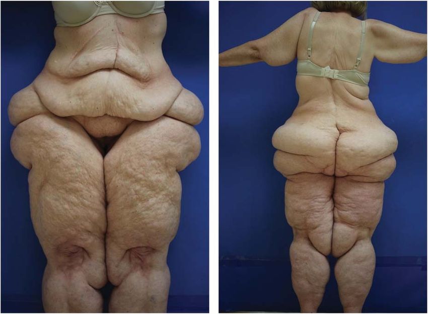
PREVENTION OF PROGRESSION OF LIPEDEMA

             only in Germany, Austria, and the Netherlands.5–7 The
             clinical presentation of lipedema has been reviewed by
             European dermatologists.4 The purpose of the
             consensus conference was to review the current
             guidelines and make recommendations especially with
             respect to long-term benefits that may occur after
             treatment of lipedema with liposuction using
             tumescent local anesthesia. Expert opinion is
             important because of limited evidence-based data on
             lipedema. The tables included in the article are
             consensus-based and do not mandate obligatory
             action.
                                                                                 Figure 1. (Front and back): A 50-year-old woman
                                                                                 exhibits the typical clinical features of Stage III
             Introduction                                                        lipedema or “two-body syndrome”. The area above the
                                                                                 waist is normal except for excess subcutaneous fat
             Lipedema, also called lipoedema in Europe, is a                     on the arms. The areas below the waist demonstrate
                                                                                 severe deformity.
             chronic progressive disorder of subcutaneous adi-
             pose tissue usually affecting the lower extremities of
             women.4 The fat accumulations are unsightly and                     lipedema patients in Sandhofer’s series had diabe-
             painful. First described by Allen and Hines8 in                     tes.9 These observations suggest that lipedema fat is
             1940, the disease is infrequently diagnosed in the                  not insulin-resistant and may protect patients from
             United States, although it is well-known in Europe.                 developing diabetes. Herbst and coworkers3
                                                                                 reported an increase in palpitations, numbness,
             Lipedema, also called “two-body syndrome,” is
                                                                                 urination, and shortness of breath in patients with
             relatively easy to diagnose by comparing the upper
                                                                                 lipedema stage III. However, the gynoid fat distri-
             and lower body during a physical examination
                                                                                 bution (i.e., thighs, buttocks, and hips) in lipedema
             (Figure 1). The circumference of the legs is pro-
                                                                                 may provide reduced cardiac risk for patients.
             portionately larger than the trunk. The feet are not
                                                                                 Thyroid stimulating hormone (TSH) should be
             affected (as in lymphedema), and the fat accumu-
                                                                                 monitored yearly in patients who have lipedema
             lations stop abruptly in the ankle region, below the
                                                                                 and obesity. The prevalence of hypothyroidism in
             knee, above the outer thigh, above the
             hips/buttocks, and below the elbow. Bruising of the
             skin in the involved areas occurs after minimal
                                                                                    TABLE 1. Lipedema is a Disease
             trauma (Table 1).
                                                                                    Uncontrolled localized fat deposition involving the
                                                                                     lower extremities (usually legs and thighs)
             Lipedema leads to multiple problems including
                                                                                    Symmetric increase in adipose tissue (“two-body
             decreased mobility and activity, secondary obesity,                     syndrome”)
             reduced general health, difficulty finding properly fit-                  Onset at puberty, pregnancy, and menopause
             ting clothing, and feelings of embarrassment. When                     Women-progressive with age (in most cases)
             obesity develops in conjunction with lipedema, the                     Tired “heavy” legs
                                                                                    Pain to touch or pressure may be mild or severe
             trunk and upper body will also demonstrate
                                                                                    Easy bruising
             enlargement.                                                           Hands and feet usually not affected (unlike
                                                                                     lymphedema)
             Comorbid Conditions With Lipedema                                      Cuffs or bulges develop around joints (e.g., ankles,
                                                                                     knees, elbows, and wrists)
             Diabetes and hypertension are uncommon in lipe-                        Negative Stemmer’s sign
                                                                                    Palpable spheroids in lipedema fat
             dema patients. Only one of 51 lipedema patients in
                                                                                    Reduced ambulation, decreased social activity
             Herbst’s series had Type 2 diabetes.3 Three of 500

   2         DERMATOLOGIC SURGERY

© 2019 by the American Society for Dermatologic Surgery, Inc. Published by Wolters Kluwer Health, Inc. Unauthorized reproduction of this article is prohibited.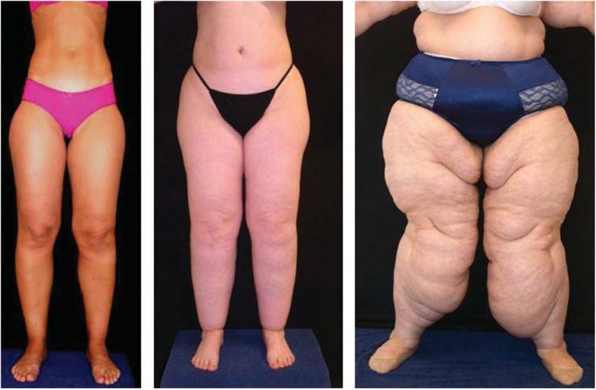
SANDHOFER ET AL

             Figure 2. A mother has Stage III lipedema and her 2 daughters have Stage II lipedema. The genetic predisposition is clear.
             (Reprinted with permission Johns Wiley and Sons, publisher) Rapprich S, Dingler A, Podda M. Liposuction is an effective
             treatment for lipedema results of a study with 25 patients. J Dtsh Dermatol Ges 2011;9(1):33–40.

             lipedema may be as high as 27%.3 In a consecutive                   Clinical Features of Lipedema
             series of 50 patients with lipedema, Herbst found
                                                                                 Clinical features are listed in Tables 2–4. The
             hypermobile joints is 50%.3 Herbst has reported
                                                                                 “cuff sign” is often present below the knee,
             that 50% of lipedema patients have hypermobile
                                                                                 above the ankles, above the elbow, and above the
             joints, suggesting an association with Ehlers Danlos
                                                                                 waist (Figure 3). The patella can disappear into
             syndrome hypermobility type.3
                                                                                 the surrounding fat, making ambulation, and
                                                                                 bending of the knee difficult (Figure 4). Forced
             Etiology
                                                                                 spread of the legs due to painful fat deposits
             The cause of lipedema is unknown. A family history of               often causes mechanical problems and decreased
             lipedema has been reported in up to 60% of cases                    mobility.
             (Figure 2). Lipedema commonly begins at puberty but
             may also become apparent with pregnancy or meno-                    The “bottleneck” fat pad or “subpatellar fat pad”
             pause. Although predominantly in women, lipedema                    on the anterior surface of the leg just below the
             has been reported to occur occasionally in hypo-                    knee is believed to be diagnostic of lipedema
             gonadal men.3 Hypertrophy and hyperplasia of adi-                   (Figure 5). It is speculated, that the deformity is
             pocytes occurs in the involved areas. Capillary                     caused by subcutaneous lymphatic stasis and
             permeability and fragility is increased causing easy                irregular subcutaneous fat accumulation. Herbst
             bruising. Long-term strain on lymphatic vessels results             and others have observed that lipedema fat
             in lymphatic stasis and degenerative changes in vessel              contains small 5- to 10-mm firm palpable bean-like
             walls. Ultimately, fibrosis of subcutaneous fat occurs               structures called spheroids. Lifestyle-induced fat
             as increasing surface deformities develop.

                                                                                    TABLE 3. Clinical Stages of Lipedema
                                                                                    (Stroessenreuther) (German S1 Guidelines)
                TABLE 2. Classification of Lipedema by
                Anatomical Site (Schrader) (German S1                               Stage I    Smooth and even skin surface; enlarged
                Guidelines)                                                                     subcutaneous fat compartment
                                                                                    Stage II   Skin surface wavy with some nodular
                Type I                         Buttocks and saddlebags                          elevations and indentations
                Type II                        Thigh (to the knee)                  Stage III Large nodules and deforming hanging fat
                Type III                       Legs (to malleoli)                              flaps, esp. on thighs and knees
                Type IV                        Arms                                 Stage IV Lipolymphedema occurs due to
                Type V                         Calves only                                     dysfunctional lymphatics

                                                                                                                          00:00:MONTH 2019                3

© 2019 by the American Society for Dermatologic Surgery, Inc. Published by Wolters Kluwer Health, Inc. Unauthorized reproduction of this article is prohibited.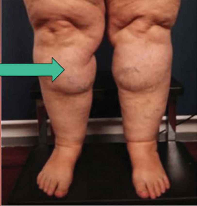
PREVENTION OF PROGRESSION OF LIPEDEMA

                TABLE 4. Differential Clinical Signs in Lipedema, Lipohypertrophy, Obesity, and Lymphedema (S1
                Guideline 037/012: Lipedema Last Updated 10/2015 Published by: AWMF Online. The Scientific Medicine
                Portal. doi:10.1111/ddg.13036)

                                                    Lipedema            Lipohypertrophy                  Obesity              Lymphedema
                Increased fat tissue                    +++                     +++                        +++                       (+)
                Disproportion                           +++                     +++                         (+)                       +
                Edema                                    (+)                     2                          (+)                     +++
                Pain on pressure                        +++                      2                          2                        2
                Easy bruising                           +++                      (+)                        2                        2

                + to +++, present; (+), possible; 2, not present.

             does not contain spheroids.3 The numbers of sphe-                   Treatment of Lipedema
             roids increase as patients progress by lipedema
                                                                                 Treatment of lipedema includes both nonsurgical
             stage. All patients with Stage III lipedema have pal-
                                                                                 and surgical methods. The goal of treatment is to
             pable spheroids in the flanks, thighs, buttocks,
                                                                                 improve the signs and symptoms of increasing leg
             medial knee, and posterior leg.3 Lipomas are not
                                                                                 volume, which include pain, swelling, limb dis-
             present in large numbers in patients with lipedema.
                                                                                 proportion, maceration, and to prevent infection,
                                                                                 erysipelas, lymphedema, and gait problems
             Associated findings in lipedema can include obesity,
                                                                                 (Tables 5–7).
             venous disease, joint pain, and hypermobile joints.

                                                                                 Manual lymphatic drainage (MLD), intermittent
             Several classifications and staging systems for
                                                                                 pneumatic compression, compression stockings,
             lipedema have been proposed (Tables 2–4 and
                                                                                 exercise, and skin care are often used to control pain
             Figure 6).
                                                                                 and symptoms. Recent studies have shown that
                                                                                 there is little or no edema in lipedema.10,11 There-
             Without treatment, lipedema will progress through
                                                                                 fore, treatment with MLD has no significant ther-
             the various stages (Figure 7).
                                                                                 apeutic effect on lipedema except perhaps making
                                                                                 the patient feel better because of the “hands-on”
                                                                                 nature of MLD. Diet is often used to prevent or treat
                                                                                 obesity when it is associated with lipedema. It is

                                                                                 Figure 4. A patient with lipedema demonstrates hooding
             Figure 3. “Cuff sign” above the ankles with sparing of feet         of the patella and the subpatellar fat pad on the anterior
             is common in lipedema patients.                                     upper leg.

   4         DERMATOLOGIC SURGERY

© 2019 by the American Society for Dermatologic Surgery, Inc. Published by Wolters Kluwer Health, Inc. Unauthorized reproduction of this article is prohibited.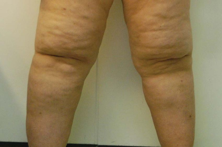
SANDHOFER ET AL

                                                                                 Figure 7. (A and B) Lipedema Progression over 13 years
                                                                                 without treatment.

                                                                                 suction techniques using general anesthesia. Lipo-
                                                                                 suction techniques using radio frequency,
                                                                                 ultrasound, or laser are not useful for lipedema
                                                                                 patients because of possible damage to lymphatic
                                                                                 vessels.

             Figure 5. Subpatellar (“bottleneck”) fat pad below the
             knee is common in lipedema patients.                                The frequency and necessity of conservative non-
                                                                                 surgical maintenance treatment were shown to be
             essential for lipedema patients to avoid weight gain.               eliminated or reduced after liposuction using tumes-
             “yo-yo” dieting and obesity have been shown to                      cent local anesthesia. The reduction in adipose
             exacerbate lipedema.10,11                                           deformities on the legs and thighs reduces skin damage
                                                                                 and facilitates improvement in mobility and gait
             Surgical treatment involves delicate, atraumatic                    (Figures 8 and 9).
             lymph-sparing liposuction using tumescent local
             anesthesia. The treatment has been proven to be safe                Treatment Planning in Liposuction
             and effective for cosmetic indications and lipe-                    for Lipedema
             dema.12–19 Examination of adipose tissue using
                                                                                 Some members of the Consensus group felt that the
             lymphoscintigraphy and immunohistochemistry
                                                                                 most painful areas of lipedema should be treated
             has demonstrated no significant damage to lym-
                                                                                 first, although patient preferences must also be
             phatic vessels from liposuction using tumescent
             local anesthesia compared with traditional lipo-

                                                                                    TABLE 5. Treatment Goals for Lipedema
                                                                                    Patients Treated With Liposuction Using
                                                                                    Tumescent Local Anesthesia

                                                                                                                               Secondary
                                                                                    Primary Goals                                Goals
                                                                                    [ Mobility and [ improvement in         Improved body
                                                                                     gait                                    contour
                                                                                    Y Excess subcutaneous fatty tissue      Y Weakness and
                                                                                     and bulkiness                           fatigue
                                                                                    Y Pain (spontaneous, continuous,     Aesthetic
                                                                                     intermittent, and painful to touch)  improvement
                                                                                    Improved quality of life                Y Edema
                                                                                    Y Bruising                              Y Skin irritation,
                                                                                                                             breakdown
                                                                                                                            [ Self-esteem
             Figure 6. Patients demonstrating lipedema (Stage I, II, III).

                                                                                                                          00:00:MONTH 2019                5

© 2019 by the American Society for Dermatologic Surgery, Inc. Published by Wolters Kluwer Health, Inc. Unauthorized reproduction of this article is prohibited.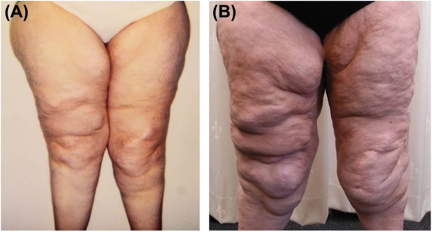
PREVENTION OF PROGRESSION OF LIPEDEMA

                TABLE 6. Lidocaine and/or Prilocaine Protocols for Tumescent Local Anesthesia

                                                     Klein12            Sattler, 1994            Hamburger, 1998                 Habbema19
                NaCl 0.9% mL                          1,000                 1,000                        1,000                       1,000
                Lidocaine, mg                         1,000                   —                           200                         400
                Prilocaine, mg                          —                    400                          200                          —
                Epinephrine, mg                          1                     1                           1                          0.8
                NaHCO3 8.4% mL                         12.5                    6                           6                          10
                Triamcinolone 10 mg/mL                 1 (0)                   1                           1                           —
                I.V. Sedation/Pain                   No/rare                 Yes                         Yes/no                       No

             taken into consideration. In general, liposuction                     Postoperative Course
             sessions should be begun proximally on the thigh
                                                                                   Lipedema patients treated with liposuction using tumes-
             with subsequent treatment sessions on the lower
                                                                                   cent local anesthesia (TLA) may drain lymph fluid for
             legs.
                                                                                   several weeks following the procedure. Swelling of the
                                                                                   treated area may last for 4 weeks or more.
             Maximum total lidocaine dose is restricted to 55
             mg/kg in young women and 45 mg/kg in women over
                                                                                   Low-dose heparin is given by some European lipo-
             60 years. Liposuction in lipedema patients is generally
                                                                                   suction surgeons as prophylaxis to prevent deep vein
             more labor-intensive in all aspects of the procedure
                                                                                   thromboses.
             compared with liposuction in aesthetic patients
             because of the large amounts of fatty tissue that are
                                                                                   Postoperative antibiotics are given for 1 to 2 weeks
             often removed.
                                                                                   postoperative.

             The venous system should be assessed preoperatively
                                                                                   Compression stockings are worn for 2 to 4 weeks
             in Stage III to IV lipedema patients, but not Stage I to II
                                                                                   postoperatively to prevent pools of lymph from
             patients unless indicated by physical examination and
                                                                                   forming in suctioned areas. After the first 4 weeks,
             symptoms (Table 8). Some experts recommend that
                                                                                   patients may prefer to wear compression stockings
             vein surgery be completed 2 months before liposuction
                                                                                   daily for comfort and support.
             to reduce phlebolymphedema and bleeding
             complications.
                                                                                   Manual lymphatic drainage can be given for as long as
                                                                                   4 to 5 weeks postoperative. Acoustic wave therapy
             One of the hallmarks of liposuction using tumescent
                                                                                   may be given for 5 to 10 weeks.
             local anesthesia is the “state of tumescence.” The state
             of tumescence is characterized by white blanching of
             the skin and a firm “watermelon” like surface that
                                                                                     TABLE 7. Technical Differences With Liposuction
             persists for more than 30 minutes. This persistent
                                                                                     in Lipedema and Lipohypertrophy
             firmness indicates that the proper amount of tumes-
             cent local anesthesia has been delivered to the target                  Lipedema                             Lipohypertrophy
             tissue.                                                                 Extensive areas                     Localized areas
                                                                                     Large volume fat                    Limited fat removal
                                                                                      removal
             Both extremities should be treated during the same
                                                                                     Circumferential                     Localized removal
             treatment session to maximize symmetry and                               treatment
             improvement. Two treatment algorithms are outlined                      Multiple sessions                   One or more
             in Table 9. Treatment plans for lipedema stages I to III                                                     limited sessions
                                                                                     Longer downtime                     Limited downtime
             are listed in Tables 10–12.

   6         DERMATOLOGIC SURGERY

© 2019 by the American Society for Dermatologic Surgery, Inc. Published by Wolters Kluwer Health, Inc. Unauthorized reproduction of this article is prohibited.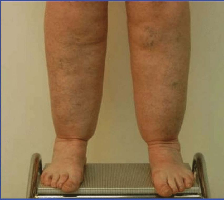
SANDHOFER ET AL

                                                                                    TABLE 8. Technical Aspects of Liposuction for
                                                                                    Lipedema Using Tumescent Local Anesthesia

                                                                                    2–6 treatment sessions may be required
                                                                                    The liposuction technique should cause the least
                                                                                     possible trauma to blood vessels, nerves, and
                                                                                     lymphatics
             Figure 8. A 42-year-old woman demonstrates typical                     Bilateral areas should be treated during the same
             lipedema with enlargement of both legs. The girth of both               treatment session to minimize asymmetry
             legs has been reduced after multiple liposuction proce-                Patients will require long-term follow-up
             dures using tumescent local anesthesia. Total volume of
                                                                                    The dripping of lymph fluid may persist for 2–4 days
             supranatant fat removed was 5.5 L.
                                                                                     postoperatively
                                                                                    The necessity of treatment for varicose veins is
                                                                                     determined on an individual basis. Small varicosities
             Low-impact active movements may begin when all                          may require no treatment, whereas large varicosities
             liposuction incisions are closed or healed. These                       are often treated in advance of liposuction.
             activities can include walking, aqua-gymnastics,
             swimming, and gymnastics.
                                                                                 vative therapy at 4 and 8 years. One-third of
             Long-Term Results in Lipedema Treated With                          patients were free of symptoms entirely after
             Liposuction Using Tumescent Local Anesthesia                        liposuction.

             Schmeller and co workers20–22 and Rapprich and co
                                                                                 The authors hypothesized that the accumulation of
             workers23–25 have reported dramatic long-term bene-
                                                                                 subcutaneous fatty tissue is the central factor in the
             fits in lipedema patients treated with liposuction using
                                                                                 pathogenesis of lipedema.22 Pain, easy bruising, and
             tumescent local anesthesia.
                                                                                 edema are secondary factors that are diminished or
                                                                                 resolved after liposuction. The authors indicated that
             In 2016, Baumgartner and coworkers 22 reported 8-
                                                                                 liposuction should be considered when symptoms
             year follow-up on 85 lipedema patients treated
                                                                                 progress during conservative therapy (Tables 13 and 14).
             with liposuction using tumescent local anesthesia.
             The same group of patients had been evaluated 4
                                                                                 Rapprich and coworkers25 assessed 85 lipedema
             years after liposuction. 20 The improvement is
                                                                                 patients who underwent liposuction using tumes-
             spontaneous pain, pressure sensitivity, bruising,
                                                                                 cent local anesthesia at 6 months after treatment.
             edema, and mobility that occurred at 4 years
                                                                                 The mean number of treatment sessions was
             postoperative was sustained at 8 years. Improve-
                                                                                 2.61/patient. After liposuction, lipedema patients
             ment in cosmetic appearance, quality of life, and
                                                                                 experienced improvement in spontaneous leg pain,
             overall impairment was also sustained at 8 years.
                                                                                 tenderness, bruising, swelling, ambulation, and
             Patients experienced a reduced need for conser-
                                                                                 quality of life.

                                                                                 Future/Research

                                                                                 Stem cells in lipedema may ultimately prove to be
                                                                                 different from those in normal fat. Much higher yields
                                                                                 of stem cells have been reported in the stromal vascular
                                                                                 fraction of lipedema fat compared with normal fat
                                                                                 cells.26 These cells may ultimately become a tissue
                                                                                 regeneration option in lipedema patients. Hematoxy-
             Figure 9. A 34-year-old woman demonstrates typical                  lin and eosin staining reveals crown-like structures in
             lipedema with enlargement of the thighs and buttocks. A
             total of 9.8 L supranatant fat removed during several               lipedema fat cells. It is not clear if the crown-like
             liposuction procedures.                                             structures are a primary cause for the growth of a

                                                                                                                          00:00:MONTH 2019                7

© 2019 by the American Society for Dermatologic Surgery, Inc. Published by Wolters Kluwer Health, Inc. Unauthorized reproduction of this article is prohibited.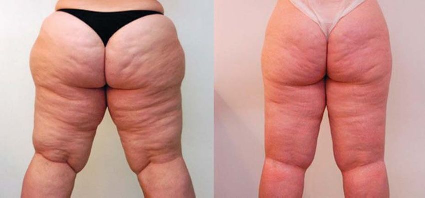
PREVENTION OF PROGRESSION OF LIPEDEMA

                TABLE 9. Two Treatment Algorithms for Lipedema Patients Treated With Liposuction Using Tumescent
                Local Anesthesia (the Minimum Interval Between Treatment Sessions is Usually 4 Weeks or More)

                First session                                    Inner thighs and knees
                Second session                                   Outer thighs and hips
                Third session                                    Lower legs (circumferential is sometimes done)
                First session                                    Outer thighs and hips
                Second session                                   Inner thighs and knees
                Third session                                    Ant upper leg and upper arms (if necessary)
                Fourth session                                   Hips and sacral area (if necessary)
                Fifth session                                    Buttocks (if necessary)

                TABLE 10. Liposuction Using Tumescent Local                         TABLE 13. Improvements in Lipedema
                Anesthesia in Stage I Lipedema Patients (Sattler                    Symptoms/Complaints After Liposuction
                2017)
                                                                                      Less Mechanical
                1–3 treatment sessions may be required                                  Problems                        Reduced Complaints
                Small varicose veins do not require treatment before
                 liposuction                                                        Y friction dermatitis               Y pain
                Most painful areas should be treated first, although                Y forced leg spread                 Y fatigue
                 there may be exceptions to this rule                               [ mobility                          Y edema and Y bruising
                Long-term follow-up is necessary                                    [ fitting of clothing               [ self-esteem
                                                                                    [ fitting of boots                  Y joint pain

                TABLE 11. Liposuction Using Tumescent Local
                Anesthesia in Stage II Lipedema Patients (Sattler                   TABLE 14. Total Lipedema Treatment Plan
                2017)
                                                                                    Patient education
                2–4 treatment sessions may be required                              Conservative therapy
                Large varicose veins may need treatment before                      Diet and exercise (to prevent secondary obesity)
                 liposuction                                                        Lymphatic drainage and compression therapy
                Most painful areas should be treated first, although                Psychotherapy/counseling
                 there may be exceptions to this rule
                                                                                    Precision, delicate lymph-sparing liposuction under
                Long-term follow-up is necessary                                     tumescent local anesthesia

                                                                                 Ehlers Danlos syndrome requires further
                TABLE 12. Liposuction Using Tumescent Local                      investigation.3
                Anesthesia in Stage III Lipedema Patients
                (Sattler 2017)
                                                                                 Lymph-sparing liposuction using tumescent local
                3–6 treatment sessions may be required                           anesthesia is the only effective treatment for patients
                Large varicose veins may need treatment before
                 liposuction
                                                                                 with lipedema.
                3–6 areas are likely to be affected
                Most painful areas should be treated first, although             References
                 there may be exceptions to this rule
                Long-term follow-up is necessary                                  1. Beninson J, Edelglass JW. Lipedema—the non-lymphatic masquerader.
                                                                                     Angiology 1984;35:506–10.

                                                                                  2. Foeldi M, Foeldi E. “Lipedema” in Foeldi’s Textbook of Lymphology.
                                                                                     Munich, Germany: Elsevier; 2006; pp. 417–427.
             different lipedema-type subset of fat cells or a sec-
                                                                                  3. Herbst KL, Mirkovskaya L, Bharhagova A, Chava Y, et al. Lipedema,
             ondary effect due to an enlarged hypodermis. Herbst’s
                                                                                     fat signs and symptoms of illness increase with advancing age. Arch
             suggestion of an association between lipedema and                       Med 2015;7:1–8.

   8         DERMATOLOGIC SURGERY

© 2019 by the American Society for Dermatologic Surgery, Inc. Published by Wolters Kluwer Health, Inc. Unauthorized reproduction of this article is prohibited.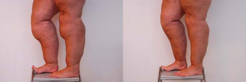
SANDHOFER ET AL

              4. Langendoen SI, Habbema L, Nijsten TEC, Neumann HAM. Lipedema                 17. Boeni R. Safety of tumescent liposuction under local anesthesia in a
                 from clinical presentation to therapy: a review of the literature. Br J          series of 4,380 patients. Dermatology 2011;222;278–81.
                 Dermatol 2009;161:980–6.
                                                                                              18. Habbema L. Safety of liposuction using exclusively tumescent local
              5. Wienert V, Walderman F, Zabel M, Rabe E, et al. Guidelines of the                anesthesia in 3,240 consecutive cases. Dermatol Surg 2009;35:1728–
                 German society of phlebology. Phlebologie 2009;38:164–7.                         35.
              6. Damstra RJ, Habbema L, Hendrickx C, Feenstra C, et al. Lipedema              19. Habbema L. Efficacy of tumescent local anesthesia with variable
                 Guidelines (g) in the Netherlands. Utrecht, the Netherlands: Dutch               lidocaine concentration in 3430 consecutive cases of liposuction. J Am
                 Soceity for Dermatology and Venereology; 2014. Available from:                   Acad Dermatol 2010;62:988–94.
                 https://www.lymfoedeem.nl/files/publicatie-r-damstra-april-2017-2.pdf.
                 Accessed July 8, 2019.                                                       20. Schmeller W, Hueppe M, Meier-Vollrath. Tumescent liposuction in
                                                                                                  lipedema yields good long-term results. Br J Dermatol 2012;166:161–8.
              7. Reich‐Schupke S, Schmeller W, Brauer WJ, Cornely ME, et al. S1
                 guidelines: lipedema. J Dtsch Dermatol Ges 2017;15:758–67.                   21. Schmeller W, Baumgartner A, Frambach Y. Tumescent Liposuction
                                                                                                  in Lipoedema in Liposuction: Principles and Practice. In: Schiffman
              8. Allen EV, Hines EAJ. Lipedema of the legs: a syndrome characterized by fat
                                                                                                  MA, DiGiuseppe A, editors. Berlin, Germany: Springer-Verlag;
                 legs and orthostatic edema. Proc Staff Meet Mayo Clin 1940;15:184–7.
                                                                                                  2016.
              9. Sandhofer M, Schauer P. Sandhofer M. Anderhuber F. Lipedema:
                                                                                              22. Baumgartner A, Hueppe M, Schmeller W. Long-term benefit of
                 Anatomic study, diagnosis and perioperative environment. J Asthet
                                                                                                  liposuction in patients with lipoedema: a follow-up study after an
                 Chir 2017;10:61–70.
                                                                                                  average of 4 and 8 years. Br J Dermatol 2016;174:1061–7.
             10. Bertsch T, Erbacher G. Lipoedema-myths and facts Part 1. Phlebologie
                 2018;2:84–92.                                                                23. Rapprich S, Koller J, Sattler G, Wörle B, et al. Liposuction—a surgical
                                                                                                  procedure in dermatology. J Dtsch Dermatol Ges 2012;10:111–3.
             11. Bertsch T, Erbacher G. Lipoedema-myths and facts Part 2. Phlebologie
                 2018;3:120–6.                                                                24. Rapprich S, Dingler A, Podda M. Liposuction is an effective treatment
                                                                                                  for lipedema-results of a study with 25 patients. J Dtsch Dermatol Ges
             12. Klein JA. The tumescent technique for Lipo-suction surgery. Am J Cos             2011;9:33–40.
                 Surg 1987;4:263–7.
                                                                                              25. Rapprich S, Baum S, Kaak I, Kottmann T, et al. Treatment of
             13. Klein JA, Jeske DR. Estimated maximal safe dosages of tumescent                  lipoedema using liposuction. Phlebologie 2015;3:1–13.
                 lidocaine. Anesth Analg 2016;122:1350–9.
                                                                                              26. Priglinger E, Wurzer C, Steffenhagen C, Maier J, et al. The adipose
             14. Hanke CW, Bernstein G, Bullock S. Safety of tumescent liposuction in             tissue- derived stromal vascular fraction cells from lipedema patients:
                 15,336 patients. National survey results. Dermatol Surg 1995;21:459–62.          are they different? Cytotherapy 2017;19:849–60.
             15. Hanke CW, Cox SE, Kuznets N, Coleman WP III. Tumescent
                 liposuction report performance measurement initiative: national survey
                 results. Dermatol Surg 2004;30:967–77; discussion 978.                       Address correspondence and reprint requests to: C.
             16. Housman TS, Lawrence N, Mellen BG, George MN, et al. The safety              William Hanke, Laser and Skin Surgery Center of Indiana,
                 of liposuction: results of a national survey. Dermatol Surg 2002;28:         8925 N. Meridian Street #200, Indianapolis, IN 46260, or
                 971–8.                                                                       e-mail: cwmhanke@thelassi.com

                                                                                                                                              00:00:MONTH 2019              9

© 2019 by the American Society for Dermatologic Surgery, Inc. Published by Wolters Kluwer Health, Inc. Unauthorized reproduction of this article is prohibited.
You can also read