The safety and efficacy of mini-percutaneous nephrolithotomy vs. retrograde intrarenal surgery for treatment of renal lithiasis in pelvic ectopic ...
←
→
Page content transcription
If your browser does not render page correctly, please read the page content below
Original Article

The safety and efficacy of mini-percutaneous nephrolithotomy
vs. retrograde intrarenal surgery for treatment of renal lithiasis in
pelvic ectopic kidney: an exploratory pilot study
Junfeng Wu, Jun Shen

Department of Urology, Qiandongnan People’s Hospital Affiliated to Guizhou Medical University, Kaili, China
Contributions: (I) Conception and design: All authors; (II) Administrative support: All authors; (III) Provision of study materials or patients: All
authors; (IV) Collection and assembly of data: All authors; (V) Data analysis and interpretation: All authors; (VI) Manuscript writing: All authors; (VII)
Final approval of manuscript: All authors.
Correspondence to: Jun Shen. Department of Urology, Affiliated Hospital of Guizhou Medical University, 28 Guiyi Street, Yunyan District, Guiyang,
China. Email: dgixbvykwqso@sina.com.

                 Background: To compare the safety and efficacy of mini-percutaneous nephrolithotomy (PCNL) and
                 retrograde intrarenal surgery (RIRS) for the treatment of renal lithiasis in patients with pelvic ectopic kidney.
                 Methods: From January 2015 to October 2017, mini-PCNL and RIRS were performed in ten patients
                 diagnosed with lithiasis in pelvic ectopic kidneys, including three cases under laparoscopy-assisted mini-
                 PCNL. Patient demographics and perioperative characteristics (age, gender, BMI, side of pelvic kidney,
                 stone size, stone number, stone location, special medical history, and ASA physical status classification),
                 and operative and post-operative related details (operation time, hospital stay, blood loss, VAS, analgesic
                 requirement, complications, and stone free outcome) were reviewed.
                 Results: Although the mean operation time of mini-PCNL (71.3 min) was shorter than RIRS (85.3 min),
                 the mean operation time of laparoscopy assisted mini-PCNL (92 min) was longer than patients without
                 laparoscopy-assisted mini-PCNL (55.8 min). However, the use of mini-PCNL allowed for larger lithiasis to
                 be dealt with (1.9 cm in laparoscopy assisted mini-PCNL and 2.4 cm in mini-PCNL without laparoscopy-
                 assist) compared with RIRS (1.2 cm). In addition, although the mean hospital-stay time, blood loss, and
                 analgesic requirement of patients undergoing RIRS were less than those receiving mini-PCNL, the success
                 rate of RIRS was only 50% (3/6) in comparison to 100% (7/7) for mini-PCNL. Except for pain and urinary
                 tract infection after the operation, there were no significant intraoperative and postoperative complications,
                 and no residual lithiasis were seen in any patient.
                 Conclusions: Although RIRS was less time-consuming and invasive, mini-PCNL can deal with the bigger
                 lithiasis and more complex situations with a higher success rate. Both mini-PCNL and RIRS are feasible and
                 safe treatments for pelvic ectopic kidney lithiasis with each carrying unique advantages. Hence in practice, an
                 appropriate individualized treatment should be selected depending on patient characteristics.

                 Keywords: Pelvic ectopic kidney; mini-percutaneous nephrolithotomy (mini-PCNL); retrograde intrarenal
                 surgery (RIRS); laparoscopy-assisted; renal lithiasis

                 Submitted Jan 14, 2021. Accepted for publication Feb 10, 2021.
                 doi: 10.21037/tau-21-77
                 View this article at: http://dx.doi.org/10.21037/tau-21-77

© Translational Andrology and Urology. All rights reserved.                Transl Androl Urol 2021;10(4):1734-1742 | http://dx.doi.org/10.21037/tau-21-77
Translational Andrology and Urology, Vol 10, No 4 April 2021                                                                             1735

Introduction                                                         patients diagnosed with lithiasis in pelvic ectopic kidneys,
                                                                     including three cases under laparoscopy-assisted mini-
Pelvic ectopic kidney is the most common form of renal
                                                                     PCNL. The mean age and BMI of the six male and four
malformation with an incidence ranging from 1 in 2,200 to
                                                                     female patients were 37.9 years (range, 27–66 years) and
1 in 3,000 (1). The condition results from a failure of ascent
                                                                     21.8 kg/m2 (range, 19.1–24.9 kg/m2), respectively (Table 1).
to the normal anatomic location in the renal fossa during
                                                                     Pelvic ectopic kidneys were located on the left side in 70%
kidney development, leaving the kidney below the pelvic
                                                                     (7/10) of patients and the final diagnosis was based on the
brim (2). Pelvic ectopic kidney is often accompanied by
                                                                     results of computed tomography urography (CTU). The
renal malrotation and a high insertion of the ureter, leading
                                                                     size, number, and location of stone are also summarized
to the inadequate evacuation of urine and formation of renal
                                                                     in Table 1. Two patients had a history of open surgery for
lithiasis (3). Furthermore, this abnormal orientation creates
                                                                     pelvic ectopic kidney lithiasis, one patient had a horseshoe
altered spatial relations with adjacent organs (as the kidney
                                                                     kidney, and another had scoliosis. The exclusion criteria for
lies anterior to the sacrum, caudal to the aortic bifurcation,
                                                                     surgery included an American Society of Anesthesiologists
and posterior to the peritoneum), abnormal calyceal                  (ASA) physical status more than grade III.
orientation, and anomalous vascular patterns (deriving                  All cases were routinely examined with laboratory
blood supply from iliac vessels or distal aorta). This makes         tests, including routine blood, blood biochemistry, blood
the active treatment of lithiasis in the pelvic ectopic kidney       coagulation function, routine urine, and urine culture.
a great challenge for surgeons (4).                                  In addition, CTU, computed tomography angiography
    Several approaches, including extracorporeal shock wave          (CTA), and three-dimensional (3D) reconstruction were
lithotripsy (SWL), laparoscopic pyelolithotomy, retrograde           implemented as routine radiologic evaluations to determine
intrarenal surgery (RIRS), percutaneous nephrolithotomy              the size, number, and location of the stone, and the organ
(PCNL), and open surgery are available for the treatment             position and vessel shape around the pelvic ectopic kidney
of renal lithiasis in anatomically normal kidneys. However,          (Figure 1).
there are no clear guidelines for the treatment of lithiasis            All operations were performed under general anesthesia
in pelvic ectopic kidney. PCNL and RIRS are the most                 in the lithotomy position. A 6 Fr ureteral catheter was first
widely used treatments for upper urinary tract lithiasis and         introduced retrograde into the pelvic ectopic kidney under
each holds unique advantages. In this study, we applied our          fluoroscopic guidance using a cystoscope. Patients were
exploratory pilot experiences of mini-PCNL and RIRS for              then adjusted to the supine position except for one patient
the treatment of lithiasis in the pelvic ectopic kidney and          who was placed in the lateral position because of scoliosis,
compared the safety and efficacy of these methods. The               in whom a roller pack was placed to lift the kidney closer to
results of this study may help clarify treatment options in          the anterior abdominal wall.
the management of patients with lithiasis and pelvic ectopic            The target calyx was determined using preoperative
kidney.                                                              CT (including CTU, CTA, and 3D reconstruction) and
    We present the following article in accordance with              intraoperative ultrasound monitoring (Figure 2A,B).
the MDAR reporting checklist (available at http://dx.doi.            After ultrasound-guided puncturing of the target calyx
org/10.21037/tau-21-77).                                             (Figure 2C), a 0.89 mm U-shaped guidewire was used to
                                                                     reconfirm access for an 18G needle and the operative
                                                                     validity of the calyx (Figure 2D), and to guide the track
Methods
                                                                     dilatation. Multi-step dilatation was used, and a 20 Fr sheath
An exploratory pilot study was undertaken in ten patients            and Karl Storz 12 Fr nephroscope were used for the mini-
diagnosed with pelvic ectopic kidney lithiasis in our hospital       PCNL procedure. Lithiasis were fragmented by Holmium
between January 2015 and October 2017. The study                     laser, and after extraction, a 6 Fr D-J stent was inserted into
conformed to the provisions of the Declaration of Helsinki           the ureteral lumen and a 16 or 18 Fr nephrostomy tube was
(as revised in 2013). The study was approved by the medical          placed where the sheath was removed.
ethics committee of the Qiandongnan People’s Hospital                   In the three patients undergoing laparoscopy-assisted
Affiliated to Guizhou Medical University. All patients               mini-PCNL the laparoscope was used to observe whether
agreed to participate in this study and signed an informed           the needle passed through other organs or major vessels,
consent form. Mini-PCNL and RIRS were performed in ten               and to monitor the process of multi-step dilatation

© Translational Andrology and Urology. All rights reserved.      Transl Androl Urol 2021;10(4):1734-1742 | http://dx.doi.org/10.21037/tau-21-77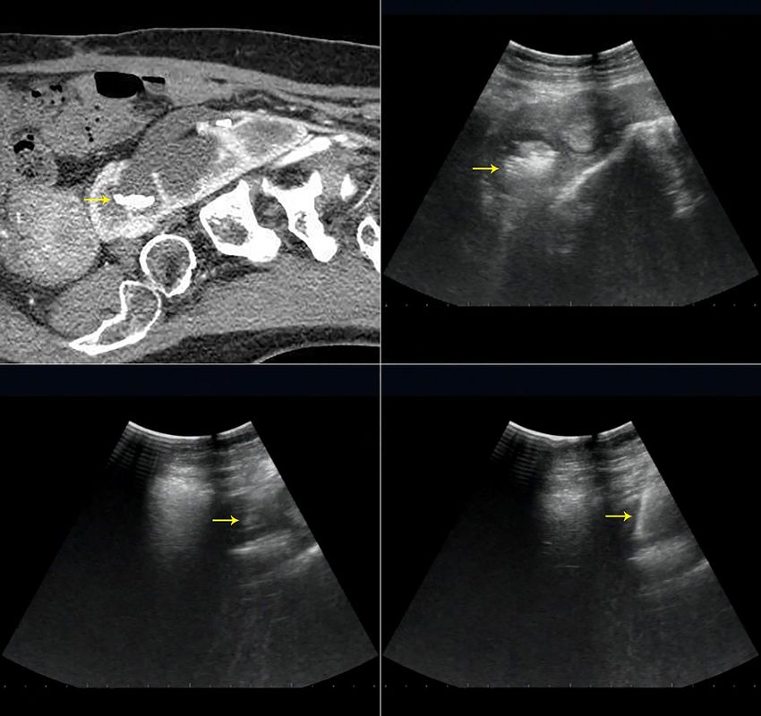
1736                                                                Wu and Shen. Treating pelvic ectopic kidney lithiasis by mini-PCNL and RIRS

 Table 1 Patient related details
                                                                            Mini-PCNL
 Variable                                                                                                                          RIRS (n=3)
                                                       With Lap (n=3)                     Without Lap (n=4)

 Mean age [range], years                                  33 [29–42]                          46 [27–67]                           32 [26–37]

 Gender (male/female)                                         1/2                                 3/1                                  2/1
                            2
 Mean BMI [range], kg/m                                19.8 [18.8–21.6]                    22.5 [20.1–23.9]                     22.8 [21.1–24.9]

 Laterality (left/right)                                      3/0                                 2/2                                  2/1

 Mean stone size [range], cm                             1.9 [1.0–3.3]                       2.4 [1.1–3.4]                        1.2 [0.8–1.5]

 Stone number

   Solitary                                                   2                                    3                                    2

   Multiple                                                   1                                    1                                    1

 Stone location

   Pelviureteric junction                                     0                                    1                                    2

   Pelvis                                                     1                                    3                                    1

   Lower calyx                                                2                                    0                                    0

 Special medical history

   Open surgery                                               1                                    1                                    0

   Horseshoe kidney                                           0                                    1                                    0

   Scoliolosis                                                0                                    1                                    0

 ASA classification

   Grade I                                                    3                                    2                                    3

   Grade II                                                   0                                    2                                    0
 PCNL, mini-percutaneous nephrolithotomy; RIRS, retrograde intrarenal surgery.

(Figure 3A,B). Two laparoscopic holes were punched, and 12                       0.6 to 1.0 J and pulse rate of 10 to 20 Hz), and the nitinol
and 5 mm trocars were placed below the umbilicus and on                          basket and grasper were used to retrieve the resultant stone
the left abdomen, respectively (Figure 3C). Once a successful                    fragments. Finally, the ureteroscope and ureteral access
puncture was established, multi-step dilatation was                              sheath were removed, and a 6 Fr D-J stent was retained.
performed under reduced abdominal pressure from 15 to                               Routine blood and biochemistry tests were performed the
10 mmHg and irrigation solutions or urine were aspirated                         morning after surgery. The catheter was removed one day
by the suction device at the same time as fragmenting                            later, and the nephrostomy tube was removed when drainage
the lithiasis. Finally, in addition to the indwelling 16 Fr                      was less than 50 mL every 24 hours. In patients undergoing
nephrostomy tube and 6 Fr D-J stent, a 14 Fr abdominal                           laparoscopy-assisted procedures, the abdominal drainage tube
drainage tube was placed into the location of the 5 mm                           was removed after bleeding or extravasation was less than
trocar.                                                                          10 mL every 24 hours. All patients were scheduled to follow-
   A Cook 12 or 14 Fr ureteral access sheath was                                 up 4 weeks later, and the D-J stent was removed once lithiasis
retrogradely placed along the guide wire in the lithotomy                        were not detected using X-ray or CT.
position for the RIRS procedure. Subsequently, an Olympus
URF-V flexible ureteroscope with 8.4 Fr tip diameter was
                                                                                 Statistical analysis
inserted to inspect the pelvicalyceal system and identify the
location of lithiasis. Lithotripsy was performed by a 200 µm                     SPSS 21.0 software (SPSS Inc., Chicago, IL, USA) was used
Holmium laser fiber with less than 20 W (energy level of                         for statistical analysis. Measurement data conforming to a

© Translational Andrology and Urology. All rights reserved.                  Transl Androl Urol 2021;10(4):1734-1742 | http://dx.doi.org/10.21037/tau-21-77
Translational Andrology and Urology, Vol 10, No 4 April 2021 1737
A B
C D
Figure 1 Radiologic evaluation using computed tomography urography (CTU), computed tomography angiography (CTA), and 3D
reconstruction. (A) Image of a patient with multiple lithiasis within the left pelvic ectopic kidney; (B,C) multiple lithiasis and collecting
system are shown in three-dimensional reconstruction of CTU; (D) vessel shape around the pelvic ectopic kidney in CTA. The yellow arrow
indicates the location of the lithiasis.
normal distribution was described by mean ± SD and an lithiasis, a single channel for mini-PCNL was used in seven
independent sample t-test was used. patients because their lithiasis were located in a limited
renal calyx or pelvis. The mean operation time of mini-
PCNL procedures (71.3 min) was shorter than RIRS
Results
(85.3 min). Due to the time taken for laparoscopy
The demographic, perioperative characteristics, and preparation and observation, the mean operation time of
operative and post-operative related results are presented laparoscopy-assisted mini-PCNL was 92 min, which was
in Tables 1 and 2. Six patients with lithiasis smaller than longer than mini-PCNL without laparoscopy-assistance
2 cm were initially treated by RIRS, but 50% of these (55.8 min) and RIRS (85.3 min). However, the use of mini-
cases (3/6) were unsuccessful due to failed insertion of PCNL with or without laparoscopy-assistance allowed us
the ureteral access sheath or abnormal calyceal structure to deal with larger lithiasis (1.9 and 2.4 cm) in comparison
hindering lithotripsy. These patients were then treated with RIRS (the largest 1.2 cm).
with mini-PCNL. Although two patients had multiple The mean operative blood loss, post-operative VAS
© Translational Andrology and Urology. All rights reserved. Transl Androl Urol 2021;10(4):1734-1742 | http://dx.doi.org/10.21037/tau-21-771738 Wu and Shen. Treating pelvic ectopic kidney lithiasis by mini-PCNL and RIRS
A B
C D
Figure 2 Preoperative computed tomography urography (CTU) and intraoperative ultrasound. (A,B) The images of lithiasis and collecting
system using preoperative CTU and intraoperative ultrasound were matched; the yellow arrow indicates the location of the lithiasis; (C,D)
the images of U-shaped guidewire in intraoperative ultrasound monitoring. The yellow arrow indicates the location of U-shaped guidewire.
score, and analgesic requirement of patients who underwent Discussion
RIRS were less than both mini-PCNL with or without
Patients with pelvic ectopic kidney are prone to chronic
laparoscopy-assistance. Except for pain and urinary tract
obstruction and renal lithiasis due to the abnormal structure
infection after the operation, no patient experienced severe
intraoperative or postoperative complications including and architecture seen in this condition. This variation in
severe bleeding, sepsis, and urine leakage after nephrostomy anatomy, including anomalous vascular patterns and altered
tube removal. One patient had a fever because of a urinary spatial relations with adjacent pelvic organs also presents
tract infection after surgery, but the infection was controlled difficult decisions for those performing nephrolithiasis
with routine antibiotics treatment. In addition, there were treatment (3).
no instances of bowel injury or abdominal infection in SWL is a first-line and established method for the
patients who underwent laparoscopy-assisted procedures. treatment of renal lithiasis less than 2 cm. SWL is also
The mean hospital stay time of RIRS (3 days) was less than the least invasive of common treatment methods used to
mini-PCNL (5.1 days) and no residual lithiasis were found treat renal lithiasis and can take place without the need for
in any patients at the four-week follow-up and the D-J stent hospitalization (5). However, the results of research on its
was removed successfully in all patients. use in treating patients with pelvic ectopic kidneys differ,
© Translational Andrology and Urology. All rights reserved. Transl Androl Urol 2021;10(4):1734-1742 | http://dx.doi.org/10.21037/tau-21-77Translational Andrology and Urology, Vol 10, No 4 April 2021 1739
A B C
Figure 3 Intraoperative laparoscopic detection. (A,B) Monitoring needle and multi-step dilatation under laparoscopy assisted surgery;
(C) nephrostomy tube and abdominal drainage tube placement. A 20 Fr sheath was placed in the location of the green arrow (nephrostomy
tube) and a 12 and 5 mm trocar were placed in the location of the yellow arrow below the umbilicus and left abdomen (abdominal drainage
tube), respectively.
Table 2 Operative and post-operative related details
Mini-PCNL
Variable RIRS (n=3)
With Lap (n=3) Without Lap (n=4)
Mean operation time [range], min 92.0 [90–95] 55.8 [51–62] 85.3 [62–98]
Mean hospital stay [range], day 5.3 [4–7] 5 [4–6] 3
Mean blood loss [range], mL 95.3 [43–158] 83 [45–109] 13.3 [5–20]
VAS score at 24h [range] 5.7 [4–8] 5.5 [4–7] 3.7 [3–4]
Analgesic requirement [range] 7.7 [5–10] 7 [6–8] 3.7 [3–4]
Complications classification
Pain (Grade I) 1 1 0
Urinary tract infection (Grade II) 0 0 1
Grade III–IV 0 0 0
Stone free, n (%) 3 (100%) 4 (100%) 3 (100%)
PCNL, mini-percutaneous nephrolithotomy; RIRS, retrograde intrarenal surgery.
with varying stone free rates, and some patients requiring fragments in normal and abnormal kidneys were 18.5% and
additional remedies. A monotherapy study showed that 37%, respectively. There are two main factors which limit
82% (9/11) of patients were stone free at the three months the therapeutic effect of SWL in pelvic ectopic kidneys.
follow-up when SWL was used to treat urinary lithiasis in Firstly, the bony pelvis and bowel cripple the effective
pelvic ectopic kidneys, although obstructive dural embolism of SWL. Secondly, the high insertion of the ureter and
was formed in two patients and only one was under an impaired pyeloureteral mobility by fibrous bands which
ancillary endourologic procedure (6). Demirkesen et al. (7) surround the kidney forming abnormal drainage patterns
compared SWL outcomes in normal and abnormal upper hinders the expulsion of residual fragments which promotes
urinary lithiasis, including eight patients with pelvic ectopic infection and stone recurrence (8).
kidney, and found those with a normal kidney had higher In comparison to PCNL, laparoscopic pyelolithotomy,
stone free rates (78%) than those with an abnormal kidney and open surgery, RIRS has become an increasingly
(56%), and the rates of clinically insignificant residual stone popular procedure to treat lithiasis due to its less invasive
© Translational Andrology and Urology. All rights reserved. Transl Androl Urol 2021;10(4):1734-1742 | http://dx.doi.org/10.21037/tau-21-771740 Wu and Shen. Treating pelvic ectopic kidney lithiasis by mini-PCNL and RIRS
nature. Using a 7.5 Fr flexible ureteroscope, holmium laser also monitor the image of lithiasis and collecting systems
lithotripsy, and nitinol baskets and graspers, six of eight in three-dimensional (3D) orientation during surgical
patients with an average 1.4 cm stone burden in anomalous procedures (13). Moreover, ultrasound guidance can image
kidneys (including four patients with pelvic ectopic kidney), intervening structures between the skin and kidney and
had complete clearance of the lithiasis, with seven being monitor bowel movement to avoid injury to the bowel
asymptomatic after the procedure (9). However, RIRS has during the puncture procedure. Despite this, there is a
its limitations in stone burden and cannot deal with the reluctance on behalf of some operators to use ultrasound as
pathologic structure of the pelvic ectopic kidney which this requires significant training, and the images produced
restricts the insertion of the ureteral access sheath and may be difficult to interpret.
removal of residual fragments. In addition, RIRS carries In addition to using intraoperative ultrasound guidance,
the problem of infection. Although no significant statistical preoperative radiologic evaluation using CTU, CTA and 3D
difference was found in the total complication rate between reconstructions also plays a crucial role during puncturing.
treatment with PCNL and RIRS for renal lithiasis, the A greater number of channels increases the risk of potential
longer operation time and higher intrarenal pressure of complications, hence the selection of the desired calyx is
RIRS compared to PCNL increased the risk of sepsis (10). crucial. A desirable access should be designed according
In our study, six patients with lithiasis less than 2 cm were to the combined outcomes of intraoperative monitoring
initially treated by RIRS, but 50% (3/6) failed because of a by ultrasound and preoperative radiologic evaluation of
failure in the insertion of the ureteral access sheath because CTU and 3D reconstruction which provide important
of the abnormal calyceal structure. The three unsuccessful information regarding stone size, number, location, the
cases then underwent a mini-PCNL procedure. Despite structure of collecting systems, and the relationship between
this, there were no significant intraoperative and lithiasis and collecting system. In addition, CTA and 3D
postoperative complications in RSIS patients and no reconstruction can display anomalous vascular patterns
residual lithiasis in successful cases. Therefore, to improve around the pelvic ectopic kidney and provide assistance to
the safety and stone clearance rate of RIRS in patients with reduce renal and perirenal vessel injury.
pelvic ectopic kidney, we recommend urinary tract infection We also do not use transabdominal access as a routine
should be controlled preoperatively, the procedures should method for PCNL. A roller pack is placed under the
only be used to treat appropriately sized lithiasis and those pelvic ectopic kidney to bring the target calyx closer to
easily accessed, and the effective application of surgical the anterior abdominal wall to facilitate access during
instruments such as nitinol baskets and graspers should be PCNL. Furthermore, even though the operation time with
used. laparoscopy-assisted PCNL is longer than other methods
PCNL is a popular and conventional surgical method because of the procedure in laparoscopy preparation
widely used to treat patients with all types of upper urinary and observation, the operation procedures especially in
tract lithiasis in the orthotopic kidney, and has a low puncture and dilatation became safer. With laparoscopic-
incidence of significant complications such as hemorrhage, assistance, blood, urine and washing solution are aspirated
sepsis, and injury of perirenal organs. However, there is by the suction device while fragmenting lithiasis, thus
dispute over the performance of PCNL on pelvic ectopic avoiding abdominal infection and irritation to the bowel
kidney lithiasis. Our experience in the treatment of pelvic by these irritants. However, laparoscopy-assisted PCNL
ectopic kidney calculus with transabdominal mini-PCNL has limitations in some patients who have a history of other
using ultrasound-guided puncture follows. disease and an unusual location of renal lithiasis.
We use fluoroscopic or ultrasound guidance puncture Lastly, standard PCNL requires a larger tract, which
methods. Although fluoroscopy has been the main tool may increase the risk of bleeding in a kidney with
to establish access in PCNL, its excessive use leads to an anomalous blood supply (14). Based on the comprehensive
increase in the risk of radiation to patients and staff (11). consideration of operation time and stone clearing
The use of ultrasound-guided puncture in PCNL was efficiency, the 12 Fr nephroscope matched with 20 Fr
first reported in the 1970s (12) and has gained increased channel is preferred in our clinical center. However, for
recognition as an alternative to fluoroscopy because of its some special cases we will also use negative pressure suction
feasibility, safety, and efficacy. Ultrasound guidance not equipment matched with the standard channel or finer
only has the advantage of low radiation exposure, it can nephroscope with the smaller channel. In addition, there
© Translational Andrology and Urology. All rights reserved. Transl Androl Urol 2021;10(4):1734-1742 | http://dx.doi.org/10.21037/tau-21-77Translational Andrology and Urology, Vol 10, No 4 April 2021 1741
is also a risk of urinary leakage in tubeless PCNL which Data Sharing Statement: Available at http://dx.doi.
may cause ileus and morbidity (15). Therefore, the use of org/10.21037/tau-21-77
tubeless PCNL as a routine procedure is debatable.
Several recent reports discussing the treatment of Conflicts of Interest: Both authors have completed the
pelvic ectopic kidney lithiasis by RIRS and PCNL, with ICMJE uniform disclosure form (available at http://dx.doi.
or without laparoscopy-assistance, have expressed results org/10.21037/tau-21-77). The authors have no conflicts of
similar to ours (4,16,17). Nevertheless, these reports have interest to declare.
not produced a systematic treatment strategy for the
disease. Overall, we summarize guidelines for the treatment Ethical Statement: The authors are accountable for all
of lithiasis in the pelvic ectopic kidney according to our aspects of the work in ensuring that questions related
experiences as follows. (I) If the size of a solitary stone to the accuracy or integrity of any part of the work
or total multiple lithiasis is less than 2 cm and each stone are appropriately investigated and resolved. The study
locates in an appropriate position (pelvis or calyx with an conformed to the provisions of the Declaration of Helsinki
uncomplicated structure of the collection system), patients (as revisedin2013). The study was approved by the medical
without ectopia of the ureteral orifice and ureterostenosis ethics committee of the Qiandongnan People’s Hospital
can be initially treated by RIRS. If this fails, the operation Affiliated to Guizhou Medical University. All patients
can then be turned into a mini-PCNL procedure. (II) If agreed to participate in this study and signed an informed
the solitary stone or total multiple lithiasis is more than consent form.
2 cm in size or the total of multiple stones is less than
2 cm with a complicated structure of the collection system, Open Access Statement: This is an Open Access article
patients without laparoscopic contraindications can be distributed in accordance with the Creative Commons
treated by laparoscopy-assisted mini-PCNL. (III) In all Attribution-NonCommercial-NoDerivs 4.0 International
other situations, mini-PCNL can be performed without License (CC BY-NC-ND 4.0), which permits the non-
laparoscopy-assistance, and open surgery can be used as commercial replication and distribution of the article with
a final method to deal with various failures. This study is the strict proviso that no changes or edits are made and the
limited by its small sample size and a larger prospective original work is properly cited (including links to both the
randomized study is recommended to verify the results. formal publication through the relevant DOI and the license).
See: https://creativecommons.org/licenses/by-nc-nd/4.0/.
Conclusions
References
Although more time-consuming and invasive than RIRS,
mini-PCNL could deal with larger lithiasis and more 1. Zafar FS, Lingeman JE. Value of laparoscopy in the
complex situations with a higher success rate. Both mini- management of calculi complicating renal malformations.
PCNL and RIRS are feasible and safe treatments for J Endourol1996;10:379-83.
pelvic ectopic kidney lithiasis with each holding unique 2. Tan YK, Cha DY, Gupta M.Management of stones in
advantages. Hence in practice, an appropriate individualized abnormal situations. Urol Clin North Am 2013;40:79-97.
treatment should be selected depending on the individual 3. Cinman NM, Okeke Z, Smith AD.Pelvic kidney: associated
characteristics of the patients. diseases and treatment. J Endourol2007;21:836-842.
4. Otaño N, Jairath A, Mishra S, et al. Percutaneous
nephrolithotomy in pelvic kidneys: is the ultrasound-
Acknowledgments
guided puncture safe? Urology 2015;85:55-8.
Funding: None. 5. Yin Z, Wei YB, Liang BL, et al. Initial experiences with
laparoscopy and flexible ureteroscopy combination
pyeloplasty in management of ectopic pelvic kidney with
Footnote
stone and ureter-pelvic junction obstruction. Urolithiasis
Reporting Checklist: The authors have completed the MDAR 2015;43:255-60.
reporting checklist. Available at http://dx.doi.org/10.21037/ 6. TalicRF.Extracorporeal shock-wave lithotripsy
tau-21-77 monotherapy in renal pelvic ectopia. Urology
© Translational Andrology and Urology. All rights reserved. Transl Androl Urol 2021;10(4):1734-1742 | http://dx.doi.org/10.21037/tau-21-771742 Wu and Shen. Treating pelvic ectopic kidney lithiasis by mini-PCNL and RIRS
1996;48:857-61. nephrolithotomy: an innovative extraction technique. J
7. Demirkesen O, Yaycioglu O, Onal B, et al. Extracorporeal Urol1977;118:671-2.
shockwave lithotripsy for stones in abnormal urinary 13. Agarwal M, Agrawal MS, Jaiswal A, et al. Safety and
tracts: analysis of results and comparison with normal efficacy of ultrasonography as an adjunct to fluoroscopy
urinary tracts. J Endourol2001;15:681-5. for renal access in percutaneous nephrolithotomy (PCNL).
8. Bush WH, Brannen GE.Extracorporeal shock-wave BJU Int 2011;108:1346-9.
lithotripsy (ESWL) of pelvic kidney calculus. Use of C-arm 14. Ganesamoni R, Sabnis RB, Mishra S, et al. Microperc for
fluoroscopy for correct patient positioning. Urology the management of renal calculi in pelvic ectopic kidneys.
1987;29:357-60. Indian J Urol2013;29:257-9.
9. Weizer AZ, Springhart WP, Ekeruo WO, et 15. Bozkurt IH, Cirakoglu A, Ozer S. Retroperitoneal
al.Ureteroscopic management of renal calculi in anomalous laparoscopic pyelolithotomy in an ectopic pelvic kidney.
kidneys. Urology 2005;65:265-9. JSLS 2012;16:325-8.
10. Zhang W, Zhou T, Wu T, et al. Retrograde Intrarenal 16. D’souza N, Verma A, Rai A. Laparoscopic-assisted mini
Surgery Versus Percutaneous Nephrolithotomy Versus percutaneous nephrolithotomy in the ectopic pelvic
Extracorporeal Shockwave Lithotripsy for Treatment of kidney: Outcomes with the laser dusting technique. Urol
Lower Pole Renal Stones: A Meta-Analysis and Systematic Ann 2016;8:87-90.
Review. J Endourol2015;29:745-59. 17. Bozkurt OF, Tepeler A, Sninsky B, et al. Flexible
11. Rao PN, Faulkner K, Sweeney JK, et al. Radiation dose to ureterorenoscopy for the treatment of kidney stone within
patient and staff during percutaneous nephrostolithotomy. pelvic ectopic kidney. Urology 2014;84:1285-9.
Br J Urol1987;59:508-12.
12. Karamcheti A, O'Donnell WF. Percutaneous (English Language Editor: B. Draper)
Cite this article as: Wu J, Shen J. The safety and efficacy of
mini-percutaneous nephrolithotomy vs. retrograde intrarenal
surgery for treatment of renal lithiasis in pelvic ectopic kidney:
an exploratory pilot study. Transl Androl Urol 2021;10(4):1734-
1742. doi: 10.21037/tau-21-77
© Translational Andrology and Urology. All rights reserved. Transl Androl Urol 2021;10(4):1734-1742 | http://dx.doi.org/10.21037/tau-21-77You can also read