Buccolingual Inclination of Canine and First and Second Molar Teeth and the Curve of Wilson in Different Sagittal Skeletal Patterns of Adults ...
←
→
Page content transcription
If your browser does not render page correctly, please read the page content below
Hindawi
International Journal of Dentistry
Volume 2020, Article ID 8893778, 8 pages
https://doi.org/10.1155/2020/8893778

Research Article
Buccolingual Inclination of Canine and First and Second Molar
Teeth and the Curve of Wilson in Different Sagittal Skeletal
Patterns of Adults Using Cone-Beam Computed Tomography

          Amin Golshah ,1 Navid Rezaei ,1 and Sara Heshmati                                    2

          1
              Department of Orthodontics, Faculty of Dentistry, Kermanshah University of Medical Sciences, Kermanshah 6715847141, Iran
          2
              Students Research Committee, Faculty of Dentistry, Kermanshah University of Medical Sciences, Kermanshah 6715847141, Iran

          Correspondence should be addressed to Navid Rezaei; navidrezaie24@gmail.com

          Received 17 July 2020; Accepted 21 October 2020; Published 4 November 2020

          Academic Editor: Sreekanth Kumar Mallineni

          Copyright © 2020 Amin Golshah et al. This is an open access article distributed under the Creative Commons Attribution
          License, which permits unrestricted use, distribution, and reproduction in any medium, provided the original work is
          properly cited.
          Objectives. This study aimed to assess the buccolingual inclination of canine and first and second molar teeth and the curve of
          Wilson in different sagittal skeletal patterns in untreated adults using cone-beam computed tomography (CBCT). Materials and
          Methods. Sixty-six CBCT scans of adults (mean age: 28.74 ± 5.25 years) were evaluated in this cross-sectional study. The images
          were standardized using the Frankfurt horizontal plane and the interorbital line. The sagittal skeletal pattern was determined using
          the ANB angle and Wits appraisal. Inclination angles were measured by NNT Viewer and Mimics software. The curve of Wilson
          was measured by connecting the tips of mesiobuccal and mesiolingual cusps of maxillary first and second molars along the buccal
          groove and measuring the formed angle. Data were analyzed using ANOVA. Results. The intraobserver agreement was 0.969. The
          mean inclination of maxillary first and second molars in class I and III patients was significantly higher than that in class II patients
          (P < 0.05). The mean inclination of mandibular first and second molars in class II patients was significantly higher than that in
          class I and III patients (P < 0.05). The difference in inclination of maxillary and mandibular canine teeth was not significant
          (P > 0.05). The mean curve of Wilson in second molars of class II patients was significantly higher than that in class I patients
          (P < 0.05). Conclusion. In different sagittal skeletal patterns, a compensatory relationship exists between the opposing teeth,
          which, along with the standards of crowns, can be used to determine the appropriate position of teeth in dental arch.

1. Introduction                                                           clearly understood considering the fact that their occlusal
                                                                          surfaces do not follow the same pattern. In 1911, George
Orthodontics is the art and science of leveling and alignment             Wilson explained this phenomenon by a compensatory
of the teeth in dental arch, which is associated with bone                curve to prevent the possible balancing interferences. This
remodeling and converts a malocclusion into a stable oc-                  curve should be concave in the mandibular arch and concave
clusion with maximum achievable order and alignment of                    or convex in the maxillary arch. Thus, the buccal and palatal
teeth and optimal function and esthetics. Orthodontic ap-                 cusps of the posterior teeth are in functional contact with
pliances are used for this purpose [1]. Occlusion is a fun-               each other [9].
damental part of orthodontic treatment [2–6]. Different                        From the frontal view, the occlusal plane is in the form of
theories have proposed different definitions for a normal                   an arch. The occlusal surfaces of maxillary posterior teeth
occlusion [7, 8]. However, the clinical application of con-               comprise the convex part of the arch while the occlusal
cepts of occlusion has not been well studied. For instance,               surfaces of the mandibular posterior teeth comprise the
the role of buccolingual position of the posterior cusps of               concave part of the arch [10]. Recently, the occlusal incli-
molar teeth in the frontal view in occlusion has not been                 nation was defined as a progressive increase in axial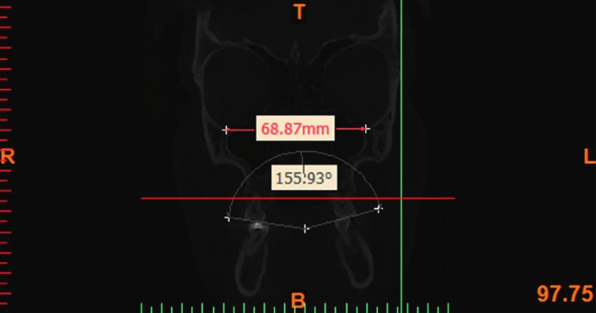
2                                                                                               International Journal of Dentistry

inclination of molars from the first molar to the third molar,       visualization of each tooth in any desired plane [29, 30].
which is a developmental feature known as the helicoid              Moreover, CBCT enables the evaluation of the entire tooth
curve [11, 12].                                                     structure. Thus, uncertainties in the longitudinal axis (in-
     From the frontal view, this curve includes the buccal          clination) of the teeth due to the use of casts with asym-
inclination of maxillary molars and the lingual inclination of      metrical wear of cusps or tooth morphology are eliminated
the mandibular molars. However, it should be noted that the         [1, 9, 26, 27]. Several techniques have been used to determine
exact amount of this inclination has not yet been quantified         the longitudinal axis and measure the inclination of teeth
[13].                                                               such as CT [26] and CBCT [9, 27, 30]. CBCT can be used to
     The buccolingual inclination of teeth has long been an         assess the position of teeth in the sagittal, axial, and coronal
interesting topic for orthodontists. Andrews described the          planes.
six keys to a normal occlusion [14], and according to him,               There is a gap of information regarding the buccolingual
the buccolingual inclination of teeth is one of the six keys to a   inclination of second molars in untreated adults [13, 27].
normal occlusion and is part of the third phase of clinical         Change in buccolingual inclination of teeth is an important
examinations according to the American Board of Ortho-              factor affecting the stability of dentition [27]. Considering
dontists (ABO) [14, 15]. The third key of Andrews is related        the significance of second molars in orthodontic treatment
to coronal inclination, which is measured at the buccal             planning and orthosurgery, and the gap of information
surface of tooth crown. The findings of Andrews revealed the         about the inclination of molar and canine teeth and the
lingual inclination of the crown of maxillary and mandibular        curve of Wilson in class I, class II, and class III patients, this
molars. A wide range of values have been reported. Andrews          study aimed to assess the buccolingual inclination of first
reported a 27° range for the maxillary first molar and 46°           and second molars and canine teeth and the curve of Wilson
range for the mandibular first molar [16]. ABO stated that in        in different sagittal skeletal patterns of untreated adults using
order to obtain a suitable occlusion, maximum inter-                CBCT.
cuspation and no balancing interferences, there should be no
significant difference between the height of buccal and               2. Materials and Methods
lingual cusps of maxillary and mandibular molars and
premolars. Thus, they tried to find a clinically acceptable          This cross-sectional study evaluated the CBCT scans of the
level for buccolingual inclination of posterior teeth by            maxilla and mandible of adults between 18 and 35 years
comparing the difference in height of buccal and lingual             (both males and females) retrieved from the archives of a
cusps [15]. The fourth key is related to the curve of Wilson,       radiology clinic. The study was approved in the ethics
that describes the inclination of maxillary posterior teeth as a    committee of Kermanshah University of Medical Sciences
concave curve that adjusts the lingual torque of molar teeth        (IR.KUMS.REC.1397.525). A written informed consent was
[9]. The curve of Wilson is a hypothetical curve that connects      obtained from all patients.
the buccal and lingual cusp tips of the right and left molar            Sample size was calculated to be 66 records (n � 22 in
and premolar teeth [17]. According to the classification             each group) according to a study by Shewinvanakitkul et al.
system introduced by the ABO, maximum intercuspation                [27] assuming the standard deviation of canine inclination
without balancing interferences was characterized by a curve        in class I and class II patients to be 3.6 and 4.5, respectively,
between the maxillary molar cusps and the mandibular arch,          accuracy (d) of 0.5, alpha � 0.05, and power of 90%.
which is slightly concave. They confirmed that the lingual               All CBCT scans had been obtained with NewTom VGi
cusps were 1-2 mm lower than the palatal cusps [9]. Studies         CBCT system (Verona, Italy) for orthodontic or surgical
on the curve of Wilson are scarce, and the available ones           treatment planning. The inclusion criteria were CBCT scans
have evaluated the changes in the curve of Wilson during            taken with 15 × 15 cm field of view in natural head position
growth and development [18] or palatal expansion [19] or its        and maximum intercuspation. The CBCT scans were se-
role as an etiologic factor in development of temporo-              lected using convenience sampling.
mandibular disorders [20].                                              CBCT scans of patients with a history of orthodontic
     Evidence shows that computed tomography (CT) is                treatment, orthognathic surgery, craniofacial syndromes
beneficial for measurement of transverse dimensions                  such as the cleft lip or palate, facial asymmetry, hemi-
[21, 22]. Several studies have used dental casts [23, 24], CT       hypertrophy of the mandible, pathologies involving the
[25, 26], and cone-beam computed tomography (CBCT)                  upper airways, upper airway infection, chronic mouth
[9, 27, 28] for assessment of inclination of teeth and the          breathing, permanent snoring, history of trauma, missing
mechanics of treatment. For instance, Tsunori et al. [25]           of more than 4 teeth in each jaw, tonsillar hypertrophy,
showed that facial type (which is correlated with the mas-          adenoids, history of tonsillectomy, and respiratory prob-
ticatory function) had a correlation with buccolingual in-          lems were excluded. CBCT scans on which the critical
clination of first and second molars. CBCT now enables               cephalometric landmarks could not be identified were also
more accurate visualization of anatomical structures and            excluded.
easier detection of pathologies.                                        The CBCT images were evaluated in axial, sagittal, and
     CBCT is commonly used in dentistry due to low ex-              frontal views. The axial view was used to assess the cross
posure dose (compared with CT) and high resolution, and is          section of teeth. The frontal view was used to assess the
frequently requested for implant and orthodontic treatment          transverse pattern of the jaws, inclination of teeth and the
planning. CBCT has a slice-by-slice mode that enables the           curve of Wilson, and the sagittal view was used to assess the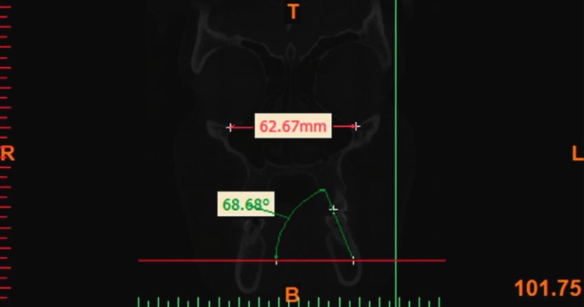
International Journal of Dentistry 3
anteroposterior relationship and the vertical relationship of
the jaws.
All CBCT images were obtained with 300 μm spatial
resolution, 110 kV and 78.59 mAs. The CBCT data were
exported in DICOM format using NNT Viewer software.
The Mimics Medical Software (version 19, Materialise,
Leuven, Belgium) was used to reconstruct lateral and
posteroanterior cephalograms. In order to standardize the
images and minimize errors in measurements, all images
were reoriented using NNT Viewer Reorientation software
(version 19, Materialise, Leuven, Belgium) such that the
Frankfurt horizontal plane and the interorbital line (a line Figure 1: Reorientation of records using the Frankfurt horizontal
connecting the inferior points of the orbital rims) were line and the interorbital line.
parallelized to the horizontal line. By doing so, the head
position was standardized in all records and all angles were
measured based on this line (Figure 1). For cephalometric
analysis, the following hard tissue reference points were
identified:
Orbitale (Or): the most inferior point of the orbital rim.
CI: the incisal edge of canine.
CA: the apex of canine.
MO: the central point of the buccolingual width of the
occlusal surface of molar tooth.
MC: the central point of the buccolingual width of the
cervical part of the anatomical crown. Figure 2: Measuring the inclination of maxillary molars.
MBM1/MBM2: the mesiobuccal cusp tip of the max-
illary first and second molars.
MLM1/MLM2: the mesiolingual cusp tip of the max-
illary first and second molars.
The cephalometric indices used for assessment of the
sagittal pattern included the ANB angle and the Wits ap-
praisal; according to which, the samples were divided into
class I (ANB: 0–4°; Wits 0 to −1), class II (ANB > 4°;
Wits > 0), and class III (ANB < 0°; Wits < −1) groups.
The measurements of inclination angles and the curve of
Wilson were made using NNT Viewer and Mimics software.
In assessment of the buccolingual inclination of molars, the
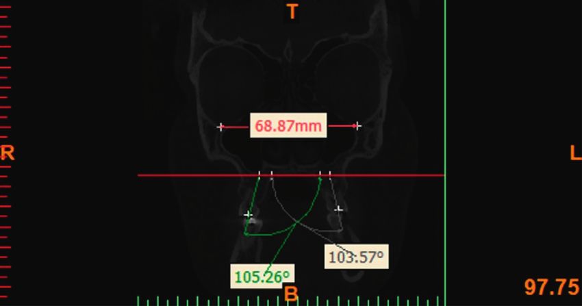
MO point was used as the reference point in order to Figure 3: Measuring the inclination of mandibular molars.
eliminate the effect of morphology of the cusp of molars. The
MC point was used as the reference point to eliminate the [31]. The Dahlberg’s formula was used to assess the method
effect of root morphology. On the frontal view, the MO-MC error. The maximum value was found to be 1.01.
line was drawn to determine the buccolingual inclination of Normal distribution of data was evaluated using the
posterior teeth (Figures 2 and 3). The CI-CA line was drawn Kolmogorov–Smirnov test. The chi-square test was used to
to determine the inclination of canine teeth (Figures 4 and compare the study groups in terms of gender. Since data
5). Its angle in the maxilla and mandible was determined by were normally distributed, ANOVA was applied for sta-
drawing a line parallel to a line connecting the two orbitale tistical analysis. For variables with nonhomogeneity of
points. variances, the Welch ANOVA was used. Tukey’s post hoc
In order to measure the curve of Wilson, the mesiobuccal test was applied for pairwise comparisons. All statistical
and mesiolingual cusp tips of the maxillary first and second analyses were carried out using SPSS version 18 (SPSS Inc.,
molars were connected along the buccal groove and the IL, USA) at 0.05 level of significance.
formed angle was measured (Figure 6).
Measurements made by an examiner and an experienced 3. Results
radiologist on 20 CBCT scans were repeated again after 2
weeks to assess the intraexaminer reliability. The lowest A total of 66 records were evaluated; out of which, 34 (51.5%)
intraclass correlation coefficient was 0.969, which was belonged to females and 32 (48.5%) belonged to males. The
considered excellent according to Cicchetti’s classification mean age of patients was 28.74 ± 5.25 years.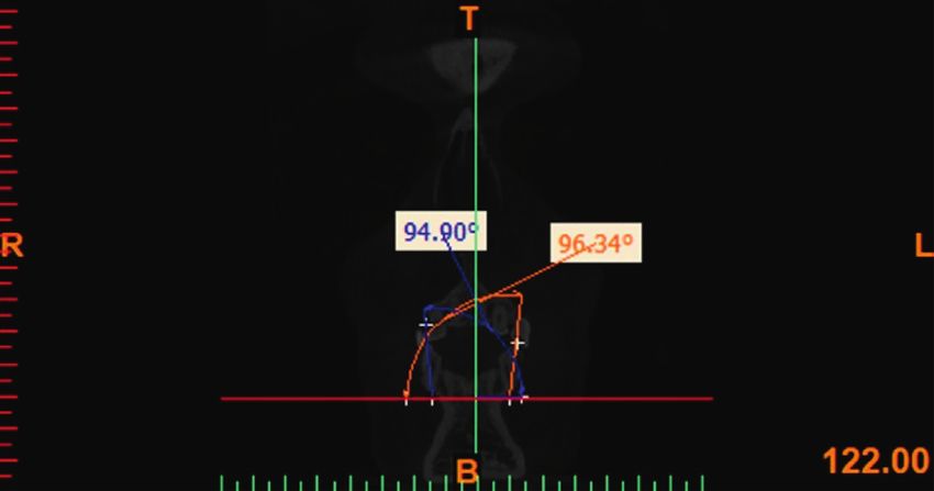
4                                                                                              International Journal of Dentistry

                                                                    Table 1: Gender distribution based on sagittal skeletal pattern.
                                                                              Cl 1           Cl 2           Cl 3
                                                                                Column         Column         Column
                                                                   Gender Count        Count          Count
                                                                                  N%              N%             N%
                                                                   Female  12     54.5  11       50.0  11       50.0
                                                                   Male    10     45.5  11       50.0  11       50.0
                                                                   Total   22    100.0  22      100.0  22      100.0

                                                                   noted in inclination of maxillary left canine between dif-
                                                                   ferent sagittal patterns (P � 0.149). A significant difference
     Figure 4: Measuring the inclination of maxillary canines.     was noted in inclination of maxillary left first molar
                                                                   (P < 0.001) such that the mean of this variable in class I and
                                                                   III was significantly higher than that in class II patients. A
                                                                   significant difference was noted in inclination of maxillary
                                                                   left second molar (P � 0.008) such that the mean of this
                                                                   variable in class I was significantly higher than that in class II
                                                                   patients. The difference in inclination of mandibular sec-
                                                                   ond molar was also significant (P < 0.001), and this variable
                                                                   in class II patients was significantly higher than that in class I
                                                                   and III patients. The difference in inclination of mandibular
                                                                   left first molar was significant as well (P � 0.002), and this
                                                                   variable in class II patients was significantly higher than that
                                                                   in class I and III patients. No significant difference was noted
                                                                   in inclination of mandibular left canine tooth among dif-
    Figure 5: Measuring the inclination of mandibular canines.     ferent sagittal patterns (P � 0.858). The difference in in-
                                                                   clination of mandibular right canine was not significant
                                                                   either (P � 0.658). The difference in inclination of man-
                                                                   dibular right first molar was significant (P < 0.001) such that
                                                                   the mean of this variable in class II was significantly higher
                                                                   than that in class I and III patients. A significant difference
                                                                   was also noted in inclination of mandibular right second
                                                                   molar (P < 0.001) such that class II patients had the highest
                                                                   and class I patients had the lowest mean of this variable with
                                                                   significant differences between all three classes.
                                                                        Table 3 compares the curve of Wilson among the three
                                                                   sagittal skeletal patterns. A significant difference was noted
                                                                   in the curve of Wilson of mandibular second molars between
                                                                   different sagittal patterns (P � 0.038) such that the mean of
            Figure 6: Measuring the curve of Wilson.               this variable in class II patients was significantly higher than
                                                                   that in class I patients. No significant difference was noted in
                                                                   the curve of Wilson of mandibular first molars between
     No significant correlation was noted between the sagittal      different sagittal patterns (P � 0.253).
skeletal pattern and gender (Table 1, chi-square test,                  Table 4 shows the mean inclination of teeth in the three
P � 0.991). The Kolmogorov–Smirnov test showed that all            sagittal skeletal patterns. Table 5 presents the mean incli-
quantitative variables had normal distribution (P > 0.05).         nation of teeth based on demographic variables of patients.
     Table 2 compares the inclination of teeth among the
three sagittal skeletal patterns. A significant difference was       4. Discussion
noted in the inclination of maxillary right second molar
among the three sagittal skeletal patterns (P � 0.002) such        This study assessed the buccolingual inclination of canine
that the mean of this variable in class I and class III patients   and first and second molar teeth and the curve of Wilson in
was significantly higher than that in class II patients. Also, a    untreated adults with different sagittal skeletal patterns using
significant difference was noted in the inclination of max-          CBCT. Of patients, 51.5% were females and 48.5% were
illary right first molar among different sagittal skeletal           males. The results showed no significant association between
patterns (P < 0.001) such that the mean of this variable in        sagittal skeletal pattern and gender. Patients between 18 and
class I and III was significantly higher than that in class II      35 years were included in this study because the inclination
patients. No significant difference was noted in the incli-          of teeth can change during the period of growth and de-
nation of maxillary right canine among different sagittal           velopment. Thus, we evaluated patients with completed
skeletal patterns (P � 0.053). No significant difference was         growth. Marshall et al. [18] reported that maxillary and
International Journal of Dentistry 5
Table 2: Comparison of the inclination of teeth among the three sagittal skeletal patterns.
Cl 1 Cl 2 Cl 3
P value
Mean SD Mean SD Mean SD
Inclination of maxillary second molar. Right 104.77b 3.45 100.20a 5.21 103.53b 4.05 0.002‡
Inclination of maxillary first molar. Right 97.42b 3.63 92.27a 5.36 98.32b 1.896 International Journal of Dentistry
Table 5: Mean inclination of teeth based on demographic variables of patients.
inc7 inc6 inc3
Standard deviation Mean Standard deviation Mean Standard deviation MeanInternational Journal of Dentistry 7
believed that when the curve of Wilson is excessively flat- compensatory relationship exists between the opposing
tened, the masticatory function is impaired [38]. ABO teeth, which along with the standards of crowns, can be used
suggests that the maxillary buccal cusps or the mandibular to determine the appropriate position of teeth in dental arch.
lingual cusps should not have more than 1 mm deviation
from the vertical axis [15]. In general, it is suggested that in Data Availability
orthodontic treatment, the curve of Wilson should be
maintained to the level that it does not impair the function of The data are available, but we cannot share them due to
mastication [13]. According to Dawson [38], the curve of patient privacy.
Wilson aims to achieve two goals: the first goal is to create an
efficient position for maximum resistance against mastica- Conflicts of Interest
tory forces. In order to achieve this goal, the buccolingual
inclination of posterior teeth should be parallel to the di- The authors declare that there are no conflicts of interest
rection of applied load and orientation of internal pterygoid regarding the publication of this paper.
muscle. The second goal is that occlusal inclination enhances
the access to food during the process of mastication. Okeson Acknowledgments
[10] explained that the curve of Wilson aims to create
maximum intercuspation. Nanda [39] stated that the This study was derived from a thesis, submitted to Ker-
presence of the curve of Wilson between the buccal surfaces manshah University of Medical Sciences, School of Den-
results in more effective occlusal function. Our results re- tistry, and was financially supported from the Kermanshah
garding the curve of Wilson showed that the mean of this University of Medical Sciences, Kermanshah, Iran.
variable in second molars of class II patients was significantly
higher than that in class I patients and the mean of this curve References
in first molars was not significantly different among different [1] H. Tong, D. Kwon, J. Shi, N. Sakai, R. Enciso, and
sagittal skeletal patterns. In order to measure the curve of G. T. Sameshima, “Mesiodistal angulation and faciolingual
Wilson in this study, the mesiobuccal and mesiolingual cusp inclination of each whole tooth in 3-dimensional space in
tips of the maxillary first and second molars along the buccal patients with near-normal occlusion,” American Journal of
groove were connected to form an angle and this angle was Orthodontics and Dentofacial Orthopedics, vol. 141, no. 5,
measured. Thus, the smaller the difference in height of the pp. 604–617, 2012.
buccal and lingual cusps of a tooth, and the higher the palatal [2] E. Motegi, H. Miyazaki, I. Ogura, H. Konishi, and M. Sebata,
inclination of that tooth, the larger the angle formed between “An orthodontic study of temporomandibular joint disorders
the two opposing teeth and the flatter the curve would be part 1: Epidemiological research in Japanese 6–18 year olds,”
(the smaller the curve of Wilson would be). Thus, the angle The Angle Orthodontist, vol. 62, no. 4, pp. 249–256, 1992.
[3] B. Thilander, G. Rubio, L. Pena, and C. de Mayorga, “Prev-
formed between the two opposing teeth had an inverse
alence of temporomandibular dysfunction and its association
correlation with the curve of Wilson. In class II patients, with malocclusion in children and adolescents: an epidemi-
considering the greater palatal inclination of molars com- ologic study related to specified stages of dental development,”
pared with class I and class III, the angle between the The Angle Orthodontist, vol. 72, no. 2, pp. 146–154, 2002.
maxillary second molars would be significantly higher and [4] B. O. Mohlin, K. Derweduwen, R. Pilley, A. Kingdon,
the curve of Wilson would be smaller. It is important to W. C. Shaw, and P. Kenealy, “Malocclusion and temporo-
determine the ideal amount of this curve and buccolingual mandibular disorder: a comparison of adolescents with
inclination of teeth for efficient function in different classes moderate to severe dysfunction with those without signs and
of occlusion. Such assessments can play a fundamental role symptoms of temporomandibular disorder and their further
in achieving the orthodontic treatment goals. Future studies development to 30 years of age,” The Angle Orthodontist,
vol. 74, no. 3, pp. 319–327, 2004.
are required to assess the correlation of curve of Spee and
[5] M. T. John, C. Hirsch, M. T. Drangsholt, L. A. Mancl, and
WALA-FA distance with the curve of Wilson in different J. M. Setz, “Overbite and overjet are not related to self-report
sagittal skeletal patterns. Also, the inclination of molar and of temporomandibular disorder symptoms,” Journal of Dental
canine teeth should be investigated with regard to the in- Research, vol. 81, no. 3, pp. 164–169, 2002.
clination of their surrounding bone. Controlled studies are [6] I. Egermark, T. Magnusson, and G. E. Carlsson, “A 20-year
also recommended to assess the effect of age on tooth in- follow-up of signs and symptoms of temporomandibular
clination and the curve of Wilson. disorders and malocclusions in subjects with and without
orthodontic treatment in childhood,” The Angle Orthodontist,
vol. 73, no. 2, pp. 109–115, 2003.
5. Conclusion [7] H. L. Beyron, “Characteristics of functionally optimal oc-
clusion and principles of occlusal rehabilitation,” The Journal
According to the results, maxillary molars have lower in-
of the American Dental Association, vol. 48, no. 6, pp. 648–
clination and mandibular molars have higher inclination in 656, 1954.
class II sagittal pattern while no significant difference was [8] C. H. Schuyler, “Correction of occlusal disharmony of the
noted in inclination of maxillary and mandibular canines in natural dentition,” The New York State Dental Journal, vol. 13,
different sagittal skeletal patterns. The curve of Wilson in no. 8, p. 445, 1947.
second molars of class II patients was significantly higher [9] J. Barrera, J. Llamas, E. Espinar, C. Sáenz-Ramı́rez, V. Paredes,
than that in class I. In different sagittal skeletal patterns, a and J. Pérez-Varela, “Wilson maxillary curve analyzed by8 International Journal of Dentistry
CBCT. A study on normocclusion and malocclusion indi- computed tomography,” Indian Journal Dental Research,
viduals,” Medicina Oral Patologı́a Oral Y Cirugia Bucal, vol. 22, no. 3, pp. 376–380, 2011.
vol. 18, no. 3, p. e547, 2013. [27] W. Shewinvanakitkul, M. Hans, S. Narendran, and J. Martin
[10] J. P. Okeson, Management of Temporomandibular Disorders Palomo, “Measuring buccolingual inclination of mandibular
and Occlusion-E-Book, Elsevier Health Sciences, Amsterdam, canines and first molars using CBCT,” Orthodontics and
The Netherlands, 2014. Craniofacial Research, vol. 14, no. 3, pp. 168–174, 2011.
[11] F. Ackermann, “The helicoid principle in human dental oc- [28] K. Kasai and A. Kawamura, “Correlation between bucco-
clusion and articulation,” International Dental Journal, lingual inclination and wear of mandibular teeth in ancient
vol. 13, pp. 532–557, 1963. and modern Japanese,” Archives of Oral Biology, vol. 46, no. 3,
[12] B. Holly Smith, “Development and evolution of the helicoidal pp. 269–273, 2001.
plane of dental occlusion,” American Journal of Physical [29] C. H. Kau, S. Richmond, J. M. Palomo, and M. G. Hans,
Anthropology, vol. 69, no. 1, pp. 21–35, 1986. “Current products and practice: three-dimensional cone
[13] R. Alkhatib and C.-H. Chung, “Buccolingual inclination of beam computerized tomography in orthodontics,” Journal of
first molars in untreated adults: a CBCT study,” The Angle Orthodontics, vol. 32, no. 4, pp. 282–293, 2005.
Orthodontist, vol. 87, no. 4, pp. 598–602, 2017. [30] J. M. Palomo, C. H. Kau, L. B. Palomo, and M. G. Hans,
[14] L. F. Andrews, “The six keys to normal occlusion,” American “Three-dimensional cone beam computerized tomography in
Journal of Orthodontics, vol. 62, no. 3, pp. 296–309, 1972. dentistry,” Dentistry Today, vol. 25, no. 11, pp. 132–135, 2006.
[15] J. S. Casko, J. L. Vaden, V. G. Kokich et al., “Objective grading [31] D. V. Cicchetti, “Guidelines, criteria, and rules of thumb for
system for dental casts and panoramic radiographs,” Amer- evaluating normed and standardized assessment instruments
ican Journal of Orthodontics and Dentofacial Orthopedics, in psychology,” Psychological Assessment, vol. 6, no. 4, p. 284,
vol. 114, no. 5, pp. 589–599, 1998. 1994.
[16] L. F. Andrews, “The straight-wire appliance,” British Journal [32] B. Yang and C.-H. Chung, “Buccolingual inclination of molars
of Orthodontics, vol. 6, no. 3, pp. 125–143, 1979. in untreated children and adults: a cone beam computed
[17] J. Daskalogiannakis and A. Ammann, Glossary of Orthodontic tomography study,” The Angle Orthodontist, vol. 89, no. 1,
Terms, Quintessence Publ., Batavia, IL, USA, 2000. pp. 87–92, 2018.
[18] S. Marshall, D. Dawson, K. A. Southard, A. N. Lee, J. S. Casko, [33] B. Sayania, M. Merchant, P. Josephs, and C.-H. Chung,
and T. E. Southard, “Transverse molar movements during “Changes in the buccolingual inclination of first molars with
growth,” American Journal of Orthodontics and Dentofacial growth in untreated subjects: a longitudinal study,” The Angle
Orthopedics, vol. 124, no. 6, pp. 615–624, 2003. Orthodontist, vol. 87, no. 5, pp. 681–687, 2017.
[19] C. S. Handelman, L. Wang, E. A. BeGole, and A. J. Haas, [34] M. Nouri, A. H. Abdi, A. Farzan, F. Mokhtarpour, and
“Nonsurgical rapid maxillary expansion in adults: report on A. A. Baghban, “Measurement of the buccolingual inclination
47 cases using the Haas expander,” The Angle Orthodontist, of teeth: manual technique vs 3-dimensional software,”
vol. 70, no. 2, pp. 129–144, 2000. American Journal of Orthodontics and Dentofacial Orthope-
[20] H. Ito, K. Okimoto, T. Mizumori et al., “A clinical study of the dics, vol. 146, no. 4, pp. 522–529, 2014.
relationship between occlusal curvature and cranio- [35] J. A. McNamaraa, “Maxillary transverse deficiency,” Ameri-
mandibular disorders,” International Journal of Prosthodon- can Journal of Orthodontics and Dentofacial Orthopedics,
tics, vol. 10, no. 1, pp. 78–82, 1997. vol. 117, no. 5, pp. 567–570, 2000.
[21] B. Podesser, S. Williams, H. P. Bantleon, and H. Imhof, [36] L. F. Andrews, “The six elements of orofacial harmony,”
“Quantitation of transverse maxillary dimensions using Andrews Journal, vol. 1, pp. 13–22, 2000.
computed tomography: a methodological and reproducibility [37] J. A. McNamara, W. L. Brudon, and V. G. Kokich, Ortho-
study,” The European Journal of Orthodontics, vol. 26, no. 2, dontics and Dentofacial Orthopedics, Needham Press, Norfolk,
pp. 209–215, 2004. MA, USA, 2001.
[22] D. G. Garib, J. F. C. Henriques, G. Janson, M. R. de Freitas, [38] P. E. Dawson, Functional Occlusion-E-Book: From TMJ to
and A. Y. Fernandes, “Periodontal effects of rapid maxillary Smile Design, Elsevier Health Sciences, Amsterdam, The
expansion with tooth-tissue-borne and tooth-borne ex- Netherlands, 2006.
panders: a computed tomography evaluation,” American [39] R. Nanda, Biomechanics and Esthetic Strategies in Clinical
Journal of Orthodontics and Dentofacial Orthopedics, vol. 129, Orthodontics, Elsevier Health Sciences, Amsterdam, The
no. 6, pp. 749–758, 2006. Netherlands, 2005.
[23] V. A. Ross, R. J. Isaacson, N. Germane, and L. K. Rubenstein,
“Influence of vertical growth pattern on faciolingual incli-
nations and treatment mechanics,” American Journal of Or-
thodontics and Dentofacial Orthopedics, vol. 98, no. 5,
pp. 422–429, 1990.
[24] G. Janson, R. Bombonatti, K. S. Cruz, C. Y. Hassunuma, and
M. Del Santo Jr., “Buccolingual inclinations of posterior teeth
in subjects with different facial patterns,” American Journal of
Orthodontics and Dentofacial Orthopedics, vol. 125, no. 3,
pp. 316–322, 2004.
[25] M. Tsunori, M. Mashita, and K. Kasai, “Relationship between
facial types and tooth and bone characteristics of the mandible
obtained by CT scanning,” The Angle Orthodontist, vol. 68,
no. 6, pp. 557–562, 1998.
[26] S. Mitra and M. S. Ravi, “Evaluation of buccolingual incli-
nation of posterior teeth in different facial patterns usingYou can also read