Distalisation of the dental arches using clear aligners and miniscrews - Exeley
←
→
Page content transcription
If your browser does not render page correctly, please read the page content below
Distalisation of the dental arches using clear
 aligners and miniscrews
 Hanshi Li,*† Li Mei,+ Yan Yang,± Xin Zhao‡ and Yu Li*
 State Key Laboratory of Oral Diseases, National Clinical Research Center for Oral Diseases, Department of Orthodontics,
 West China Hospital of Stomatology, Sichuan University, Chengdu, Sichuan, China,* Private orthodontic practice,
 Chengdu, China,† Discipline of Orthodontics, Department of Oral Sciences, Sir John Walsh Research Institute, Faculty of
 Dentistry, University of Otago, New Zealand,+ State Key Laboratory of Oral Diseases, National Clinical Research Center
 for Oral Diseases, Department of Maxillofacial Surgery, West China Hospital of Stomatology, Sichuan University, Chengdu,
 Sichuan, China± and Department of Pediatric Dentistry, Stomatological Hospital, Chongqing Medical University, China‡

 Purpose and methods: The distalisation of molars using clear aligners has been found to be achievable; however, the distalisation
 of an entire arch is still an orthodontic challenge and reported as only clinically possible using fixed appliances. To date,
 there has been no study on the distalisation of both dental arches using a clear aligner system. In the present report, a case is
 described in which the entire maxillary and mandibular arches were successfully distalised using clear aligners and miniscrews
 for the treatment of a bimaxillary protrusion malocclusion in a 22-year-old female patient. Pre- and post-treatment records as well
 as 1.5-year follow-up records are presented.
 Results: The distalisation of both dental arches was achieved using a V-pattern staging process by moving the second molars,
 then the first molars, followed by the premolars and the anterior teeth, as well as by elastics applied between precision cutouts
 in the aligners and the miniscrews. The dental arches were efficiently distalised by approximately 3 millimeters in the first molar
 areas after 13 months of treatment. A review 1.5 years post-treatment showed that the outcome was stable without significant
 relapse observed in the facial profile and occlusion.
 Conclusion: The distalisation of both dental arches is achievable using clear aligner systems by applying elastics between
 miniscrews and precision aligner cutouts in the treatment of a bimaxillary protrusion malocclusion.
 (Aust Orthod J 2021; 37: 128 - 138. DOI: 10.21307/aoj-2021-014)

 Received for publication: February 2020
 Accepted: January 2021

 Hanshi Li: doralihanshi@hotmail.com; Li Mei: li.mei@otago.ac.nz; Yan Yang: 1127152151@qq.com; Xin Zhao: 240074637@qq.com\;
 Yu Li: yuli@scu.edu.cn

 Background                                                           The aetiology of a bimaxillary protrusion has been
 A bimaxillary protrusion is characterised by protrusive              found to be complex, and may consist of a genetic
 and proclined maxillary and mandibular incisors                      component superimposed on environmental factors
 accompanying increased procumbency of the lips                       related to mouth breathing, tongue and lip malfunc-
 and is commonly seen in African-American and                         tion, and enlarged tongue volume.4 The orthodontic
 Asian populations.1 The malocclusion can negatively                  treatment of the malocclusion often involves the ini-
 impact on a patient’s facial aesthetics, self-esteem,                tial extraction of the four premolars followed by re-
 psychological well-being and quality of life.2 Patients              traction and retroclination of the anterior teeth using
 presenting with a bimaxillary protrusion malocclusion                maximum anchorage mechanics, ideally resulting in a
 usually seek treatment for a chief complaint related to              decrease in lip procumbency.5,6
 their prominent dentition and lips and a desire for an               An alternative treatment option to manage a
 improvement of their profile.3 These patients not only               bimaxillary protrusion is the distalisation of both
 demand but also need orthodontic treatment.                          dental arches using temporary anchorage devices

128      Australasian Orthodontic Journal Volume 37 No. 1 May 2021                             © Australian Society of Orthodontists Inc. 2021
DISTALISATION OF THE DENTAL ARCHES USING CLEAR ALIGNERS AND MINISCREWS

(TADs). Although distalisation of an entire maxillary               and mandibular dentitions using clear aligners and
and/or mandibular arch is generally considered                      miniscrews for a patient presenting with a bimaxillary
to be extremely difficult compared with molar                       protrusion.
distalisation,7 it has been demonstrated that an entire
mandibular dentition may be successfully distalised
with the aid of miniscrews in the correction of a Class             Case report
III malocclusion.8 The distalisation of maxillary and               A 22-year-old female presented with the main
mandibular dentitions was first reported in 2004                    complaint of “protrusive lips” and said she “would like
for the treatment of Class I bimaxillary protrusion                 to have treatment with clear aligners” because of her
and anterior crowding using fixed appliances and                    work as an actress. A facial analysis showed that there
miniscrews.9 The miniscrew-anchored orthodontics10                  was a symmetrical face, a convex profile and a decreased
has been shown to be an effective approach for                      nasolabial angle (Figure 1). No temporomandibular
distalising dentitions in patients presenting with Class            joint disorder was noted during the consultation and
I bimaxillary protrusion and in cases of Class II and               clinical examination. An intraoral assessment revealed
Class III malocclusions; however, this technique has                that there was an Angle Class I molar relationship on
only been reported in association with fixed appliance              her left side and a Class III relationship on the right
systems. To date, there is no report describing the                 side, a 1 mm overjet and overbite, and symmetrical
distalisation of both dentitions using a clear aligner              dental arches with no crowding. Both upper and lower
system.                                                             midlines were 1 mm to the left of the facial midline.
The aim of this case report was to introduce a clinically           No mandibular functional shift was observed (Figures
effective approach for distalising both maxillary                   1 and 2).

Figure 1. Pretreatment facial and intraoral photographs.

                                                                          Australasian Orthodontic Journal Volume 37 No. 1 May 2021   129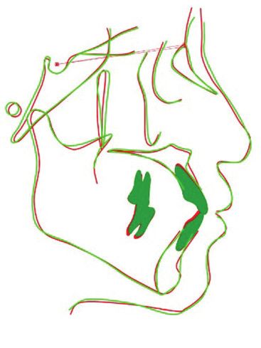
LI, MEI, YANG, ZHAO AND LI

 Figure 2. Pretreatment dental casts.

 Panoramic radiography showed that the 46 was a
 crowned tooth with previous root canal treatment.
 The four third molars were present (Figure 3). A
 cephalometric analysis indicated a skeletal Class I
 pattern with an average mandibular plane angle. Both
 the maxillary and mandibular incisors were proclined
 (Figure 3).

 Treatment alternatives
 The objectives of orthodontic treatment were to
 retract the upper and lower anterior teeth, decrease
 the lip prominence and improve the profile.
 Due to the patient’s request for clear aligner treatment,
 the following two treatment options were discussed.
 Option one: Invisalign treatment (Align Technology,
 Inc., CA, USA) involving the extraction of the four
 first premolars, the retraction and retroclination of the
 upper and lower incisors to decrease lip prominence.
 Option two: Invisalign treatment to distalise both
 dental arches, retract and retrocline the upper and
 lower incisors, and to decrease the lip prominence.                  Figure 3. Pretreatment cephalometric radiograph (top left) and tracing
 Miniscrews would be required to reinforce posterior                  (top right) and panoramic radiograph (bottom).

130       Australasian Orthodontic Journal Volume 37 No. 1 May 2021
DISTALISATION OF THE DENTAL ARCHES USING CLEAR ALIGNERS AND MINISCREWS

anchorage. Elastics applied between the miniscrews                               traditional fixed appliances with ceramic brackets
and precision cutouts in the clear aligners would be                             were discussed as treatment alternatives. However,
used to achieve the distalisation of both arches (Figure                         the patient persisted in a non-extraction treatment
4). The third molars 28, 38 and 48 would be extracted;                           program requiring the distalisation of the entire
the 18 was not extracted but monitored during                                    dentitions using clear aligners.
treatment because it was affected by microdontia and
in a high position with an expected inconsequential
impact on the distalisation of the dentition in the
                                                                                 Treatment progress
respective quadrant. The extraction of the deeply                                The ClinCheck (Align Technology, Inc., CA, USA)
impacted 18 was likely to involve risks of tearing of                            was used to visualise the treatment procedures and
tissue flaps and the excessive removal to bone and                               detail the required tooth movements.
overlying soft tissue (Figure 3).                                                In the first trial, a total of 30 aligners (19 upper and
The second option was chosen because the patient                                 11 lower aligners involving six months of treatment)
was against removing healthy premolar teeth and the                              were used to level and align both dentitions and to
possible compromise in aesthetics by the use of virtual                          distalise both arches. No interproximal reduction was
pontics during retraction and space management.                                  planned during this stage. A miniscrew (length of 10
                                                                                 mm and diameter of 2 mm; Ormco Corporation, CA,
During the planning discussion regarding treatment
                                                                                 USA) was inserted between the second premolar and
alternatives, the patient was informed of a previous                             first molar in each quadrant. Precision cutouts (hooks
study that indicated there was no significant outcome                            on canines) were prescribed in the aligners (Figure
difference between second premolar extraction and                                4). According to Align Technology’s instructions, the
distalisation with interproximal reduction (IPR)                                 patient was required to wear each aligner with elastics
treatment,11 and that first premolar extraction might                            (200g force on each side, size 1/8, 3.5 oz) between the
be more effective in improving the bimaxillary                                   precision cutouts and miniscrews for at least 22 hours
protrusion. In addition, lingual fixed appliances and                            per day for 10–14 days in order to distalise both dental
                                                                                 arches (Figure 4). However, no clinically significant
                                                                                 improvement was observed in the retraction of the
                                                                                 lips nor improvement in the molar relationships after
                                                                                 the first 100 days of treatment. The upper and lower
                                                                                 midlines remained 1 mm and 2 mm to the left of
                                                                                 the facial midline, respectively (Figure 5). Therefore,
                                                                                 a refinement was required before attempting further
                                                                                 distalisation of the dentitions.
                                                                                 In a second phase, 60 aligners (30 for each arch, 10
                                                                                 months treatment) were prescribed. In the ClinCheck
                                                                                 review, the distalisation of the dentitions was revised
                                                                                 to a “V pattern”,12 requiring the distalisation of the
                                                                                 second molars, then the first molars, followed by
                                                                                 the premolars and the anterior teeth. The planned
                                                                                 magnitude of molar distalisation in the maxillary arch
                                                                                 was 0.9 mm (right side) and 1.1 mm (left side) and
                                                                                 1.0 mm (right) and 0.9 mm (left) in the mandibular
                                                                                 arch in order to improve the dental midlines and the
                                                                                 molar relationship on the right side (Figure 5). No
                                                                                 IPR was performed in this stage.
                                                                                 After the completion of the first refinement, IPR was
                                                                                 performed in both arches (1.2 mm in the upper and
                                                                                 1.4 mm in the lower), and an additional 10 aligners
Figure 4. Intraoral photographs showing the elastics running from the
                                                                                 were provided for a second refinement to achieve a
precision-cuts to the miniscrews.                                                normal overjet and overbite (Figures 6 and 7).

                                                                                       Australasian Orthodontic Journal Volume 37 No. 1 May 2021   131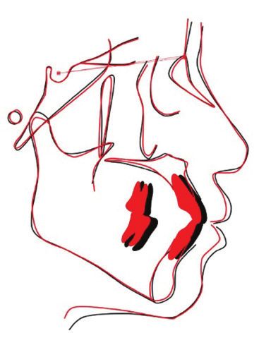
LI, MEI, YANG, ZHAO AND LI

 Figure 5. Facial and intraoral photographs before the first refinement during treatment.

 The active treatment duration was 13 months, during                               E-plane was 2.1 mm for the upper lip and 2.9 mm for
 which time the miniscrews remained stable. Vacuum                                 the lower lip). The S-Go/N-Me showed little change,
 formed retainers were provided for retention and the                              indicating an unaltered facial height (Figure 10). The
 patient was followed up for 1.5 years (Figure 8).                                 value of SN-MP increased by 1.5° indicating a slight
                                                                                   clockwise rotation of the mandible. The Ptm-U6
                                                                                   indicated that the upper first molars moved posteriorly
 Treatment results                                                                 by 3.0 mm (Table I). Considering that IPR was
 The post-treatment examination demonstrated that                                  performed only on teeth in front of the first molars
 the treatment objectives were achieved (Figures 6 and                             (i.e., incisors, canines and premolars), the 3.0 mm
 7). The facial photographs showed an improved profile                             distalisation of the upper first molar was attributed to
 and an aesthetic smile (Figure 6). The panoramic                                  the distalisation of the entire maxillary dentition.
 radiograph showed paralleled roots with no significant                            At a 1.5 year review, the treatment results were stable
 root resorption or alveolar bone recession (Figure 9).                            and no significant relapse was observed in the profile
 A cephalometric analysis and superimposition (Figures                             nor occlusion (Figures 8 and 12). The two maxillary
 10, 11 and Table I) revealed that the maxillary and                               miniscrews were planned to remain in situ for two
 mandibular incisors were retracted (the reduction                                 years after debonding to assist retention, during
 of U1-AP and L1-AP was 2.2 mm and 3.2 mm,                                         which time the patient was asked to wear elastics in
 respectively). The SNA and SNB values decreased                                   the maxilla between the miniscrews and the cutouts
 by 1.6° and 2.2° respectively, indicating a posterior                             (hooks on canines) in the clear retainers at night. It
 remodelling of A and B points following upper and                                 was considered that the retained upper incisors would
 lower incisor retraction. The lip prominence was                                  naturally hold the lower incisors due to the corrected
 reduced (the reduction of the distance from lips to                               overjet and overbite.

132       Australasian Orthodontic Journal Volume 37 No. 1 May 2021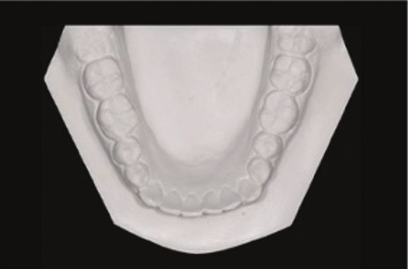
DISTALISATION OF THE DENTAL ARCHES USING CLEAR ALIGNERS AND MINISCREWS
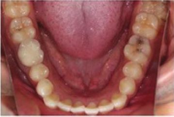
Figure 6. Post-treatment facial and intraoral photographs.
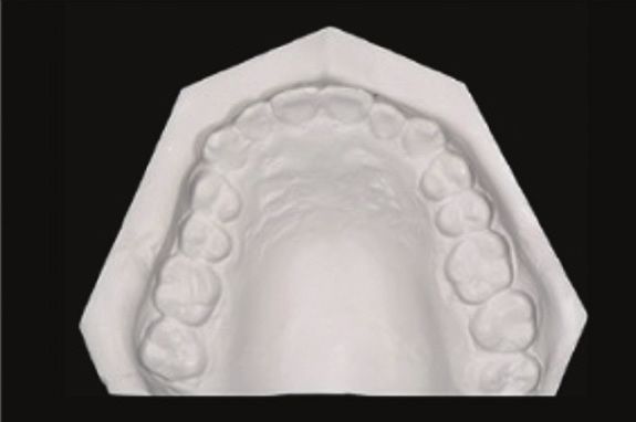
Figure 7. Post-treatment dental casts.
Australasian Orthodontic Journal Volume 37 No. 1 May 2021 133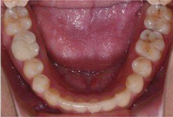
LI, MEI, YANG, ZHAO AND LI

 Table I. Cephalometric analyses before and after the treatment.

  Measurement                                                                      Normal43, 44             Before           After          Changes*
  Skeletal
  SNA (Sella-Nasion-point A, °)                                                    83.6 ± 3.6               84.8             83.2             -1.6
  SNB (Sella-Nasion-point B, °)                                                    79.7 ± 3.6               81.5             79.3             -2.2
  ANB (Point A-Nasion-point B, °)                                                   3.9 ± 1.8                3.0              3.9             -0.9
  SN-MP (SN plane-mandibular plane, °)                                             32.9 ± 4.0               34.4             35.9              1.5
  S-Go/N-Me (Sella-gonion/nasion-menton, %)                                        65.9 ± 4.0               67.3             66.1             -1.2
  Dental
  U1-L1 (Axial inclination of upper and lower incisors, °)                        117.6 ± 8.8              116.7            115.8             -0.9
  U1-SN (U1-sella nasion, °)                                                       65.0 ± 7.5               67.9             66.9             -1.0
  U1-AP (Protrusion of maxillary incisors, mm)                                      6.7 ± 2.0               10.8              8.6             -2.2
  L1-AP (Protrusion of mandibular incisors, mm)                                     3.0 ± 2.0                8.3              5.1             -3.2
  FMIA (Frankfort horizontal plane-L1, °)                                          57.0 ± 7.0               57.5             61.6              4.1
  U1-PP (U1-palatal plane, mm)                                                     27.5 ± 2.0               30.5             29.7             -0.8
  U6-PP (U6-palatal plane, mm)                                                     21.5 ± 2.0               26.6             25.6             -1.0
  Ptv-U6 (Pterygomaxillary fissure-U6, mm)                                         18.0 ± 3.0               22.8             19.8             -3.0
  Profile
  Upper lip to E-plane (mm)                                                        -1.5 ± 2.4                1.0              -1.1            -2.1
  Lower lip to E-plane (mm)                                                         0.9 ± 2.5                4.5               1.6            -2.9
 * + (-) indicate the values increased (decreased) after the treatment.

 Figure 8. Facial and intraoral photographs after 1.5 years retention. The upper two miniscrews remained in the oral cavity for retention
 (elastics from the cuts in the retainer to the miniscrews).

134        Australasian Orthodontic Journal Volume 37 No. 1 May 2021
DISTALISATION OF THE DENTAL ARCHES USING CLEAR ALIGNERS AND MINISCREWS

Discussion                                                                      treatment option of premolar extraction, even with
The present case report has described a young female                            virtual pontics for aesthetic compensation due to
adult who presented with a bimaxillary protrusion                               her work as an actress, was not accepted. Therefore,
malocclusion. The patient was concerned about                                   treatment involved the extraction of third molars and
her smile aesthetics, hence the preference for clear                            the distalisation of both arches using clear aligners
aligners rather than fixed appliances. In addition, a                           supported by miniscrews.
                                                                                Bimaxillary protrusion is commonly seen among black
                                                                                and Asian populations.13 These patients usually have
                                                                                an acceptable occlusion but a prominent lip profile.
                                                                                The orthodontic treatment for protrusive patients
                                                                                often involves the extraction of premolars to retract
                                                                                the anterior teeth and reduce the prominence of the
                                                                                lips.14 However, this option usually takes an extended
                                                                                period of time, and some patients may refuse the
                                                                                extraction of premolars for reasons related to reduced
                                                                                smile aesthetics during treatment. An alternative
                                                                                option is arch distalisation, which may involve the
                                                                                extraction of the third molars only and thereby avoid a
                                                                                compromise in aesthetics induced by extractions. The
                                                                                treatment outcomes of a second premolar extraction
                                                                                program or arch distalisation options (including facial
                                                                                attractiveness, age appearance and soft-tissue measures)
                                                                                do not reportedly show significant differences in
                                                                                the long term.15,16 The extraction of first premolars
                                                                                provides more space for the retraction of the anterior
                                                                                teeth. Moreover, additional advantages of distalising
                                                                                the dentitions rather than premolar extractions
                                                                                include the preservation of a complete dentition,
                                                                                the ease of root position control, the convenience of
Figure 9. Post-treatment cephalometric radiograph (top left) and tracing        controlling the amount of anterior tooth retraction, as
(top right) and panoramic radiograph (bottom).                                  well as good patient acceptance.17

Figure 10. Superimposition of the pretreatment      Figure 11. Superimposition of the pretreatment    Figure 12. Superimposition of the post-treatment
(black line) and post-treatment (red line)          (black line) and post-treatment (red line)        (red line) and the 1.5-year retention (green line)
cephalometric radiographs.                          cephalometric radiographs (maxilla and            cephalometric radiographs.
                                                    mandible alone).

                                                                                       Australasian Orthodontic Journal Volume 37 No. 1 May 2021      135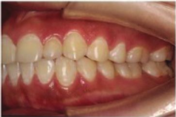
LI, MEI, YANG, ZHAO AND LI

 The Invisalign system has become increasingly                       first molar areas after one year of treatment. A much
 popular, especially for adult patients,18-20 since it               greater amount of first molar distalisation (0.9 mm
 was first introduced in 1997, due to the advantages                 and 1.0 mm on the right upper and lower; 1.1 mm and
 of aesthetics and comfort in comparison with fixed                  0.9 mm on the left side) was achieved, as prescribed
 appliances.21 The clinical efficiency of clear aligner              in the ClinCheck prediction, which suggests that
 treatment, including molar distalisation, has been                  separate molar and whole arch distalisation can occur
 confirmed and it has been further suggested that                    at the same time. Compared with the synchronous
 tooth movement, such as incisor torque, premolar                    arch distalisation modality, the present approach
 derotation and molar distalisation, can be effectively              may have advantages related to the ease of achieving
 performed using the Invisalign system.22,23 A high                  separate tooth distalisation, even with lighter forces;
 accuracy (88%) of bodily movement of the upper                      and the bone remodelling which is activated in situ, to
 molars could be achieved using aligners when a mean                 facilitate the entire arch distalisation.
 distalisation movement of 2.7 mm was prescribed                     When distalising a dental arch, the opposing arch
 in the pretreatment planning.23 The maxillary first                 may serve as an anchorage source. When distalising
 molars can be successfully distalised by 2.25 mm                    both dental arches, TADs remain a positive option for
 without significant tipping using aligners.22
                                                                     anchorage support. The diameter, length, insertion
 However, the Invisalign system, as well as other                    angle and position of the TADs are important. The
 clear aligner systems, may not be as effective as fixed             diameter of a miniscrew for arch distalisation was
 appliances for treating a bimaxillary protrusion with the           recommended to be between 1.2 mm26,27 and 2 mm,28,29
 accompanying extraction of four premolars followed                  and the minimum length of the miniscrew should
 by anterior tooth retraction. Clear aligners are usually            be at least 6 mm according to a systematic review.30
 associated with tipping (instead of bodily) movement                Although interradicular sites are common areas for
 of the teeth, resulting in tilting of the posterior teeth           TAD placement, the maxillary infrazygomatic region
 and lingual crown torque of the incisors towards the                and mandibular buccal shelf have been recommended
 extraction spaces that incompletely close. Therefore,               for upper and lower arch distalisation to avoid the
 arch distalisation serves as an optional treatment                  potential interference of the TADs with the tooth
 plan in clear aligner treatment, especially for mild                roots.31 However, the placement of TADs at these two
 to moderate bimaxillary protrusion. However, the                    sites is technically difficult and so, in recent years, it
 treatment modality for the distalisation of both arches             has been recommended that the TADs be placed at
 in clear aligner systems has not yet been reported.                 interradicular sites at a higher position and at a greater
 During arch distalisation using fixed appliances, all of            angle to support anchorage.32,33 This was found to be
 the teeth are forced synchronously, without separate                appropriate in the present case. The insertion angle
 distalisation, which has been found to be clinically                of 55° to 70° relative to the maxillary occlusal plane
 successful.16,24,25 The first phase of treatment used this          has been found to enhance primary stability for
 strategy to distalise both arches using clear aligners              the distalisation of the maxillary dentition but may
 supported by miniscrews without clinical success.                   increase the risk of sinus perforation in the maxillary
 The different outcomes may be explained by the                      molar region.28 The optimal position of the TADs for
 imprecision between the slot and the arch wire in fixed             maxillary dentition distalisation has been reported
 appliance systems, which allows easy simultaneous                   to be the region between the first and second molars
 distal tipping of all the teeth; whereas, in clear aligner          because of the relatively thicker buccal alveolar bone
 systems, the entire arch is tightly wrapped by the                  compared with other sites.28,34 This insertion position,
 plastic, which prevents easy tipping movement. In the               however, is still not ideal because of an associated
 second phase of treatment, the teeth were separately                potential risk of interfering with root movement,
 distalised in a staged V-pattern12 by moving the                    and therefore requiring a later repositioning of the
 second molars, then the first molars, followed by the               miniscrews. In the present case, the upper dentition
 premolars and the anterior teeth, and at the same                   was distalised by 3.0 mm according to the Ptv-U6
 time the whole arch was distalised using miniscrews.                measurement, resulting in a clinically asymptomatic
 As a result, both dental arches were successfully and               but a radiologically close position of the miniscrews
 efficiently distalised approximately 3.0 mm at the                  to the roots of the second premolars after treatment.

136      Australasian Orthodontic Journal Volume 37 No. 1 May 2021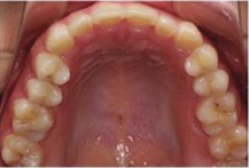
DISTALISATION OF THE DENTAL ARCHES USING CLEAR ALIGNERS AND MINISCREWS

The molars were successfully distalised using the                succeeding fixed appliance treatment after an average
clear aligners without clinically significant adverse            molar distalisation movement of about 5.1 mm.42
molar extrusion and a mandibular clockwise rotation              The treatment results of entire dentition distalisation
determined by the cephalometric superimpositions.                using miniscrews appeared relatively stable after a
This may be a result of the lack of extrusive forces             long period of review (up to five years’ retention).24,25
generated by the interarch elastics, but rather, the             The risk of relapse in both arch distalisations is
forces from the precision cutouts to the miniscrews              theoretically higher than that of a single arch, since
were relatively higher than the centre of resistance of          there is no retention force from intercuspation due to
the molars, which minimised any potential adverse                the simultaneous relapse tendency in both arches. In
molar extrusion. In addition, the aligner coverage               the present case, the patient was asked to wear elastics
over the teeth could also effectively prevent possible           in the maxilla between the miniscrews and the cutouts
molar extrusion. Therefore, the application of elastics          (hooks on canines) in the clear retainers at night. The
from the TADs to the precision cutouts of the aligners           miniscrews in the mandible were removed after active
is recommended, rather than to the individual teeth,             treatment. After 1.5 years of retention, the treatment
during entire arch distalisation.                                result remained stable; however, long-term stability
                                                                 still requires further observation.
The impact of distalisation treatment (either only
molars or the entire dentition) on the third molars is
still unclear. It has been reported that the distalisation       Summary and conclusion
of maxillary molars using a pendulum appliance does
                                                                 The distalisation of both dental arches may be achieved
not influence root formation nor the position of the
                                                                 by clear aligner systems using elastics between the
third molars.35 However, an alternative study suggested
                                                                 miniscrews and precision cutouts in the treatment of
that orthodontically-treated patients may develop a
                                                                 a bimaxillary protrusion malocclusion.
higher likelihood of impacted third molars, which
potentially leads to local clinical morbidities, related
to pericoronitis or caries on adjacent teeth.36 A study          Corresponding author
using a pendulum appliance for molar distalisation               Professor Yu Li
suggested that there may be unwanted tipping of                  State Key Laboratory of Oral Diseases
the second molar if the distalisation of the first and           National Clinical Research Center for Oral Diseases
second molars was carried out simultaneously without             Department of Orthodontics
germectomy of the third molar.37 There is a report               West China Hospital of Stomatology
regarding external root resorption of the second molars          Sichuan University
using cervical traction for first molar distalisation.           14#, 3rd Section, South Renmin Road
Such devices may apply a relatively high force over a            Chengdu 610041
long duration and an unerupted, developing second                China
molar may be affected.38 There is no report regarding            Email: yuli@scu.edu.cn
external root resorption of the second molars caused
by the third molars during distalisation. In the present
case, the impacted, undersized 18 was not extracted              References
                                                                 1.   Bills D, Handelman C, BeGole E. Bimaxillary dentoalveolar
but was closely monitored. It did not cause significant
                                                                      protrusion: traits and orthodontic correction. Angle Orthod
root resorption of the 17 nor a negative impact on the                2005;75:333-9.
distalisation of the dentition, perhaps due to the small         2.   Almutairi T, Albarakati S, Aldrees A. Influence of bimaxillary
size and the high position of the tooth.                              protrusion on the perception of smile esthetics. Saudi Med J
                                                                      2015;36:87-93.
The long-term stability of arch distalisation is still           3.   Sundareswaran S, Vijayan R. Profile changes following orthodontic
                                                                      treatment of class I bimaxillary protrusion in adult patients of
unclear. It has been found that the stability of treatment
                                                                      Dravidian ethnicity: A prospective study. Indian J Dent Res
results achieved by headgear and an improved                          2017;28:530-7.
Nance appliance for dentition distalisation were                 4.   Lamberton C, Reichart P, Triratananimit P. Bimaxillary protrusion as
equivocal.39-41 A longitudinal study of the pendulum                  a pathologic problem in the Thai. Am J Orthod 1980;77:320-9.
                                                                 5.   Tan TJ. Profile changes following orthodontic correction of
appliance for molar distalisation showed that almost                  bimaxillary protrusion with a preadjusted edgewise appliance. Int J
half of the patients experienced relapse during the                   Adult Orthodon Orthognath Surg 1996;11:239-51.

                                                                        Australasian Orthodontic Journal Volume 37 No. 1 May 2021     137
LI, MEI, YANG, ZHAO AND LI
6. Bravo L. Soft tissue facial profile changes after orthodontic treatment 27. Park H, Kwon T, Sung J. Nonextraction treatment with microscrew
with four premolars extracted. Angle Orthod 1994;64:31-42. implants. Angle Orthod 2004;74:539-49.
7. Bechtold T, Kim J, Choi T, Park Y, Lee K. Distalization pattern of the 28. Liu H, Wu X, Yang L, Ding Y. Safe zones for miniscrews in
maxillary arch depending on the number of orthodontic miniscrews. maxillary dentition distalization assessed with cone-beam computed
The Angle Orthod 2013;83:266-73. tomography. Am J Orthod Dentofacial Orthop 2017;151:500-6.
8. Jing Y, Han X, Guo Y, Li J, Bai D. Nonsurgical correction of a Class III 29. Poggio P, Incorvati C, Velo S, Carano A. “Safe zones”: a guide for
malocclusion in an adult by miniscrew-assisted mandibular dentition miniscrew positioning in the maxillary and mandibular arch. Angle
distalization. Am J Orthod Dentofacial Orthop 2013;143:877-87. Orthod 2006;76:191-7.
9. Park H, Kwon T, Sung J. Nonextraction Treatment with Microscrew 30. Chen Y, Kyung H, Wen T, Yu W. Critical factors for the success
Implants. The Angle Orthod 2004;74:539-49. of orthodontic mini-implants: a systematic review. Am J Orthod
10. Deguchi T, Kurosaka H, Oikawa H, Kuroda S, Takahashi I, Dentofacial Orthop 2009;135:284-91.
Yamashiro T et al. Comparison of orthodontic treatment outcomes 31. Chang C, Liu S, Roberts W. Primary failure rate for 1680 extra-
in adults with skeletal open bite between conventional edgewise alveolar mandibular buccal shelf miniscrews placed in movable
treatment and implant-anchored orthodontics. Am J Orthod mucosa or attached gingiva. Angle Orthod 2015;85:905-10.
Dentofacial Orthop 2011;139:S60-8. 32. Bechtold T, Kim J, Choi T, Park Y, Lee K. Distalization pattern of the
11. Jung M. A comparison of second premolar extraction and mini- maxillary arch depending on the number of orthodontic miniscrews.
implant total arch distalization with interproximal stripping. Angle Angle Orthod 2013;83:266.
Orthod 2013;83:680-5. 33. Lee S, Abbas N, Bayome M, Baik U, Kook Y, Hong M et al. A
12. Graber LW, Vanarsdall RL, Vig KWL, Huang GJ. Orthodontics: comparison of treatment effects of total arch distalization using
Current Principles and Techniques. E-Book. Elsevier Health modified C-palatal plate vs buccal miniscrews. Angle Orthod
Sciences; 2016. 2017;88:45-51.
13. Zhang J, Qiao M. [Cephalometric analysis of the patients with 34. Oh Y, Park H, Kwon T. Treatment effects of microimplant-aided
bimaxillary protrusion in south China]. Hua Xi Kou Qiang Yi sliding mechanics on distal retraction of posterior teeth. Am J
Xue Za Zhi = West China Journal of Stomatology 2001;19:32-4. Orthod Dentofacial Orthop 2011;139:470-81.
Chinese. 35. Ghosh J, Nanda R. Evaluation of an intraoral maxillary molar
14. Dandajena T, Nanda R. Bialveolar protrusion in a Zimbabwean distalization technique. Am J Orthod Dentofacial Orthop
sample. Am J Orthod Dentofacial Orthop 2003;123:133-7. 1996;110:639-46.
15. Janson G, Junqueira C, Mendes L, Garib D. Influence of premolar 36. Miclotte A, Franco A, Guerrero M, Willems G, Jacobs R. The
extractions on long-term adult facial aesthetics and apparent age. Eur association between orthodontic treatment and third molar position,
J Orthod 2016;38:272-80. inferior alveolar nerve involvement, and prediction of wisdom tooth
16. Verma SL, Sharma VP, Tandon P, Singh GP, Sachan K. Comparison eruption. Surg Radiol Anat 2015;37:333-9.
of esthetic outcome after extraction or non-extraction orthodontic 37. Kinzinger G, Fritz U, Sander F, Diedrich P. Efficiency of a pendulum
treatment in class II division 1 malocclusion patients. Contemp Clin appliance for molar distalization related to second and third molar
Dent 2013;4:206-12. eruption stage. Am J Orthod Dentofacial Orthop 2004;125:8-23.
17. Kang S, Kim H, Hwang H, Lee K. Immediate changes in the 38. Langford SR, Sims MR. Upper molar root resorption because of
mandibular dentition after maxillary molar distalization using distal movement. Report of a case. Am J Orthod 1981;79:669-79.
headgear. Korean J Orthod 2017;47:142-7. 39. Yoshida N, Jost-Brinkmann PG, Koga Y, Mimaki N, Kobayashi
18. Kesling H. Coordinating the predetermined pattern and tooth K. Experimental evaluation of initial tooth displacement, center
positioner with conventional treatment. Am J Orthod Oral Surg of resistance, and center of rotation under the influence of an
1946;32:285-93. orthodontic force. Am J Orthod Dentofacial Orthop 2001;120:190-
19. Rossini G, Parrini S, Castroflorio T, Deregibus A, Debernardi C. 7.
Efficacy of clear aligners in controlling orthodontic tooth movement: 40. Oosthuizen L, Dijkman J, Evans W. A mechanical appraisal of the
a systematic review. Angle Orthod 2015;85:881-9. Kloehn extraoral assembly. Angle Orthod 1973;43:221-32.
20. Melsen B. Northcroft lecture: how has the spectrum of orthodontics 41. Baldini G. Unilateral headgear: lateral forces as unavoidable side
changed over the past decades? J Orthod 2011;38:134-43; quiz 45. effects. Am J Orthod 1980;77:333-40.
21. Rosvall MD, Fields HW, Ziuchkovski J, Rosenstiel SF, Johnston 42. Caprioglio A, Fontana M, Longoni E, Cozzani M. Long-term
WM. Attractiveness, acceptability, and value of orthodontic evaluation of the molar movements following Pendulum and fixed
appliances. Am J Orthod Dentofacial Orthop 2009;135:276.e1-12; appliances. Angle Orthod 2013;83:447-54.
discussion -7.
22. Ravera S, Castroflorio T, Garino F, Daher S, Cugliari G, Deregibus
A. Maxillary molar distalization with aligners in adult patients: a
multicenter retrospective study. Prog Orthod 2016;17:12.
23. Simon M, Keilig L, Schwarze J, Jung BA, Bourauel C. Treatment
outcome and efficacy of an aligner technique--regarding incisor
torque, premolar derotation and molar distalization. BMC Oral
Health 2014;14:68.
24. Chen K, Cao Y. Class III malocclusion treated with distalization
of the mandibular dentition with miniscrew anchorage: A 2-year
follow-up. Am J Orthod Dentofacial Orthop 2015;148:1043-53.
25. Kuroda S, Hichijo N, Sato M, Mino A, Tamamura N, Iwata M,
et al. Long-term stability of maxillary group distalization with
interradicular miniscrews in a patient with a Class II Division 2
malocclusion. Am J Orthod Dentofacial Orthop 2016;149:912-22.
26. Park H, Lee S, Kwon O. Group distal movement of teeth using
microscrew implant anchorage. Angle Orthod 2005;75:602-9.
138 Australasian Orthodontic Journal Volume 37 No. 1 May 2021You can also read