Clinical characteristics of oral mucosal lesions in patients with systemic lupus erythematosus and their association with clinical and laboratory ...
←
→
Page content transcription
If your browser does not render page correctly, please read the page content below

Clinical characteristics of oral mucosal lesions in patients with systemic lupus
erythematosus and their association with clinical and laboratory parameters
Lilly Esquivel-Pedraza 1, 2, Laura Fernández-Cuevas 3, Alba Cicero-Casarrubias 4, *, Sergio Ponce de León-
Rosales 5, Mónica Fernández-Sánchez 1, 6, Rocío Orozco-Topete 1, 7, Carla Archer-Dubon 1, Ana Lilia Ruelas-
Villavicencio 1, Marcela Saeb-Lima 8, Ma. Guadalupe Ortíz-Pedroza 1, Linda García-Hidalgo 1, Ma. Josefina
Carbajosa-Martínez 1, María del Pilar Milke-García 9, Judith Domínguez-Chérit 1 and Silvia Méndez-Flores 1
1 Department   of Dermatology, Instituto Nacional de Ciencias Médicas y Nutrición “Salvador Zubirán”, Mexico City, Mexico.
2 Department of Health Care, Universidad Autónoma Metropolitana. Mexico City, Mexico.
3 Oral Pathology Department, Centro Dermatológico “Dr. Ladislao de la Pascua”, Mexico City, Mexico.
4 Department of Immunology and Rheumatology, Instituto Nacional de Ciencias Médicas y Nutrición “Salvador Zubirán”,

Mexico City, Mexico.
5 Department of Education, Instituto Nacional de Ciencias Médicas y Nutrición “Salvador Zubirán”, Mexico City, Mexico.
6 Centro de Investigación de Enfermedades Infecciosas. Instituto Nacional de Enfermedades Respiratorias, Mexico City,

Mexico.
7 Hospital Médica Sur
8 Pathology Department, Instituto Nacional de Ciencias Médicas y Nutrición “Salvador Zubirán”, Mexico City, Mexico.
9 Nutrition Division. Instituto Nacional de Ciencias Médicas y Nutrición “Salvador Zubirán”, Mexico City, Mexico.

GSC Advanced Research and Reviews, 2021, 06(02), 001–012

Publication history: Received on 22 January 2021; revised on 30 January 2021; accepted on 02 February 2021

Article DOI: https://doi.org/10.30574/gscarr.2021.6.2.0013

Abstract
Introduction. Systemic lupus erythematosus (SLE) is an autoimmune disease that includes a broad spectrum of
mucocutaneous manifestations.
Objectives. To characterize the clinical spectrum of oral mucosal lesions in patients with SLE and to analize their
association with clinical and laboratory parameters.
Methods. We performed a cross-sectional study with systematic oral evaluations in SLE adult patients. Systemic and
cutaneous lupus activities were recorded. We collected epidemiologic, clinical, and laboratory data. Statistical analysis
included the kappa coefficient, X2 test, Fisher’s exact test and Mann-Whitney U-test, adjusting for multiple comparisons
according to Bonferroni’s method.
Results. A total of 181 patients (92.8% females) were included, with a median age of 37 (range 16-76) years. Cutaneous,
systemic, and oral manifestations of lupus erythematosus (LE) activity were found in 31.5%, 23.8% and 18.8% of
patients, respectively. Higher titres of anti-double-stranded (ds) DNA antibodies were detected in patients with LE-
related oral lesions (LEOL) when compared to those without LEOL [356 (82-1083) UI vs 45 (0-417) UI; p=0.02). LEOL
did not correlate to cutaneous (k=0.380) nor systemic (k=0.228) LE-activity (p

GSC Advanced Research and Reviews, 2021, 06(02), 001–012

1. Introduction
Systemic lupus erythematosus (SLE) is an autoimmune disease characterized by the production of various
autoantibodies such as antinuclear and anti-ds DNA (anti-dsDNA) [1,2]. Lupus erythematosus (LE) includes a broad
spectrum of manifestations, from a cutaneous-limited type (CLE) to a systemic type. SLE can affect virtually every organ.
Lesions of the oral mucosa can be found in both SLE and CLE [3,4]. In LE, the mouth is usually affected both by the
disease and by its treatment [5].

There are currently few studies addressing the oral mucosal lesions in patients with SLE, and the reported prevalence
of LE-related oral lesions (LEOL) vary widely [3,5]. In previous reports, methodological aspects such as the involved
specialists, study population, study objective, and operational definitions have resulted in an inconsistent frequency
and in different descriptions of LEOL [6-9].

Thus, the objective of this study was to characterize the clinical spectrum of LEOL and its relation with certain clinical
and laboratory parameters in SLE patients seen in a tertiary health care center.

2. Methods
A cross-sectional and analytic study was performed in our center from 2006-2014. Inclusion criteria to the study were
subjects >18 years old with a confirmed diagnosis of SLE according to international criteria [10,11], who attended to
the Dermatology Clinic and willingly signed an informed consent form. Exclusion criteria included cutaneous LE without
SLE, HIV-infected patients, pharmacologic immunosuppression for any reason other than LE management, and inability
to perform a complete mouth inspection. The study was approved by the research and ethics committee of our
institution.

A systematic oral examination was performed by an oral pathologist. We used standardized clinical criteria for the
diagnosis of mouth diseases. The aforementioned diagnoses were confirmed by cytology, histopathology, or laboratory,
as applicable. We routinely took smears from oral lesions with clinical suspicion of candidal or herpetic origin; the
samples were fixed in alcohol and stained with periodic acid Schiff or Papanicolaou, respectively [12,13]. The clinical
diagnosis of LEOL was based on previously published descriptions [4,14] and differential diagnosis was emphasized.
Pictures of the oral findings were taken.

We registered local irritating factors in the mouth such as use of dentures; presence of dental, orthodontic, or prosthetic
cutting edges; daily use of mouthwash, smoking, and alcohol consumption. Heavy alcohol drinking was defined as daily
alcohol ingestion or intoxication >once weekly. Oral hygiene status was determined according to the simplified oral
hygiene index [15]; we considered an index 1.0 as poor oral hygiene.

We recorded the reason for dermatologic consultation, initial manifestation of LE, time elapsed from LE onset to
diagnosis, number of physicians consulted prior to LE diagnosis, and history of LEOL. Cutaneous LE lesions were
simultaneously evaluated by dermatologists, according to reported criteria [16-18].

From the medical chart, we obtained data of current LE systemic activity (
GSC Advanced Research and Reviews, 2021, 06(02), 001–012

Kappa test was used to appraise the concordance among LEOL and cutaneous or systemic activity. The non-parametric
Mann-Whitney’s U test was used for the comparison of laboratory values in patients with and without active LE;
statistical significance was set at alpha 0.05, two-tailed.

We used X2 or Fisher’s exact test to determine association between LE activity (oral, cutaneous, or systemic) and other
variables, adjusting for multiple comparisons according to Bonferroni’s method [21] to estimate the strength of the
association in which the alpha value was established at 0.003.

3. Results
A total of 181 SLE patients were included; 168 (92.8%) females and 13 (7.2%) males, with a median age of 37 (range
16-76) years. The clinical and laboratory characteristics of the patients are described in table 1.

Table 1 Clinical and laboratory characteristics of 181 patients with lupus erythematosus.

                                                                                          n            (%)
              Diagnosis of lupus:
                Systemic                                                           159            (82.4)
                Systemic & discoid                                                 22             (11.4)
              Current treatment for lupus*                                         136            (76.8)
               Prednisone (n= 178)                                                 91             (51.1)
               Antimalarial drugs (n=156)                                          70             (44.9)
               Azathioprine (n=174)                                                51             (29.3)
               Methotrexate (n=180)                                                7              (3.9)
              Local irritating factors
               Tobacco habit (n=177)                                               60             (33.9)
               Alcohol consumption (n=176)
                  Occasional                                                       46             (26.1)
                  Strong                                                           3              (1.7)
               Oral rinses use (n=173)                                             59             (34.1)
               Prosthetic tools (n= 145)
                  Fixed                                                            43             (29.7)
                  Removable                                                        18             (12.4)
                  Both                                                             8              (5.5)
               Cutting edges (n= 120)                                              49             (40.8)
               Oral hygiene (n=141)
                  Good hygiene                                                     91             (64.5)
                  Poor hygiene                                                     50             (35.5)
              Laboratory values:                                                   Median         Range
               Leukocytes [K/mL] (n=165)                                           4.9            (2.7-13.8)
               Hemoglobin [g/dL] md (n=165)                                        13.4           (4.5-17)
               Platelets [K/mL] (n=163)                                            240            (17-491)
               Anti-dsDNA [UI] (n=68)                                              111            (0-2927)
               C3 [UI] (n=70)                                                      75.8           (1.5-141)
               C4 [UI] (n=67)                                                      11.3           (1.7-511)
                       * Includes azathioprine, prednisone, hydroxichloroquine/chloroquine and/or methotrexate

A total of 51 (29.5%) patients (n=173) consulted for CLE manifestations; 19 (11.0%) consulted for CLE and other
dermatoses, and 103 (59.5%) had a variety of other skin conditions. The initial manifestation of SLE (n=174) was
cutaneous in 55 (31.6%) cases, arthritis in 51 (29.3%), oral lesions in 23 (13.2%), hematologic in 11 (6.3%), renal in

                                                                                                                     3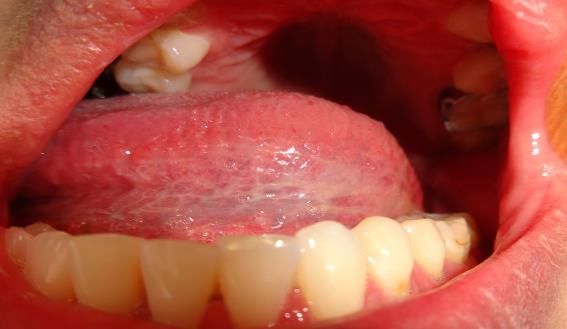
GSC Advanced Research and Reviews, 2021, 06(02), 001–012
seven (4.0%), neurologic in seven (4.0%), and serositis in five (2.9%) patients. LE diagnosis (n=110) coincided with
pregnancy in 20 (18.2%) patients. The median delay to LE diagnosis (n=158) was six (range 0-288) months and a
median of three (range 1-30) physicians (n=104) were consulted before the diagnosis was established. According to the
medical records, patients recalled a (n=177) previous history of LEOL in 111 (62.7%) cases.
Lupus-related manifestations are described in Table 2. Oral mucosal abnormalities were found in 174 (96.1%) patients;
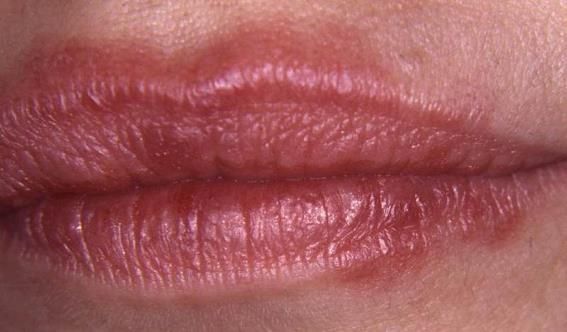
34 (18.8%) of them coursed with LEOL. The most common presentations of LEOL are depicted in Figure 1A-G.
Telangiectasias were the most frequently observed subtype of LEOL (n=14), they were characterized by multiple flat,
millimetric, round, red lesions, sometimes spidery in appearance, distributed on the palate (n=9), lip mucosa (n=3),
and/or buccal (n=2) mucosa (Figure 1A).
Table 2 Lupus erythematosus-related manifestations in 181 patients.
n (%)
Skin 57 (31.5)
Specific
discoid lupus 21 (11.6)
lupus profundus 5 (2.8)
subacute lupus 3 (1.7)
chilblain 1 (0.5)
bullous 1 (0.5)
Non-specific
photosensitivity 16 (8.8)
vasculitis 15 (8.3)
diffuse alopecia 3 (1.7)
pustular lesions 1 (0.5)
not specified 9 (5.0)
Systemic 43 (23.8)
articular 22 (12.1)
hematologic 10 (5.5)
renal 8 (4.4)
(3.3)
other* 6
(1.7)
not specified 3
Mouth 34 (18.8)
telangiectasia 14 (7.7)
erosion 11 (6.1)
ulcer 6 (3.3)
white reticular patch 7 (3.9)
erythematous plaque 7 (3.9)
other** 5 (2.8)
* Includes neurologic (3 patients), immunologic (2 patients), serositis (1 patient)
** Includes erythema (2 patients), atrophy (2 patients) and edema (1 patient)
Erosive lesions were observed in eleven patients, and were located on hard palate (n=5), gingiva (n=4), and buccal
mucosa (n=4), vermilion border (n=3), lip mucosa (n=2), and retromolar (n=1) area (Figure 1B). Oral ulcers were seen
in six subjects, and were superficial, well-delimited, clean-based, with angled irregular borders, surrounded or not by
erythema or erosive slightly keratinized margins; they affected mainly the central area of hard palate (n=4), labial
mucosa (n=4), distal area of the buccal mucosa (n=2), and vermilion (n=1) border; (Figure 1C).
4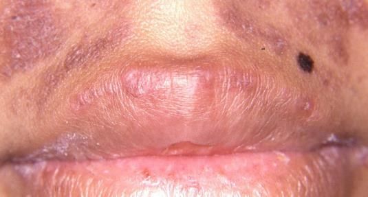
GSC Advanced Research and Reviews, 2021, 06(02), 001–012

         Figure 1A Multiple telangiectasias on labial mucosal surface and vermillion border of the lower lip.

   Figure 1B Erosions affecting lower labial mucosa and vermillion border of the lower lip, surrounded by a white
                                                 reticular margin.

Figure 1C Typical ulceration in lupus erythematosus, with irregular erythematous margins and fine scales, involving
                                            the central hard palate area.

White reticulate patches were seen in seven patients, and were located on buccal mucosa (n=4), ventral tongue (n=3),
and lip mucosa (n=2); these lesions were mainly bilateral, with marked or discrete millimetric white lines in the buccal
area (Figure 1D), but with an apparent fibrous pattern when these were located on the ventral surface of the tongue
(Figure 1E).

                                                                                                                      5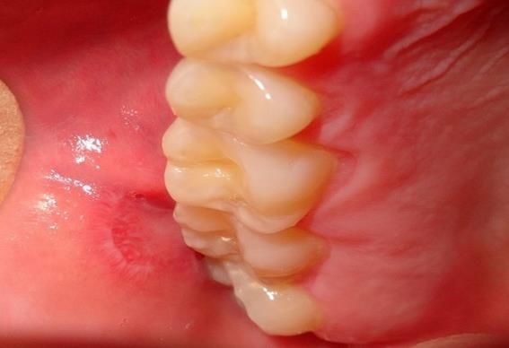
GSC Advanced Research and Reviews, 2021, 06(02), 001–012

    Figure 1D White reticular plaque located                   Figure 1E Reticular plaque with a cicatricial
            in right buccal mucosa.                          aspect, affecting left ventral surface of the tongue.

Erythematous plaques (n=7) were red inflamed patches (Figure 1F); some of them with scaling, crusty appearance
(Figure 1G), and exclusively involving the vermilion borders of the lip. Other oral mucosal manifestations of lupus
included erythema on the hard palate and retromolar buccal mucosa (n=2); vermilion atrophy (n=2) and lip edema
(n=1). The frequency of all oral findings is detailed in Table 3.

                Figure 1F Erythematous plaques distributed on vermillion borders and skin of lips.

              Figure 1G Red-rose plaques with fine scale, located on upper vermillion border of the lip.

                                                                                                                     6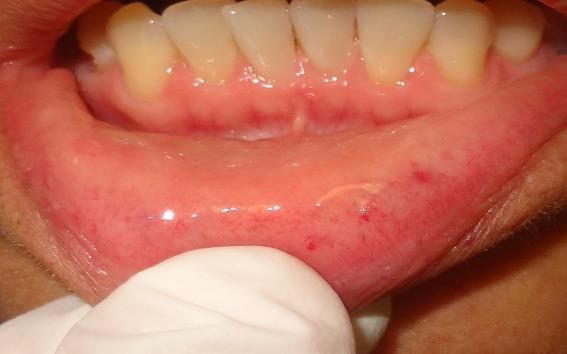
GSC Advanced Research and Reviews, 2021, 06(02), 001–012

Table 3 Frequency of oral mucosal conditions in 181 patients (pts) with lupus erythematosus.

                          Oral lesion                                                               n           (%)
                          Lupus erythematosus-related lesions                                       34         (18.8)
                          Infections by causal agent
                                 Candidosis1                                                        50         (27.6)
                                 HPV2                                                               3          (1.7)
                                 Fistula3                                                           3           (1.7)
                                 Hairy leukoplakia                                                  2          (1.1)
                                 Herpes simplex virus                                               1          (0.5)
                                 Herpes zoster virus                                                1          (0.5)
                          Miscelaneous
                                 Hyperpigmentation                                                  100        (55.2)
                                   racial/physiologic                                               62         (34.2)
                                   diffuse (drug related)                                           19         (10.5)
                                   melanocytic macules                                              19         (10.5)
                                 Coated tongue                                                      44         (24.3)
                                 Paleness                                                           40         (22.1)
                                 Xerostomia                                                         39         (21.5)
                                 Fissured tongue                                                    26         (14.4)
                                 Exfoliative cheilitis                                              23         (12.7)
                                 Traumatic erosion                                                  21         (11.6)
                                 Erythema migrans4                                                  16         (8.8)
                                 Frictional keratosis                                               13         (7.2)
                                 Scar                                                               13         (7.2)
                                 Fibrous hyperplasia                                                8          (4.4)
                                 Fordyce’s spots                                                    8          (4.4)
                                 Varix                                                              7          (3.9)
                                 Leukoedema                                                         7          (3.9)
                                 Traumatic ulcer                                                    7          (3.9)
                                 White occlusal line (linea alba)                                   6          (3.3)
                                 Mucosal exfoliation (chemical burns)                               6          (3.3)
                                 Mucous retention phenomenon5                                       5          (2.8)
                                 Tongue indentation                                                 4          (2.2)
                                 Morsicatio buccarum/labiorum                                       4          (2.2)
                                 Tongue atrophy6                                                    3          (1.7)
                                 Hairy tongue                                                       3          (1.7)
                                 Recurrent aphthous ulcers7                                         2          (1.1)
                                 Intramucous nevus                                                  1          (0.5)
                                 Idiopatic leukoplakia                                              1          (0.5)
                                 Smoker’s palate                                                    1          (0.5)
                                 Amalgam tattoo                                                     1          (0.5)
                     1 Includes erythematous (38 pts), denture-related (9 pts), angular cheilitis (6 pts), pseudomembranous
                  (4 pts), related to inflammatory papillary hyperplasia (2 pts) and chronic hyperplastic (1 patient) candidosis.
                    2 Includes Heck’s disease (2 pts) & squamous cell papilloma (1 patient); 3 Dental and periodontal origin.
     4 Includes tongue (15 pts), buccal and tongue (1 patient); 5 Includes mucocele (3 pts), ranula (1 pt) & maxillary sinus cyst (1 patient).
                      6 Nutritional deficiency related. 7 Includes minor & major (1 patient), minor (1 patient) presentation.

                                                                                                                                                 7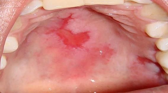
GSC Advanced Research and Reviews, 2021, 06(02), 001–012

Table 4 Laboratory values in patients with and without lupus erythematosus activity.

  Laboratory                                                                    Lupus erythematosus activity
    values
                                       Mouth                                                     Skin                                                       Other*
                        Activity          No activity                         Activity                No activity                           Activity              No activity
                 n   Md      (Q1-Q3)    n Md (Q1-Q3)            p       n     Md (Q1-Q3 )         n     Md (Q1-Q3)           P       n      Md (Q1-Q3)        n    Md (Q1-Q3)      p

Leukocytes      36 4.4     (3.7-4.9)   136 5.1 (4.3-7.0) 0.001         57    4.6 (3.8-5.1) 114 5.1 (4.3-7.4) 0.03                  39       4.6   (4.3-6.6) 133 4.9 (3.9-6.2) 0.2
(K/mL)

C3 (UI)         17 45      (30-53)     55 82 (62-96)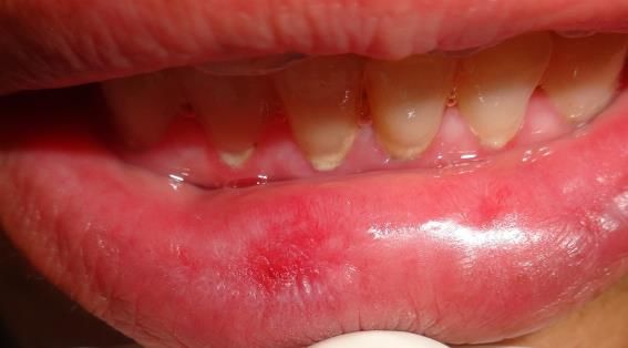
GSC Advanced Research and Reviews, 2021, 06(02), 001–012

The presence of LEOL showed a moderate and mild concordance with presence of cutaneous (k=0.380; p0.003) with the rest of the analyzed variables (gender, age
GSC Advanced Research and Reviews, 2021, 06(02), 001–012

patients with LEOL showed a higher correlation with serological markers in comparison to patients with cutaneous or
systemic activity, with the exception of articular disease. In a recent study including cutaneous and systemic LE-patients
[26], erosions, white reticular plaques and telangiectasias displayed associations with anti-dsDNA antibodies but no
strong relationship was observed with the presence of LE-ulcers, similar to our findings [26, 35]. Further research
focused on LEOL is required to allow the characterization of LEOL as a potentially useful clinical parameter in the
prediction and evaluation of serologic and clinical activity in LE disease, as our results suggest. In addition, it is
important to consider that anti-dsDNA antibodies have been implicated in cellular damage in SLE [37, 38], but their lack
of correlation with disease activity has also been described [32].

We must underscore that clinicians must be skilled in the differential diagnosis of oral mucosal lesions in order to
presume serum activity in LE patients through detection of LEOL. In this study, we also reported the prevalence of
numerous mucosal findings that are not directly related to LE.

We observed a high percentage of oral candidosis, mainly the erythematous variety. LE- patients present frequently
with one or several predisposing factors for the development of oral candidosis, such as the use of immunosuppressive
drugs (e.g. steroids), antibiotics, oral prosthetics, smoking, and the immunosuppression caused by LE itself. These may
explain the high frequency of observed cases in our patients. On the other hand, it is possible that in LE this clinical
presentation of oral candidosis has been underdiagnosed, as it is commonly unrecognized by the physician and
consequently underrated, despite its high prevalence, similar to what has been observed in HIV- infected subjects [39].

We also observed a high frequency of oral melanosis, another finding scarcely mentioned in the literature. This finding
could be attributed to the ethnic characteristics of our study population [40,41]. Among Venezuelan LE patients, who
are ethnically similar to the Mexican population, these lesions have been attributed to the use of antimalarials [24].
Other authors found a relationship with LE activity and pigmented macules [26], but such lesions could also be
explained as a consequence of postinflammatory damage on oral mucosa. In our analysis, after adjusting for multiple
variables, we did not find an association between oral melanosis and antimalarials, LE activity or smoking (data not
shown). Further studies are required to determine the possible etiological factors of oral melanosis in LE patients,
especially in those patients susceptible to hyperpigmentation.

An important limitation of this study was that it included only ambulatory-LE patients and few were naïve to treatment;
consequently, the prevalence of LEOL could be modified. To minimize this bias, we included a considerable number of
variables that potentially affect the development of oral lesions.

5. Conclusion
In this study, we present a compilation of oral lesions in patients with LE. The frequency of LEOL was high and strongly
correlated with serologic disease markers, independently of cutaneous or systemic activity; although more research is
mandatory in order to confirm our findings and to evaluate the potential relevance of LEOL in the follow-up and early
detection of disease flares in LE patients. It would be of great value for all physicians involved in the care of LE patients
to become acquainted with these lesions and their differential diagnoses, and to participate in adequately designed
research to increase our knowledge regarding the pathogenesis of these lesions.

Compliance with ethical standards

Acknowledgments
Authors would like to thank Romina Idalia Hernández-Ramírez, D.D.S, & Jenny Maldonado Molina, Dietitian, for their
technical assistance.

Disclosure of conflict of interest
None reported.

Statement of informed consent
Informed consent was obtained from all individual participants included in the study.

                                                                                                                          10
GSC Advanced Research and Reviews, 2021, 06(02), 001–012
Author contributions
All authors had full access to all the data in the study and take responsibility for the integrity of the data and the accuracy
of the data analysis. Study concept and design: LEP, LFC, ROT, ACC and CAD. Acquisition of data: LEP, LFC, SMF, MFS,
MJCG, MGOP, LGH and SMF. Analysis and interpretation of data: LEP, LFC, ROT, CAD, MFS, AHS, SMF, SPL, ACC and MSL.
Drafting of the manuscript: LEP, SMF, LFC, ACC, CAD, SPL, MFS and MSL. Critical revision of the manuscript for important
intellectual content: LEP, LFC, SMF, MSL, ACC, MPMG, ROT, JDC and SPL. Administrative, technical and material support:
LEP, LFC, ACC, SMF, MSL, MFS, MJCG, LGH, MPMG, JDC and MGOP. Study supervision: LEP, LFC, CAD, SMF, ACC, and ROT.
References
[1] Ghiggeri GM, D´Alessandro M, Bartolomeo D. An update on antbibodies to nucleosome components as
biomarkers of systemic lupus erythematosus and of lupus flares. Int J Mol Sci 2019; 20: 1-14.
[2] Li W-g, Ye ZZ, Yin Z-h, Zhang K. Clinical and immunological characteristics in 552 systemic lupus erythematosus
patients in a southern province of China. Int J Rheum Dis. 2017; 20: 68-75.
[3] Andreasen JO. Oral manifestations in discoid and systemic lupus erythematosus. Acta Odontol Scand. 1964; 22:
295-310.
[4] Schiodt M. Oral manifestations of lupus erythematosus. Int J Oral Surg. 1984; 13(2): 101-147.
[5] Hsu S, Dickinson D. A new approach to managing oral manifestations of Sjogren’s syndrome and skin
manifestations of lupus. J Biochem Mol Biol. 2006; 39(3): 229-239.
[6] Nico MMS, Vilela MAC, Rivitti EA, Lourenço SV. Oral lesions in lupus erythematosus: correlation with cutaneous
lesions. Eur J Dermatol. 2008; 18 (4): 376-81.
[7] Sultan SM, Ioannou Y, Isenberg DA. A review of gastrointestinal manifestations of systemic lupus erythematosus.
Rheumatology. 1999; 38(10): 917-932.
[8] Aliko A, Alushi A, Tafaj A, Lela F. Oral mucosa involvement in rheumatoid arthritis, systemic lupus erythematosus
and systemic sclerosis. Int Dent J. 2010; 60(5): 353-358.
[9] Nico MMS, Bologna SB, Lourenço SV. The lip in lupus erythematosus. Clin Exp Dermatol. 2014; 39(5): 563-569.
[10] Hochberg MC. Updating the American College of Rheumatology revised criteria for the classification of systemic
lupus erythematosus. Arthritis Rheum. 1997; 40(9): 1725.
[11] Tan EM, Cohen AS, Fries JF, et al. The 1982 revised criteria for the classification of systemic lupus erythematosus.
Arthritis Rheum 1982; 25(11): 1271-1277.
[12] Ellepola ANB, Samaranayake, LP. Oral candidal infections and antimycotics. Crit Rev Oral Biol Med 2000; 11(2):
172-198.
[13] Barrett AP, Buckley DJ, Greenberg ML, Earl MJ. The value of exfoliative cytology in the diagnosis of oral herpes
simplex infection in immunosuppressed patients. Oral Surg Oral Med Oral Pathol. 1986; 62(2): 175-178.
[14] Burge SM, Frith PA, Juniper RP, Wojnarowska F. Mucosal involvement in systemic and chronic cutaneous lupus
erythematosus. Br J Dermatol. 1989; 121(6): 727-741.
[15] Greene JC, Vermillion JR. The simplified oral hygiene index. J Am Dent Assoc. 1964; 68(1): 7-13.
[16] Albrecht J, Berlin JA, Braverman IM, et al. Dermatology position paper on the revision of the 1982 ACR criteria
for systemic lupus erythematosus. Lupus. 2004; 13(11): 839-849.
[17] Yell JA, Mbuagbaw J, Burge SM. Cutaneous manifestations of systemic lupus erythematosus. Br J Dermatol. 1996;
135(3): 355-362.
[18] Lipsker D. The need to revisit the nosology of cutaneous lupus erythematosus. Lupus. 2010; 19: 1047-1049.
[19] Liang MH, Stern S, Esdaile JM. Systemic lupus erythematosus activity. An operational definition. Rheum Dis Clin
North Am. 1988; 14: 57-66.
[20] Bombardieri S, Vitali C, Caponi L, Manca L, Bencivelli W. Activity criteria in systematic lupus erythmeatosus. Clin
Exp Rheumatol. 1994; 12: 45-48.
[21] Aickin M, Gensler H. Adjusting for multiple testing when reporting research results: the Bonferroni vs Holm
methods. Am J Public Health. 1996; 86(5): 726-728.
11GSC Advanced Research and Reviews, 2021, 06(02), 001–012
[22] Arredondo A, Aviles R. Costs and epidemiological changes of chronic diseases: implications and challenges for
health systems. PLoS One. 2015; 10(3): e0118611.
[23] Isenberg D, Rahman A. Systemic lupus erythematosus - 2005 annus mirabilis? Nat Clin Pract Rheumatol. 2006;
2(3): 145-152.
[24] López-Labady J, Villarroel-Dorrego M, González N, et al. Oral manifestations of systemic and cutaneous lupus
erythematosus in a Venezuelan population. J Oral Pathol Med. 2007; 36(9): 524-527.
[25] Khatibi M, Shakoorpour AH, Moosavian Jahromi Z, Ahmadzadeh A. The prevalence of oral mucosal lesions and
related factors in 188 patients with systemic lupus erythematosus. Lupus. 2012; 21(12): 1312-1315.
[26] Del Barrio-Díaz P, Reyes-Vivanco C, Cifuentes-Mutinelli M, Manríquez J, Vera-Kellet C. Association between oral
lesion and disease activity in lupus erythematosus. J Eur Acad Dermatol Venereol. 2020; 34: 349-356.
[27] Schiodt M. Oral discoid lupus erythematosus. III. A histopathologic study of sixty-six patients. Oral Surg Oral Med
Oral Pathol. 1984; 57(3): 281-293.
[28] Marques ERMC, Lourenço SV, Lima DM, Nico MMS. Oral lesions in lupus erythematosus-cytokines profiles of
inflammatory infiltrate. J Cutan Pathol. 2010; 37(4): 439-445.
[29] Hochberg MC. Updating the American College of Rheumatology revised criteria for the classification of systemic
lupus erythematosus. Arthritis Rheum. 1997; 40: 1725.
[30] Chiewchengchol D, Murphy R, Edwards SW, Beresford MW. Mucocutaneous manifestations in juvenile-onset
systemic lupus erythematosus: a review of literature. Pediatr Rheumatol Online J. 2015; 13: 1.
[31] Tug S, Helmig S, Menke J, et al. Correlation between cell free DNA levels and medical evaluation of disease
progression in systemic lupus erythematosus patients. Cell Immunol. 2014; 292(1): 32-39.
[32] Illei GG, Tackey E, Lapteva L, Lipsky PE. Biomarkers in systemic lupus erythematosus. II. Markers of disease
activity. Arthritis Rheum. 2004; 50(7): 2048-2065.
[33] Quatrocchi P, Barrile A, Bonanno D, et al. Ruolo degli anticorpi anti-nucleosomi nel lupus eritematoso sistemico.
Risultati di uno studio condotto in pazienti con lupus eritematoso sistemico ed alter connettiviti sistemiche.
Reumatismo. 2005; 57(2): 109-113.
[34] Schiodt M, Permin H, Wiik A, Manthorpe R. Autoantibodies in patients with oral lupus erythematosus, lichen
planus and leukoplakia. An aid in diagnosis. Acta Pathol Microbiol Scand Immunol. 1983; 91(1): 59-63.
[35] Urman JD, Lowenstein MB, Abeles M, Weinstein A. Oral mucosal ulceration in systemic lupus erythematosus.
Arthritis Rheum. 1978; 21(1): 58-61.
[36] Jacob L, Lety MA, Choquette D, et al. Presence of antibodies against a cell-surface protein, cross-reactive with
DNA, in systemic lupus erythematosus: a marker of the disease. Proc Natl Acad Sci USA. 1987; 84(9): 2956-2959.
[37] Rahman A. Autoantibodies, lupus and the science of sabotage. Rheumatology. 2004; 43(11): 1326-1336.
[38] Mackworth-Young CG, Chan JKH, Bunn CC, et al. Complement fixation by anti-dsDNA antibodies in SLE:
measurement by radioimmunoassay and relationship with disease activity. Ann Rheumatic Dis. 1986; 45: 314-
318.
[39] Cruz GD, Lamster IB, Begg MD, et al. The accurate diagnosis of oral lesions in human immunodeficiency virus
infection: Impact on medical staging. Arch Otolaryngol Head Neck Surg. 1996; 122(1): 68-73.
[40] Heidin CA, Axéll T. Oral melanin pigmentation in 467 Thai and Malaysian people with special emphasis on
smoker’s melanosis. J Oral Pathol Med. 1991; 20(1): 8-12.
[41] Banderas JA, Toshikasu O, González M. Oral mucosa lesions in Mazahua Indian adolescents. Acta Odontol
Latinoam. 1999; 12(1): 11-20.
12You can also read