Persistent Symptoms in Adult Patients 1 Year After Coronavirus Disease 2019 (COVID-19): A Prospective Cohort Study
←
→
Page content transcription
If your browser does not render page correctly, please read the page content below
Clinical Infectious Diseases
    Major Article

Persistent Symptoms in Adult Patients 1 Year After
Coronavirus Disease 2019 (COVID-19): A Prospective
Cohort Study
Jessica Seeßle,1 Tim Waterboer,2 Theresa Hippchen,1 Julia Simon,2,3 Marietta Kirchner,4 Adeline Lim,1 Barbara Müller,5 and Uta Merle1,

                                                                                                                                                                                                Downloaded from https://academic.oup.com/cid/advance-article/doi/10.1093/cid/ciab611/6315216 by guest on 11 September 2021
1
 Department of Internal Medicine IV, University Hospital Heidelberg, Heidelberg, Germany; 2Infections and Cancer Epidemiology, German Cancer Research Center (Deutsches
Krebsforschungszentrum [DKFZ]), Heidelberg, Germany; 3Faculty of Biosciences, Heidelberg University, Heidelberg, Germany; 4Institute of Medical Biometry and Informatics, University Hospital
Heidelberg, Heidelberg, Germany; and 5Department of Infectious Diseases, Virology, University Hospital Heidelberg, Heidelberg, Germany

   Background. Long COVID is defined as the persistence of symptoms beyond 3 months after severe acute respiratory syndrome
coronavirus 2 (SARS-CoV-2) infection. To better understand the long-term course and etiology of symptoms we analyzed a cohort
of patients with COVID-19 prospectively.
   Methods. Patients were included at 5 months after acute COVID-19 in this prospective, noninterventional, follow-up study.
Patients followed until 12 months after COVID-19 symptom onset (n = 96; 32.3% hospitalized, 55.2% females) were included in this
analysis of symptoms, quality of life (based on an SF-12 survey), laboratory parameters including antinuclear antibodies (ANAs),
and SARS-CoV-2 antibody levels.
   Results. At month 12, only 22.9% of patients were completely free of symptoms and the most frequent symptoms were reduced
exercise capacity (56.3%), fatigue (53.1%), dyspnea (37.5%), and problems with concentration (39.6%), finding words (32.3%), and
sleeping (26.0%). Females showed significantly more neurocognitive symptoms than males. ANA titers were ≥1:160 in 43.6% of pa-
tients at 12 months post–COVID-19 symptom onset, and neurocognitive symptom frequency was significantly higher in the group
with an ANA titer ≥1:160 versus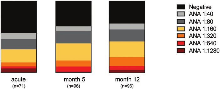
follow-up of a cohort of patients with COVID-19, with the              anxiety, palpitations, hair loss, and (only at 9 and 12 months)
aim to analyze the development of symptoms over follow-up              difficulty finding words.
time and with a special focus on differences between sub-
groups defined by gender, age, severity of acute COVID-19              Assessment of Life Quality by the 12-Item Short Form Survey
illness, and presence of antinuclear antibody (ANA) titers re-         For life quality assessment, study participants answered a
flecting autoimmunity.                                                 standardized 12-item Short Form Survey (SF-12) question-
                                                                       naire at the 5-, 9-, and 12-month time points. The SF-12 is
                                                                       a general health-related quality-of-life survey that measures
METHODS                                                                general health status. Two scales are calculated according to
Study Population                                                       a standardized evaluation with the help of an examination

                                                                                                                                         Downloaded from https://academic.oup.com/cid/advance-article/doi/10.1093/cid/ciab611/6315216 by guest on 11 September 2021
All patients with COVID-19 treated for acute COVID-19 as               tool: the Physical Component Scale (PCS) and the Mental
out- or inpatients at the Department of Internal Medicine IV           Component Scale (MCS). All scores were normalized against
of the University Hospital Heidelberg and with symptom onset           the 2009 US population reference scores. Although it has not
between 22 February 2020 and 18 April 2020 were invited 10 to          been validated for patients post–COVID-19, the SF-12 has
18 weeks after COVID-19 symptom onset to participate in this           been used extensively and is demonstrated to be reliable and
prospective, noninterventional COVID-19 long-term follow-up            well validated [9, 10].
study (Ethics Committee of University of Heidelberg: reference
                                                                       Serological Analyses
number: S-546/2020; DRKS00025089) (Supplementary Figure
1, Supplementary Table 1). In this study the long-term course          The laboratory parameters and ANA titers were determined
of symptoms, laboratory and immunological parameters, and              in the accredited central laboratory and in the Department
quality of life was analyzed at longitudinal follow-up time points     of Infectious Diseases of the Heidelberg University Hospital
starting 5 months after acute COVID-19 symptom onset.                  using standard operating procedures according to the manu-
   Written informed consent according to the Declaration of            facturers’ instructions. The ANA titers were determined
Helsinki was obtained from all patients and the local ethics           during acute disease (until 16 days post–symptom onset;
committees approved data collection and analysis. Inclusion            measurements were available for 71 of 96 patients) and at
criteria were status after polymerase chain reaction (PCR)–con-        follow-up visits using an immunofluorescence testing kit
firmed SARS-CoV-2-infection, prior out- or inpatient treatment         (Euroimmun AG, Lübeck, Germany). Immunoglobulin (Ig) G
(with live discharge) for acute COVID-19 at the Department of          (IgG) antibody levels against the S1 protein of SARS-CoV-2
Internal Medicine IV, and age 18 years or older. The timeframes        were determined using an enzyme-linked immunosorbent
for the follow-up visits were as follows: 5 months (20–22 weeks        assay (ELISA) kit (EI 2606–9601 G; Euroimmun AG) run on
post–symptom onset), 9 months (33–40 weeks post–symptom                an Analyzer I instrument (Euroimmun AG). Multiplex SARS-
onset), and 12 months (50–54 weeks post–symptom onset).                CoV-2 serology was performed as recently described, and
From the 96 patients seen at 5 and 12 months, only 80 had an           briefly, N and S1-RBD antibody levels measured in median
intermediate follow-up examination at our outpatient clinic at         fluorescence intensity (MFI) units [11]. Competition of sera
9 months post–COVID-19. Therefore, analysis of the develop-            with S1-ACE2 interaction was measured using the cPass sur-
ment of symptoms focused on the comparison of the 5- and               rogate SARS-CoV-2 neutralization test kit (L00847; Genscript,
12-month time points.                                                  Piscataway, NJ, USA).
   Disease severity of acute COVID-19 was divided into 4
categories: (1) mild, (2) moderate, (3) severe, and (4) critical di-   Statistical Analysis
sease, as previously described [8]. Symptom questionnaire and          Variables are described as median (interquartile range [IQR])
laboratory values from acute phase disease were available for          or absolute (relative, %) frequencies for metric or categorical
all 146 patients initially included and analyzed retrospectively.      variables, respectively. Comparisons between months 5 and
                                                                       12 were performed by Wilcoxon signed-rank test for metric
Assessment of Symptoms                                                 or McNemar test for categorical variables and restricted to
The following symptoms were recorded in a structured, paper-           patients who participated in both measurement times ob-
based questionnaire with additional grading of the following           served. Comparisons of symptom frequencies between sub-
present symptoms in 3 subjective grades (mild, moderate, se-           groups were performed with a chi-square test (or Fisher’s
vere): fever, sore throat, vomiting/nausea, diarrhea, decrease         exact test for symptoms with low absolute frequencies [
for independent variables. P values < .05 were considered Ninety-six patients who had completed follow-up until
statistically significant. Statistical analyses were performed 12 months post–COVID-19 were included in the analysis.
with IBM SPSS Statistics for Windows, version 24.0 (IBM Co-existing conditions at the time of acute COVID-19 are sum-
Corporation, Armonk, NY, USA). marized in Table 1. Until 12 months post-COVID the following
diagnoses had been newly established in the study cohort: hy-
pertension (n = 7), diabetes mellitus type 2 (n = 3), asthma
RESULTS
(n = 1), cardiovascular disease (n = 1), obstructive sleep apnea
Baseline Characteristics syndrome (n = 1), neuritis of nervus trigeminus (n = 1),
Patient characteristics are outlined in Table 1. Of 146 patients perimyocarditis (n = 1), depression (n = 1), postural ortho-
initially consenting to study participation and seen at the static tachycardia syndrome (n = 1), perianal abscess (n = 1),
Downloaded from https://academic.oup.com/cid/advance-article/doi/10.1093/cid/ciab611/6315216 by guest on 11 September 2021
5-month time point, 50 were lost to follow-up at the 12-month reactivation of varicella zoster virus (n = 1), autoimmune hepa-
follow-up visit and were therefore excluded from this 12-month titis (n = 1), and nonalcoholic steatohepatitis (n = 2).
long-term analysis (Supplementary Figure 1 and Supplementary
Table 1). Laboratory Findings
Laboratory findings of the study cohort are outlined in Table 2.
At 5, 9, and 12 months after acute infection, inflammatory
parameters (C-reactive protein [CRP], leukocytes, ferritin)
Table 1. Demographic Characteristics of the Study Population (n = 96) at
the Time Point of Acute COVID-19 were in the normal range for most of the patients.
The distribution of ANA titers within the study group during
Post–COVID-19 % acute COVID-19, as well as at 5 and 12 months post–symptom
Age, median (IQR), y 57 (50–63) onset, is outlined in Figure 1 and Supplementary Table 2.
Age group The proportion of women who showed elevated ANA titers
80 y 1 1.0
Patients answered a symptom questionnaire during acute
Gender (male) 43 44.8
Hospitalized 31 32.3 COVID-19 and at follow-up visits at 5, 9, and 12 months
Coexisting conditions (Figure 2). Symptoms already present before COVID-19 onset
Asthma 12 12.5 were not included in the analysis. The most frequent symp-
Hypertension 35 35.1 toms 5 months post–COVID-19 were reduced exercise ca-
Cardiovascular disease 4 4.2
pacity (53.1%), fatigue (41.7%), sleeping problems (32.3%),
Diabetes mellitus type 2 7 7.3
concentration problems (31.3%), and dyspnea (27.1%). At 5
Active malignancy 4 4.2
Autoimmune disease 5 5.2 and 12 months post–symptom onset, only 22.9% of patients
Depression 7 7.3 were completely free of long-COVID symptoms. Between
Neurocognitive disease 0 0 5 months and 12 months after symptom onset, the reported
Adipositas (BMI >30 kg/m2) 23 24.0 symptom frequency decreased significantly only for hair loss
Oxygen-support category
(26.1% vs 10.4%, P = .022), but increased for fatigue (from
Supplemental oxygen via nasal tube 21 21.9
Noninvasive ventilation via high-flow nasal 3 3.1
41.7% to 53.1%, P = .043) and dyspnea (from 27.1% to 37.5%,
oxygen P = .041). For all other symptoms there were no significant
Invasive ventilation 4 4.2 changes in reported symptom frequencies between 5 and
Disease severity 12 months of follow-up. At 12 months, the most frequently
Mild 15 15.6
reported symptoms were reduced exercise capacity, fatigue,
Moderate 53 55.2
Mild/moderate, ≥60 y 29 30.2
dyspnea, concentration problems, problems finding words,
Mild/moderate, male 25 26.0 and sleeping problems.
Severe 24 25.0 The number of positive acute symptoms was associated with
Critical 4 4.2 long COVID at 12 months: patients with at least 1 persisting
Severe/critical, ≥60 y 15 15.6
long-COVID symptom at 12 months post–COVID-19 had
Severe/critical, male 18 18.8
experienced significantly more positive symptoms during the
Data are presented as n (%) for categorical variables.
acute phase (8; IQR, 6–11) than patients who were free of long-
Abbreviations: BMI, body mass index; COVID-19, coronavirus disease 2019; IQR, inter-
quartile range. COVID symptoms (7; IQR, 4–9) (P = .009).
Long-COVID 1-Year Follow-up • cid 2021:XX (XX XXXX) • 3Table 2. Laboratory and Clinical Findings of Study Population 5, 9, and 12 Months Post–Symptom Onset of COVID-19
Limits of Post–COVID-19 Post–COVID-19 Month Post–COVID-19
Normal Month 5 (n = 96) 9 (n = 80) Month 12 (n = 96) P
Hemoglobin, g/dL 13–17 14.2 (13.3–14.8) 14.0 (13.3–14.9) 14.3 (13.4–14.9) .13
Leukocytes, nL 4–10 6.0 (5.2–7.6) 6.1 (5.1–7.5) 5.9 (5.1–7.5) .24
Lymphocytes, nL 1.0–4.8 1.7 (1.4–2.2) 1.7 (1.4–2.1) 1.8 (1.4–2.2) .04
Creatinine, mg/dL 0.6–1.2 0.8 (0.7–0.9) 0.8 (0.6–0.9) 0.8 (0.7–0.9) .85
CK, U/L 60 92.0 (85.5–104.1) 91.4 (79.7–103.3) 92.0 (82.3–101.8) .17
mL/min/1.73 m2
LDH, U/LDownloaded from https://academic.oup.com/cid/advance-article/doi/10.1093/cid/ciab611/6315216 by guest on 11 September 2021
Figure 2. Frequencies of symptoms (%) in the study cohort at acute COVID-19, as well as at 5, 9, and 12 months post–COVID-19 symptom onset. P values for the group
differences between 5- and 12-month time points are based on McNemar test for dependent samples. Symptoms with significant differences are marked with an asterisk
(*P < .05). Abbreviations: COVID-19, coronavirus disease 2019; n.d., not determined.
Assessment of Life Quality and S1-RBD at all time points, all other patients were positive at
To assess quality of life of the study cohort, the SF-12 question- the 12-month follow-up for S1-RBD IgG measured with multi-
naire was used to evaluate the physical and mental health at 5, plex serology, while 5.7% of patients showed sero-reversion for
9, and 12 months post–symptom onset. anti-nucleocapsid antibodies.
At 12 months, patients with at least 1 long-COVID symptom Comparison of patient groups stratified by disease severity
had a significantly reduced physical and mental life quality revealed a significant difference between mild/moderate and
compared with patients without symptoms (PCS, P = .006; severe/critical patient groups, with a higher level in the severe/
MCS, P = .031) (Figure 4). critical group 5, 9, and 12 months post–COVID-19 for anti-
The results of the PCS and MCS for the whole group and bodies against S1 and S1-RBD, as well as for ACE2 competition
stratified by gender and disease severity are presented in efficiency, but not for nucleocapsid-specific antibodies at all 3
Supplementary Figure 4A–F. follow-up time points (Supplementary Figure 6). No relevant
differences in antibody levels were observed when participants
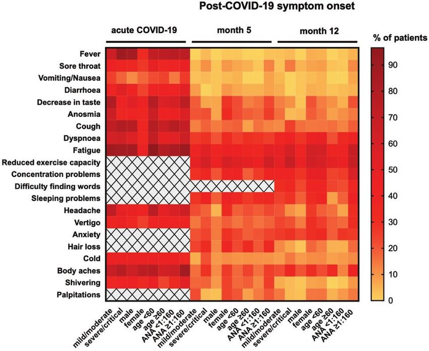
SARS-CoV-2–Specific Antibody Levels were stratified by ANA titers (Downloaded from https://academic.oup.com/cid/advance-article/doi/10.1093/cid/ciab611/6315216 by guest on 11 September 2021 Figure 3. Frequencies of symptoms (%) among the study population presented as a heatmap for the time of acute COVID-19 and at 5 and 12 months post–symptom onset. Symptom frequencies are stratified by disease severity (mild/moderate and severe/critical disease), gender (male and female), age (
Downloaded from https://academic.oup.com/cid/advance-article/doi/10.1093/cid/ciab611/6315216 by guest on 11 September 2021
Figure 4. The PCS and MCS assessed by the SF-12 questionnaire for the study population subgrouped for patients with at least 1 symptom or no symptoms at 12 months
post–COVID-19 symptom onset. Data are presented as medians and interquartile ranges, and P values for the group differences are based on the Mann-Whitney U test for
independent samples. Significant differences are marked with an asterisk (*P < .05, **P < .01). Abbreviations: COVID-19, coronavirus disease 2019; MCS, Mental Component
Scale; PCS, Physical Component Scale; SF-12, 12-item Short Form Survey.

   It is currently unclear why some patients experience long-                          immunity-relevant anti–S1 antibodies declined only mod-
term symptoms after COVID-19. Potential causes for different                           estly and all patients remained positive in the ACE2 compe-
outcomes of infection are viral dose as well as host-dependent                         tition assay at 12 months. As an earlier report suggests that
factors—for example, genetic susceptibility or induction of an-                        vaccination against SARS-CoV-2 alleviates long-COVID
ti-inflammatory cells and proteins. For acute SARS-CoV-2 in-                           symptoms [24, 25], we analyzed for differences in antibody
fection, the development of ANA IgA autoantibodies has been                            titer levels between patients stratified for the presence of
shown [14] and the presence of high-titer serum IgG antibodies                         symptoms at 12 months after symptom onset. We did not
targeting the GD1b ganglioside has been demonstrated in cer-                           observe any differences between the groups in antibody
tain neurologically affected patients [13]. In a cohort of 31 pa-                      levels for all analyzed antibodies.
tients with long COVID, functionally active autoantibodies                                The strength of our study is the long-term follow-up of pa-
targeting G-protein–coupled receptors were detected in a high                          tients with examination of all we report on the patients exam-
proportion of patients experiencing a variety of post–COVID-                           ined at 5 and 12 months, although more patients were initially
19 symptoms [15]. Although the nature of the self-antigens                             included at 5 months. In addition, our study is, to date, the
recognized by autoimmune-like antibodies is diverse, a general                         longest follow-up of patients post–COVID-19. Like for all ob-
characteristic shared by infections is the generation of ANAs,                         servational studies, the increased willingness of symptomatic
which appear during acute infection and may remain at lower                            patients to take part in a follow-up study is a potential con-
levels during chronic infections [23]. Therefore, we analyzed our                      founding factor. In line with this, reduced exercise capacity
follow-up cohort for the presence of ANAs and the correlation                          was reported, with a significantly lower frequency in the co-
of ANA positivity and long-COVID symptoms. We observed                                 hort of 50 patients lost to the 12-month follow-up than in the
that patients with ANA titer elevations ≥1:160 at 12 months                            remaining group. We acknowledge this potential selection
after acute infection had a significantly higher frequency of sev-                     bias. The generalizability of the results may be limited by the
eral long-COVID symptoms. Thus, we speculate that there is                             single-center, unblinded, and nonrandomized design and the
an underlying autoimmune component to the post–COVID-19                                relatively small cohort size.
syndromes reported here, including neurocognitive symptoms
and dyspnea. As expected [12], more women showed ANA                                   Conclusions
positivity than men and the group of women with ANA titer                              Neurocognitive long-COVID symptoms can persist at least
elevations ≥1:160 displayed significantly higher symptom fre-                          until 1 year after COVID-19 symptom onset and reduce
quencies than the group of women with lower titers. As this                            quality of life significantly. Several neurocognitive symptoms
observation was significant for female but not for male partici-                       were associated with ANA titer elevations, rendering autoim-
pants, autoimmune reactions might, in part, be responsible for                         munity a potential cofactor in the etiology of long COVID.
the female predominance in long-COVID syndromes [6, 7].                                As the disease is poorly understood, it is too early to specu-
   Analysis of SARS-CoV-2 antibody titers showed a sig-                                late about disease etiology and prognosis. Our results provide
nificant decline, especially for anti-nucleocapsid anti-                               information useful to clinicians caring for patients post–
bodies from 5 to 12 months after acute infection, while                                COVID-19 disease.

                                                                                             Long-COVID 1-Year Follow-up • cid 2021:XX (XX XXXX) • 7
Supplementary Data 8. Gandhi RT, Lynch JB, Del Rio C. Mild or moderate Covid-19. N Engl J Med 2020;
383:1757–66.
Supplementary materials are available at Clinical Infectious Diseases online.
9. Brazier J, Roberts J, Tsuchiya A, Busschbach J. A comparison of the EQ-5D and
Consisting of data provided by the authors to benefit the reader, the posted
SF-6D across seven patient groups. Health Econ 2004; 13:873–84.
materials are not copyedited and are the sole responsibility of the authors, so 10. Johnson JA, Coons SJ. Comparison of the EQ-5D and SF-12 in an adult US
questions or comments should be addressed to the corresponding author. sample. Qual Life Res 1998; 7:155–66.
11. Butt J, Murugan R, Hippchen T, et al. From multiplex serology to serolomics—a
Notes novel approach to the antibody response against the SARS-CoV-2 proteome.
Viruses 2021; 13:749. doi: 10.3390/v13050749.
Author Contributions. J. Seeßle, T. W., U. M., and B. M. were involved in
12. Quintero OL, Amador-Patarroyo MJ, Montoya-Ortiz G, Rojas-Villarraga A,
the study concept and design, drafting of the manuscript, and study super- Anaya JM. Autoimmune disease and gender: plausible mechanisms for the fe-
vision. J. Seeßle, T. W., T. H., J. Simon, A. L., B. M., and U. M. were involved male predominance of autoimmunity. J Autoimmun 2012; 38:J109–19.
in acquisition of data. J Seeßle, T. W., M. K., J. Simon, B. M., and U. M. were 13. Guilmot A, Maldonado Slootjes S, Sellimi A, et al. Immune-mediated neu-
involved in interpretation of data, statistical analysis, and revision of the rological syndromes in SARS-CoV-2-infected patients. J Neurol 2021;
Downloaded from https://academic.oup.com/cid/advance-article/doi/10.1093/cid/ciab611/6315216 by guest on 11 September 2021
manuscript for intellectual content. All authors read and approved the final 268:751–7.
version of the manuscript. 14. Sacchi MC, Tamiazzo S, Stobbione P, et al. SARS-CoV-2 infection as a trigger of
Acknowledgments. The authors acknowledge Jessica Langel, Petra autoimmune response. Clin Transl Sci 2021; 14:898–907.
15. Wallukat G, Hohberger B, Wenzel K, et al. Functional autoantibodies against
Klöters-Plachky, Jutta Mohr, and Alexandra Hof for patient-related and
G-protein coupled receptors in patients with persistent long-COVID-19 symp-
technical support; Markus Zorn for laboratory analyses; and Sylvia Parthé,
toms. J Transl Autoimmun 2021; 4:100100.
Paul Schnitzler, Maria Anders-Össwein, and Stefanie Wolf for support with 16. Tan EM, Feltkamp TE, Smolen JS, et al. Range of antinuclear antibodies in
serological analyses. The data will be made publicly available no later than “healthy” individuals. Arthritis Rheum 1997; 40:1601–11.
the time of online publication. 17. Carvalho-Schneider C, Laurent E, Lemaignen A, et al. Follow-up of adults with
Financial support. The work of T. W. was supported by a donation from noncritical COVID-19 two months after symptom onset. Clin Microbiol Infect
the Dieter Morszeck Foundation. 2021; 27:258–63.
Potential conflicts of interest. The authors: No reported conflicts of 18. Arnold DT, Hamilton FW, Milne A, et al. Patient outcomes after hospitalisation
interest. All authors have submitted the ICMJE Form for Disclosure of with COVID-19 and implications for follow-up: results from a prospective UK
cohort. Thorax 2020; 76:399–401. doi: 10.1136/thoraxjnl-2020–216086.
Potential Conflicts of Interest.
19. Moreno-Pérez O, Merino E, Leon-Ramirez JM, et al; COVID19-ALC Research
Group. Post-acute COVID-19 syndrome. Incidence and risk factors: a
Mediterranean cohort study. J Infect 2021; 82:378–83.
References 20. Halpin SJ, McIvor C, Whyatt G, et al. Postdischarge symptoms and rehabilitation
1. NICE COVID-19 rapid guidelines. PharmacoEcon Outcomes News 2021;877:33. needs in survivors of COVID-19 infection: a cross-sectional evaluation. J Med
doi: 10.1007/s40274-021-7682-3. Virol 2021; 93:1013–22.
2. Nalbandian A, Sehgal K, Gupta A, et al. Post-acute COVID-19 syndrome. Nat 21. Jacobs LG, Gourna Paleoudis E, Lesky-Di Bari D, et al. Persistence of symptoms
Med 2021; 27:601–15. doi: 10.1038/s41591-021-01283-z. and quality of life at 35 days after hospitalization for COVID-19 infection. PLoS
3. Dennis A, Wamil M, Alberts J, et al; COVERSCAN Study Investigators. One 2020; 15:e0243882.
Multiorgan impairment in low-risk individuals with post-COVID-19 syndrome: 22. Garrigues E, Janvier P, Kherabi Y, et al. Post-discharge persistent symptoms and
a prospective, community-based study. BMJ Open 2021; 11:e048391. health-related quality of life after hospitalization for COVID-19. J Infect 2020;
4. Marx V. Scientists set out to connect the dots on long COVID. Nat Methods 2021; 81:e4–6.
18:449–53. 23. Rivera-Correa J, Rodriguez A. Divergent roles of antiself antibodies during infec-
5. Carfì A, Bernabei R, Landi F; Gemelli Against COVID-19 Post-Acute Care Study tion. Trends Immunol 2018; 39:515–22.
Group. Persistent symptoms in patients after acute COVID-19. JAMA 2020; 24. Mishra PK, Bruiners N, Ukey R, et al. Vaccination boosts protective responses and
324:603–5. counters SARS-CoV-2-induced pathogenic memory B cells. medRxiv [Preprint].
6. Huang C, Huang L, Wang Y, et al. 6-Month consequences of COVID-19 in pa- April 14, 2021. 2021. doi:10.1101/2021.04.11.21255153. Accessed 4 May 2021.
tients discharged from hospital: a cohort study. Lancet 2021; 397:220–32. 25. Arnold DT, Milne A, Samms E, et al. Symptoms after COVID-19 vaccination in
7. Sudre CH, Murray B, Varsavsky T, et al. Attributes and predictors of long COVID. patients with persistent symptoms after acute infection: a case series. Ann Intern
Nat Med 2021; 27:626–31. Med 2021. doi:10.7326/M21-1976.
8 • cid 2021:XX (XX XXXX) • Seeßle et alYou can also read