Effects of cardiac pacemakers on left ventricular volumes and function assessed by 3D echocardiography, Doppler method, and global longitudinal ...
←
→
Page content transcription
If your browser does not render page correctly, please read the page content below

Dawood et al. The Egyptian Heart Journal
https://doi.org/10.1186/s43044-021-00138-9
                                                     (2021) 73:16
                                                                                                                                        The Egyptian Heart
                                                                                                                                                   Journal

 RESEARCH                                                                                                                                            Open Access

Effects of cardiac pacemakers on left
ventricular volumes and function assessed
by 3D echocardiography, Doppler method,
and global longitudinal strain
Moustafa Dawood , Eman Elsharkawy, Mohamed Ayman Abdel-Hay and Moustafa Nawar*

  Abstract
  Background: Many previous studies reported the negative effects of right ventricular (RV) pacing on the left
  ventricular (LV) structure and ejection fraction. Studying pacing hemodynamics is essential to understand these
  detrimental effects. In this study, we tried to understand RV pacing effects on LV volumes and function using
  advanced tools like 3D echo and global longitudinal strain (GLS). This was a prospective study of 175 consecutive
  patients (LVEF>50%) presented permanent pacing. Of 175 patients, only 50 patients met study criteria, divided into
  two groups (single or dual pacing). LV volumes and function were assessed by full-volume 3D echocardiography
  and GLS before pacing, at 1-week and 6-month post-pacing. Cardiac output (COP) was calculated by pulsed wave
  Doppler method and 3D echo.
  Results: Doppler method results were similar to 3D echo in calculating SV and COP. At 1-week post pacing, both
  groups showed a significant decrease in SV due to a drop in EDV while ESV did not change significantly. Despite
  the drop in SV, there was a significant increase in cardiac output (COP) due to achieving higher heart rates post-
  pacing. There was a significant drop in EF and GLS in both groups.
  At 6 months, SV continued to decrease with a corresponding decrease in COP and LVEF. This drop in SV was due
  to a significant increase in ESV while EDV did not show a significant change at a 6-month follow-up. Also, the drop
  EF and GLS became more significant.
  There were no significant differences between both groups regarding the changes in LV volumes (EDV, ESV, SV),
  LVEF or GLS throughout the study (pre-pacing, at 1-week and 6-months post pacing). However, dual-chamber
  pacing group provided higher heart rates and as a result higher COP than the single-chamber group.
  Conclusions: RV pacing led to a significant drop in LV COP, ejection fraction (EF), and GLS over short- and long-
  term duration. Dual chamber pacing provided higher COP than a single chamber pacing. This was due to tracking
  the S. A node with pacing at higher heart rates not due to an increase in SV and preserving atrioventricular
  synchrony. Both Doppler method and 3D echo can be used to calculate SV and COP.
  Keywords: Cardiac output, 3D echocardiography, Global longitudinal strain, Single chamber, Dual chamber cardiac
  pacemakers, Pacing hemodynamics

* Correspondence: mosnawar@yahoo.com
Alexandria Faculty of Medicine, Alexandria, Egypt

                                        © The Author(s). 2021 Open Access This article is licensed under a Creative Commons Attribution 4.0 International License,
                                        which permits use, sharing, adaptation, distribution and reproduction in any medium or format, as long as you give
                                        appropriate credit to the original author(s) and the source, provide a link to the Creative Commons licence, and indicate if
                                        changes were made. The images or other third party material in this article are included in the article's Creative Commons
                                        licence, unless indicated otherwise in a credit line to the material. If material is not included in the article's Creative Commons
                                        licence and your intended use is not permitted by statutory regulation or exceeds the permitted use, you will need to obtain
                                        permission directly from the copyright holder. To view a copy of this licence, visit http://creativecommons.org/licenses/by/4.0/.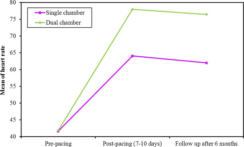
Dawood et al. The Egyptian Heart Journal   (2021) 73:16                                                           Page 2 of 11

Background                                                       presented to our university hospitals for device
Cardiac pacing is the established treatment of the heart         implantation.
block since more than 50 years [1]. At first, pacemakers           The exclusion criteria included the presence of
were capable of pacing only one chamber of the heart,            more than mild valvular heart disease, left ventricular
usually the right ventricle. With more advances, dual-           ejection fraction less than 50%, presence of ischemic
chamber pacemakers were developed to sense and pace,             heart disease, and recent cardiac surgery during the
both the atrium and the ventricle and thus are able to           last 3 months before enrollment. Patients with poor
achieve atrioventricular synchrony [2]. A single-chamber         echo windows, patients with slow atrial fibrillation or
pacemaker has only one lead implanted (usually in the            other types of arrhythmia that can affect stroke vol-
right ventricle) so it controls the activity of the ventricles   ume measurement, debilitated or cancer patients with
regardless the condition of the atria whether in systole         expected survival less than 1 year and patients with
or in diastole which means that the atria may contract           previously implanted devices were excluded. From
against closed AV valves while the ventricles are in sys-        175 patients, 124 patients were excluded. Sixty-five
tole causing pacemaker syndrome (dyspnea and symp-               patients were presented with reduced LV systolic
toms of pulmonary congestion) [3]. However, a dual               function for cardiac resynchronization therapy. Six-
chamber has two leads, one in the right atrium and one           teen patients were assigned for ICD devices as a pri-
in the right ventricle resembles the normal activities of        mary or secondary prevention with no indication for
the heart and reflects intrinsic depolarization. It gives        permanent pacing. Twenty-two patients had previ-
time to the atria to empty in the ventricles preserving          ously implanted devices at elective replacement period
the atrial kick and was quickly classified as physiologic        and were scheduled for battery replacement. Seven
pacing mode [2].                                                 patients were subjected to reoperation due to device
   As a result, dual-chamber pacing, as compared with            related complications (4 with twiddler syndrome and
single-chamber         ventricular     pacing,      improves     3 for device extraction due to bloodstream or device-
hemodynamic function [4–6], but the clinical benefit             related infection). Eight patients presented with slow
is uncertain. Many studies suggest that dual-chamber             atrial fibrillation or SSS. Two patients had poor echo
pacing has better outcomes regarding symptoms, ex-               views, and 4 patients had significant valvular heart
ercise tolerance, and ambulatory blood pressure moni-            disease. One patient died before the second follow-up.
toring. It was associated with a lower incidence of              The remaining 50 patients were distributed to have
atrial fibrillation, stroke, and heart failure than single-      single chamber (27 patients) or dual chamber pace-
chamber pacing [7]. There was also evidence of                   maker (23 patients).
improved survival [8]. Some current guidelines recom-
mend dual-chamber pacing except in patients with
atrial flutter or fibrillation [9].                              Patient characteristics
   On the other hand, a number of randomized studies             Patient’s demographic data and indications for pacing
suggested no significant differences between single-             were collected and revised 24 h before pacing. Patient’s
chamber and dual-chamber pacing in the rates of atrial           age ranged from 12 to 97 years with mean age of 63.12
fibrillation, heart failure, or a composite of stroke, ische-    ± 16.85 years. According to gender, 27 patients were
mic attack or death [10–12]. In this study, we aimed to          males (54%) and 23 were females (46%). All the patients
study pacing hemodynamics, effects on left ventricular           had structurally normal heart confirmed by echocardiog-
(LV) volumes, and function over a 6-month interval. We           raphy (Table 1).
used the standard Doppler wave methods in addition to
3D echo to calculate SV and COP. Being more superior
to 2D echo, the remaining LV volumes and ejection frac-          Table 1 Summary of patients’ characteristics
tion were calculated using 3D echocardiography.                  Predisposing                     Total (n=50)
                                                                 factors
                                                                                                  No.                   %
Methods                                                          Age (years)                      63.12 ± 16.85
The study was approved by our faculty of medicine eth-           Mean ± SD.
ics committee. All patients provided written informed            Sex
consent.
                                                                   Male                           27                    54.0
                                                                   Female                         23                    46.0
Patients’ selection
                                                                 DM                               13                    26.0
Patient recruitment started from October 2017 to Au-
                                                                 HTN                              21                    42.0
gust 2018. During this period, 175 consecutive patients
Dawood et al. The Egyptian Heart Journal   (2021) 73:16                                                                   Page 3 of 11

Data collection: 2D echocardiography and PW Doppler          inferior to RV septal pacing [13, 14], RV apex was se-
method                                                       lected as the site of RV lead implantation in all can-
A full 2D echocardiographic study was done to ex-            didates. Patients with single-chamber pacemakers
clude patients with significant valvular heart disease,      were programmed to VVIR mode. Patients with dual-
IHD, or reduced LVEF. The baseline heart rate was            chamber pacing were programmed to DDDR mode.
recorded. Our study used two different methods to            Rate responsive mode was selected as it resulted in
calculated SV; full volume 3D Echo and Doppler               better outcomes regarding patient quality of life and
method by calculating VTI at LVOT by PW Doppler.             exercise tolerance [15]. Suggested settings for dual-
Both methods were applied before pacing and at               chamber pacemakers were lower and upper rate limits
follow-up visits. In order to avoid any confounding          of 60 beats per minute and 130 beats per minute, re-
factors during SV and COP estimation, pre-pacing             spectively. For single-chamber pacemakers, the sug-
LVOT diameter was recorded and reused at follow-up           gested lower and upper rate limits were 60 beats per
for each patient. Also, to avoid confounding factors         minute and 130 beats per minute, respectively. In
between both methods during COP calculation, the             order to maximize the contribution of the atrial kick
same heart rate was multiplied by 3D and Doppler             to SV in the DDDR group, dynamic AV time delay
SV at every visit.                                           [16] was selected with resting paced/sensed AV time
                                                             delay adjusted to 200/150 ms [16, 17]. Patients were
Full-volume 3D Echo acquisition and GLS analysis             recruited 7–10 days post-pacing and after 6 months.
A 3D full-volume acquisition of the left ventricle was       Device interrogation was done to check the adjusted
done using the Philips Medical iE33 echocardiography         pacing parameters and acquire ventricular pacing per-
system with X5-1 transthoracic probe. For 3D full-           centage. Also, 3D echo and GLS were calculated at
volume acquisitions, ensuring adequate frame rate            follow-up visits as described before.
and packet size and capture of the full cardiac cycle,
the system obtained a 30°–30° pyramid over 4 to 6 al-        Statistical analysis
ternate gated cardiac cycles. The resulting dynamic          The database was maintained and analyzed by an in-
3D full volume sector was reviewed and navigated             dependent data-management group. To assess the dis-
through immediately to ensure all areas of interest          tribution of the data derived from this study, we
have been captured. All the acquisitions were ECG            calculated the standardized skewness and kurtosis of
gated and patients were told to hold their breath dur-       each of the variables. Normally distributed values
ing acquisition to avoid stitch artifacts. After comple-     were expressed as mean and skewed values as the
tion of the study, 3D acquired data were transferred         median (interquartile range). Paired two-tailed group
to Q-lab 10 for off-line analysis. In order to calculate     comparisons were made with Student’s t (parametric)
LV volumes and EF, both the long and short LV axes           or Wilcoxon signed-rank (non-parametric) tests as ap-
were adjusted to get maximum LV dimensions and to            propriate. p values of less than 0.05 were regarded as
avoid foreshortening then five landmarks were chosen         significant.
to initiate edge detection by semi-automated quantifi-
cation software. Four landmarks were placed at mitral        Results
annulus, and the fifth was placed at the apex in apical      It was found that Doppler method was not inferior to
four or apical two views. The software delineated LV         3D echo in calculating SV and COP (Table 2). There
boundaries automatically, but it allowed manual mod-         was no significant difference between both methods
ifications to include or exclude any part for more           in both groups pre-pacing or at follow-up visits.
accurate adjustment of LV borders. After borders de-
lineation, the software automatically calculated EDV,        Table 2 Comparison between 3D and PW methods according
ESV, SV, COP, and EF.                                        to COP in each group
   For GLS calculation, 2D gated acquisition of the apical   COP                             3D            PW             W
                                                                                                                           X      P
views (apical four, two, and three) were done according      Single chamber
to the standard techniques. Patients were told to hold
                                                               Post-pacing (7–10 days)       3.69 ± 1.13   3.68 ± 1.11    0.141   0.888
their breath during acquisition, and foreshortening was
avoided. GLS was calculated for each of the three apical       Follow up after 6 months      3.27 ± 1.12   3.40 ± 1.03    1.830   0.067
view, and then, mean GLS was calculated automatically.       Dual chamber
                                                               Post-pacing (7–10 days)       4.48 ± 1.14   4.35 ± 1.04    1.511   0.131
Pacemaker implantation and programming                         Follow up after 6 months      3.96 ± 1.04   3.90 ± 0.83    0.070   0.944
Implantation was performed according to the operator’s       WX, p: Wx and P values for Wilcoxon signed-rank test for comparing between
preference. Being easily accessible, more stable and non-    single and dual chamber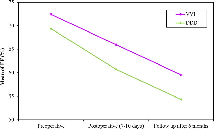
Dawood et al. The Egyptian Heart Journal      (2021) 73:16                                                           Page 4 of 11

Many studies compared 2D and 3D Echo to CMR as                           decrease at 6 months (p value < 0.001,
Dawood et al. The Egyptian Heart Journal (2021) 73:16 Page 5 of 11
Fig. 2 Comparison between the different periods according to 3D COP in each group
Table 3 Comparison between single- and dual-chamber pacing groups including all the measured parameters at the three time
intervals (pre-pacing, at 1 week and 6 months post pacing)
Measurement Pre-pacing p value Post pacing at 1 week p value Post pacing p value
At 6 months
EDV (ml)
Single-chamber 102.6 ± 25.95 0.360 86.81 ± 25.09 0.259 88.93 ± 30.39 0.098
Dual-chamber 109.0 ± 22.91 93.43 ± 18.80 97.04 ± 19.62
ESV (ml)
Single-chamber 28.48 ± 9.67 0.055 28.96 ± 11.25 0.080 36.52 ± 21.87 0.143
Dual-chamber 34.43 ± 11.75 34.91 ± 12.24 44.65 ± 15.66
SV (ml)
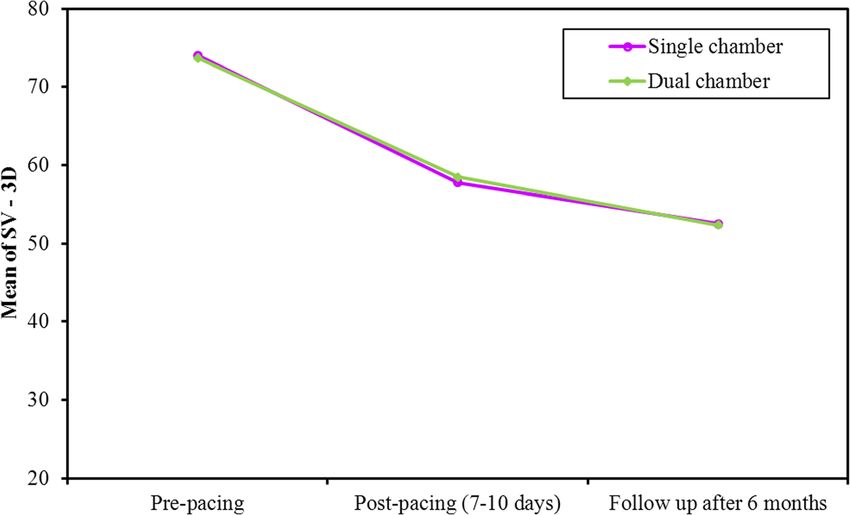
Single-chamber 74.07 ± 20.20 0.944 57.78 ± 17.47 0.880 52.56 ± 17.54 0.962
Dual-chamber 73.7 ± 17.35 58.52 ± 17.02 52.35 ± 13.13
HR (bpm)
Single-chamber 41.63 ± 8.45 0.938 64.04 ± 9.79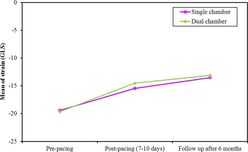
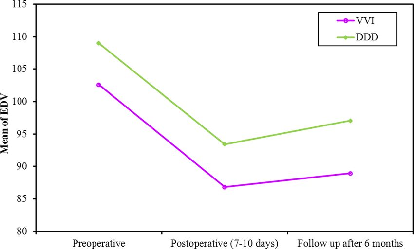
Dawood et al. The Egyptian Heart Journal     (2021) 73:16                                                        Page 6 of 11

 Fig. 3 Comparison between the different periods according to 3D SV in each group

no difference between both types except for their ef-                   rate response in both groups, dual-chamber pacing
fects on HR and COP. Dual-chamber pacemakers                            group managed to provide higher resting HR by tra-
provided higher COP through pacing at higher HR                         cing the patients’ intrinsic HR. Both groups showed
not due to achieving AV synchrony and preserving                        short- and long-term detrimental effects on LV vol-
the atrial kick. Despite the implementation of the                      umes and function. At 1 week, SV reduction was

 Fig. 4 Comparison between the different periods according to EDV in each group
Dawood et al. The Egyptian Heart Journal     (2021) 73:16                                                             Page 7 of 11

 Fig. 5 Comparison between the different periods according to ESV in each group

due to a significant drop in left ventricular EDV.                      increased myocardial stretching forces led to more effi-
Several factors contributed to the decrease in LV                       cient contraction and enhanced pre-pacing EF. Both
EDV during RV pacing:                                                   groups showed a significant increase in COP post-pacing
                                                                        despite the drop in SV as it was compensated by pacing
   The drop in EDV could be a result of pacing at                      at higher heart rates.
    higher heart rates which shortened cardiac cycle                      Few published data were found on the acute effects of
    duration mainly diastole leaving less time to the                   RV apical pacing on LV volumes and function. Our
    ventricles to fill with blood and decreasing EDV and,               results matched with the previously published studies:
    according to Frank-Starling law, decreasing the myo-
    cardium stretching forces leading to SV drop.                          In 2006, Lieberman et al. [21] compared the effects
   Normal LV diastolic filling may be impaired by the                      RV, LV, and biventricular (BiV) pacing in patients
    loss of normal atrioventricular conduction in single-                   with preserved LV systolic function. It was found
    chamber pacing group with subsequent decrease in                        that RV apical pacing was associated with reduction
    left atrial contribution to diastolic filling.                          in LVEF (from 51±12% to 48±14%, P=NS), without
   In addition, the earlier activation of the RV may                       affecting LV dimensions.
    hamper LV filling that is accomplished by the                          In 2008, Liu et al. [22] studied acute effects of RV
    shared inter-ventricular septum [18–20]. All these                      apical pacing on LV function in 35 patients with sick
    mechanisms resulted in a decrease in LV preload                         sinus syndrome. There was a decrease in LV EDV
    during RV apical pacing and resulted in a lower LV                      (from 79±22 to 76±20 mL, P=0.07) and LVEF (from
    stroke volume.                                                          57±8% to 54±8%, P=0.01) during RV pacing.
                                                                           In 2009, Delgado et al. [23] studied a group of 25
   This drop in SV had a corresponding reduction in EF                      patients during EPS for AVNRT ablation. Left
at 1 week. However, the pre-pacing EF was overesti-                         ventricular synchrony, volumes, and function were
mated because lower heart rates increased diastolic                         measured. There was a significant decrease in LV
filling time and EDV. According to Frank-Starling law,                      end-diastolic diameter and volume during RV apical
Dawood et al. The Egyptian Heart Journal (2021) 73:16 Page 8 of 11 Table 4 Pacing related changes in all the measured parameters at the three time intervals (pre-pacing, at 1 week and 6 months post pacing) Measurement Pre-pacing Post pacing at 1 week Post pacing at 6 months Test P value EDV (ml) Fr Single-chamber 102.6 ± 25.95 86.81 ± 25.09 88.93 ± 30.39 30.882*
Dawood et al. The Egyptian Heart Journal     (2021) 73:16                                                                 Page 9 of 11

 Fig. 6 Comparison between the different periods according to EF (%) in each group

     pacing, whereas LV end-systolic diameter and vol-                      The multicenter, randomized UK-PACE Trial [12] in-
     ume did not change. Consequently, LVEF decreased                    cluded 2021 patients presented for permanent pacing.
     significantly from 56±8% to 48±9% (P
Dawood et al. The Egyptian Heart Journal (2021) 73:16 Page 10 of 11
Fig. 7 Comparison between the different periods according to strain (GLS) in each group
0.001). The study concluded that RV apical pacing Supplementary Information
induced acute LV dyssynchrony. The online version contains supplementary material available at https://doi.
org/10.1186/s43044-021-00138-9.
A recent study conducted in 2017 evaluated the value
of GLS as a predictor for pacing induced LV dyssyn- Additional file 1. SV by PW, 3D.
chrony. In 93 patients followed for 5 years, cardiomyop- Additional file 2. 3D Echo.
athy developed more prevalently in dyssynchrony group Additional file 3. GLS.
(group 1: 20% vs. group 2; 3.1%, pDawood et al. The Egyptian Heart Journal (2021) 73:16 Page 11 of 11
Competing interests 19. Leclercq C, Gras D, Le Helloco A, Nicol L, Mabo P, Daubert C (1995)
The authors declare that they have no competing interest. Hemodynamic importance of preserving the normal sequence of
ventricular activation in permanent cardiac pacing. Am Heart J 129(6):1133–
Received: 27 May 2020 Accepted: 4 February 2021 1141
20. Bleasdale R, Turner M, Mumford C, Steendijk P, Paul V, Tyberg J, Morris-
Thurgood J et al (2004) Left ventricular pacing minimizes diastolic
ventricular interaction, allowing improved preload-dependent systolic
References performance. Circulation. 110(16):2395–2400
1. Members ATF, Brignole M, Auricchio A, Baron-Esquivias G, Bordachar P, 21. Lieberman R, Padeletti L, Schreuder J, Jackson K, Michelucci A, Colella A,
Boriani G, Breithardt O-A et al (2013) 2013 ESC Guidelines on cardiac pacing Eastman W et al (2006) Ventricular pacing lead location alters systemic
and cardiac resynchronization therapy: the Task Force on cardiac pacing hemodynamics and left ventricular function in patients with and without
and resynchronization therapy of the European Society of Cardiology (ESC). reduced ejection fraction. J Am Coll Cardiol 48(8):1634–1641
Developed in collaboration with the European Heart Rhythm Association 22. Liu WH, Chen MC, Chen YL, Guo BF, Pan KL, Yang CH, Chang H-W (2008)
(EHRA). Eur Heart J 34(29):2281–2329 Right ventricular apical pacing acutely impairs left ventricular function and
2. Buckingham TA, Janosik DL, Pearson AC (1992) Pacemaker hemodynamics: induces mechanical dyssynchrony in patients with sick sinus syndrome: a
clinical implications. Prog Cardiovasc Dis 34(5):347–366 real-time three-dimensional echocardiographic study. J Am Soc
3. Nielsen JC, Bøttcher M, Nielsen TT, Pedersen AK, Andersen HR (2000) Echocardiogr 21(3):224–229
Regional myocardial blood flow in patients with sick sinus syndrome 23. Delgado V, Tops LF, Trines SA, Zeppenfeld K, Marsan NA, Bertini M, Holman
randomized to long-term single chamber atrial or dual chamber ER et al (2009) Acute effects of right ventricular apical pacing on left
pacing—effect of pacing mode and rate. J Am Coll Cardiol 35(6):1453–1461 ventricular synchrony and mechanics clinical perspective. Circ Arrhythm
4. Kruse I, Arnman K, Conradson T, Ryden L (1982) A comparison of the acute Electrophysiol 2(2):135–145
and long-term hemodynamic effects of ventricular inhibited and atrial 24. Sweeney MO, Hellkamp AS, Ellenbogen KA, Greenspon AJ, Freedman RA,
synchronous ventricular inhibited pacing. Circulation. 65(5):846–855 Lee KL, Lamas GA (2003) Adverse effect of ventricular pacing on heart
5. Boon N, Frew A, Johnston J, Cobbe S (1987) A comparison of symptoms failure and atrial fibrillation among patients with normal baseline QRS
and intra-arterial ambulatory blood pressure during long term dual duration in a clinical trial of pacemaker therapy for sinus node dysfunction.
chamber atrioventricular synchronous (DDD) and ventricular demand (VVI) Circulation. 107(23):2932–2937
pacing. Heart. 58(1):34–39 25. Schmidt M, Brömsen J, Herholz C, Adler K, Neff F, Kopf C, Block M (2007)
6. Lau CP, Wong CK, Leung WH, Liu WX (1990) Superior cardiac Evidence of left ventricular dyssynchrony resulting from right ventricular
hemodynamics of atrioventricular synchrony over rate responsive pacing at pacing in patients with severely depressed left ventricular ejection fraction.
submaximal exercise: observations in activity sensing DDDR pacemakers. Europace. 9(1):34–40
Pacing Clin Electrophysiol 13(12):1832–1837 26. Akerström F, Arias MA, Pachón M, Jiménez-López J, Puchol A, Juliá-Calvo J
7. Tang CY, Kerr CR, Connolly SJ (2000) Clinical trials of pacing mode selection. (2013) The importance of avoiding unnecessary right ventricular pacing in
Cardiol Clin 18(1):1–23 clinical practice. World J Cardiol 5(11):410
27. Dreger H, Maethner K, Bondke H, Baumann G, Melzer C (2011) Pacing-
8. Lamas GA, Pashos CL, Normand S-LT, McNeil B (1995) Permanent
induced cardiomyopathy in patients with right ventricular stimulation for >
pacemaker selection and subsequent survival in elderly Medicare
pacemaker recipients. Circulation. 91(4):1063–1069 15 years. Europace. 14(2):238–242
28. Babu NS, Srinath SC, Lahiri A, Chase D, John B, Roshan J (2018) Three-
9. Group E (1991) Recommendations for pacemaker prescription for
dimensional echocardiography with left ventricular strain analyses helps
symptomatic bradycardia. Br Heart J 66(2):185
earlier prediction of right ventricular pacing-induced cardiomyopathy. J
10. Lamas GA, Orav EJ, Stambler BS, Ellenbogen KA, Sgarbossa EB, Huang SKS,
Saudi Heart Assoc 30(2):102–107
Marinchak RA et al (1998) Quality of life and clinical outcomes in elderly
29. Wilkoff BL, Cook JR, Epstein AE, Greene HL, Hallstrom AP, Hsia H, Kutalek SP
patients treated with ventricular pacing as compared with dual-chamber
et al (2002) Dual-chamber pacing or ventricular backup pacing in patients
pacing. N Engl J Med 338(16):1097–1104
with an implantable defibrillator: the dual chamber and VVI implantable
11. Castelnuovo E, Stein K, Pitt M, Garside R, Payne E. The effectiveness and
defibrillator (DAVID) trial. Jama. 288(24):3115–3123
cost-effectiveness of dual-chamber pacemakers compared with single-
30. Steinberg JS, Fischer A, Wang P, Schuger C, Daubert J, Mcnitt S, Andrews M
chamber pacemakers for bradycardia due to atrioventricular block or sick
et al (2005) The clinical implications of cumulative right ventricular pacing
sinus syndrome: systematic review and economic evaluation. NIHR Health
in the multicenter automatic defibrillator trial II. J Cardiovasc Electrophysiol
Technology Assessment programme: Executive Summaries: NIHR Journals
16(4):359–365
Library; 2005.
31. Ha S, Song Y, Lee W, Bang W, Yoo S, Cheong S (2017) P1676Global
12. Toff WD, Camm AJ, Skehan JD (2005) Single-chamber versus dual-chamber
longitudinal strain improve prediction of right ventricular pacing induced
pacing for high-grade atrioventricular block. N Engl J Med 353(2):145–155
left ventricular dyssynchrony in patients with permanent pacemaker. Eur
13. Kaye GC, Linker NJ, Marwick TH, Pollock L, Graham L, Pouliot E, Poloniecki J
Heart J 38(1):366
et al (2014) Effect of right ventricular pacing lead site on left ventricular
function in patients with high-grade atrioventricular block: results of the
Protect-Pace study. Eur Heart J 36(14):856–862 Publisher’s Note
14. Nikoo M, Ghaedian M, Kafi M, Jorat M, Fakhrpour A, Pakfetrat M, Ostovan M Springer Nature remains neutral with regard to jurisdictional claims in
et al (2011) Effects of right ventricular septal versus apical pacing on plasma published maps and institutional affiliations.
natriuretic peptide levels. J Cardiovasc Dis Res 2(2):104–109
15. Sulke N, Chambers J, Dritsas A, Sowton E (1991) A randomized double-blind
crossover comparison of four rate-responsive pacing modes. J Am Coll
Cardiol 17(3):696–706
16. Mehta D, Gilmour S, Ward DE, Camm AJ (1989) Optimal atrioventricular
delay at rest and during exercise in patients with dual chamber
pacemakers: a non-invasive assessment by continuous wave Doppler. Heart.
61(2):161–166
17. Wish M, Fletcher RD, Gottdiener JS, Cohen AI (1987) Importance of left atrial
timing in the programming of dual-chamber pacemakers. Am J Cardiol
60(7):566–571
18. Rosenqvist M, Isaaz K, Botvinick EH, Dae MW, Cockrell J, Abbott JA, Schiller
NB et al (1991) Relative importance of activation sequence compared to
atrioventricular synchrony in left ventricular function. Am J Cardiol 67(2):
148–156You can also read


