Diagnostic utility of low hemoglobin density to detect iron deficiency in patients with inflammatory bowel disease
←
→
Page content transcription
If your browser does not render page correctly, please read the page content below
ORIGINAL ARTICLE                                                                                Annals of Gastroenterology (2021) 34, 1-7

Diagnostic utility of low hemoglobin density to detect iron
deficiency in patients with inflammatory bowel disease

Karima Farraga,b, Krenare Ademaja,b, Eleni Leventib,c, Aysegül Aksanb,d*, Jürgen Steina,b*
DGD Kliniken Sachsenhausen, Frankfurt am Main; Interdisziplinäres Crohn Colitis Centrum Rhein-Main, Frankfurt
am Main; Klinikum Hanau; Institute of Nutritional Science, Justus-Liebig University Giessen, Germany

Abstract                             Background In the absence of a feasible noninvasive gold standard, iron deficiency (ID) anemia
                                     (IDA) is best measured using multiple indicators. However, the choice of an appropriate single
                                     iron biomarker for ID screening continues to be debated. Low hemoglobin density (LHD%) from
                                     Coulter counters has been suggested as a useful tool to detect ID. This study investigated the
                                     reliability of LHD% for the assessment of iron status in patients with inflammatory bowel disease
                                     (IBD) and IDA, anemia of chronic disease (ACD) or mixed anemia (MIX).
                                     Methods The study population consisted of 143 patients with IBD (aged 39.03±12.53 years,
                                     61.5% female). Blood count, transferrin saturation, serum ferritin, and C-reactive protein were
                                     determined by routine assays. Patients with anemia were divided into 3 groups: IDA, ACD
                                     and MIX, according to specific criteria. Receiver operator characteristic (ROC) curves were
                                     constructed.
                                     Results ROC analysis for LHD% in the detection of ID yielded a cutoff value of 3.8%. In
                                     anemic patients, LHD% values did not differ statistically significantly between groups (IDA,
                                     ACD, MIX) and no significant difference in LHD% values was observed between patients with
                                     IDA and ID.
                                     Conclusions These results demonstrate that LHD% is a reliable biomarker for the detection
                                     of iron deficiency in patients with IBD and anemia, regardless of whether inflammation
                                     is present. Our findings indicate that LHD% can provide added value in diagnosing iron
                                     deficiency.
                                     Keywords Inflammatory bowel disease, iron deficiency, low hemoglobin density
                                     Ann Gastroenterol 2021; 34 (1): 1-7

a
 Gastroenterology and Clinical Nutrition, DGD Kliniken                   Introduction
Sachsenhausen, Frankfurt am Main (Karima Farrag, Krenare Ademaj,
Jürgen Stein); bInterdisziplinäres Crohn Colitis Centrum Rhein-Main,
Frankfurt am Main (Karima Farrag, Krenare Ademaj, Eleni Leventi,             The prevalence of iron deficiency (ID) in patients with
Aysegül Aksan, Jürgen Stein); cDepartment of Gastroenterology,           inflammatory bowel disease (IBD) has been reported at 36-
Klinikum Hanau (Eleni Leventi); dInstitute of Nutritional Science,       76%, with approximately 50% of patients having isolated ID
Justus-Liebig University Giessen (Aysegül Aksan), Germany
                                                                         without anemia. Since ID, even without manifest anemia, can
Conflicts of Interest: KF: speakers’ honoraria Immundiagnostik AG.       substantially impact quality of life and healthcare costs, IBD-
KA, EL: No conflicts of interest. AA: Research funding, consulting and   associated ID requires appropriate diagnostic and therapeutic
congress fees from Immundiagnostik AG and Vifor. JS: consultancy         management [1-4].
fees from Immundiagnostik AG, payment for lectures from
Immundiagnostik AG
                                                                             Two different types of impaired iron homeostasis may be
*Contributed equally to this work                                        differentiated: absolute ID and functional ID. In absolute ID,
                                                                         iron stores are depleted as a result of insufficient dietary iron
Correspondence to: Karima Farrag, Dept. of Gastroenterology and
                                                                         intake, iron malabsorption and/or chronic gastrointestinal
Clinical Nutrition, DGD Kliniken Sachsenhausen, Schulstrasse 31,
Frankfurt am Main, Germany, e-mail: farrag@gmx.de                        blood loss [5,6]. ID anemia (IDA) occurs when iron stores are
                                                                         fully depleted and iron supply is insufficient for hemoglobin
Received 30 November 2020; accepted 31 December 2020;                    (Hb) synthesis [7]. In functional ID, iron supply is inadequate
published online 2 April 2021
                                                                         to ensure sufficient hemoglobinization of reticulocytes and
DOI: https://doi.org/10.20524/aog.2021.0622                              mature erythrocytes. This imbalance between erythroid

© 2021 Hellenic Society of Gastroenterology                                                                          www.annalsgastro.gr
2 K. Farrag et al marrow iron requirements and actual iron supply inhibits am Main, Germany) who had been diagnosed with IBD hemoglobinization, resulting in hypochromic mature red according to standard clinical, endoscopic, radiological cells and reticulocytes [8,9]. Despite a plethora of iron status and pathological criteria were retrospectively analyzed. The parameters, screening for ID remains challenging. Serum control group consisted of healthy adults attending the same ferritin (s-ferritin) indirectly estimates body iron stores center for routine consultations. Inclusion criteria were age and is the most specific biomarker of absolute ID. Absolute 18-65 years and body mass index (BMI)
Diagnostic utility of LHD% in patients with IBD 3

analysis was used to evaluate the diagnostic performance of                             Baseline laboratory parameters of the IBD group according
LHD% for the assessment of iron status. Statistical significance                    to iron status are shown in Table 2. Of the 143 patients with
was predetermined as P
4 K. Farrag et al

Table 2 Laboratory markers according to iron status in patients with IBD

  Markers                 Absolute ID (n=79)          Functional ID (n=26)           P‑value1        IDA (n=44)           ACD or MIX (n=21)             P‑value2

  Hb (g/dL)                  12.5 (7.4‑16.2)                 11.4 (9.5‑130)           0.012*        11.0 (7.4‑13.3)           11.8 (9.2‑12.9)            0.311
  RDW (%)                   14.6 (11.9‑22.5)              14.8 (11.7‑21.2)            0.894        15.2 (12.5‑20.8)          15.0 (11.7‑21.2)            0.776
  MCHC (g/dL)               32.8 (28.6‑35.7)              32.3 (29.9‑34.3)            0.190        31.9 (28.6‑34.0)          32.2 (29.9‑34.3)            0.424
  Ferritin (ng/mL)           15.1 (5.0‑29.1)             94.1 (30.5‑1028.0)
Diagnostic utility of LHD% in patients with IBD 5
120.0
100.0
80.0
60.0
40.0
20.0
0.0
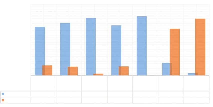
Functional Not iron
Control
ID (n=46) IDA (n=33) ACD (n=13) MIX (n=8) deficient
ID (n=5) (n=49)
(n=38)
Elevated LHD 82.3 88.5 97.0 84.6 100.0 21.1 3.9
LHD < 3.8% 17.1 14.9 3.0 15.4 0.0 78.9 96.1
Figure 1 Percentage of elevated LHD% in the study population
ID, iron deficiency; IDA, iron deficiency anemia; ACD, anemia of chronic disease; MIX, mixture of IDA and ACD; LHD, low hemoglobin density
alone, particularly in patients with chronic inflammatory To our knowledge, this is the first study to highlight
disease [29-31]. However, TfR-F has limited availability and the value of LHD% as an additional parameter to aid ID
cost constraints [32]. Moreover, current assays are hampered by diagnosis in patients with IBD by evaluating iron status while
a lack of external quality controls and limited availability within incorporating the differentiation of absolute and functional
the test repertoire of hospital-based laboratories [27,33,34]. ID. The results of our study confirmed that LHD% is a reliable
Hypochromic red blood cells (%HYPO), a measure of the diagnostic marker of iron status in patients with IBD. LHD%
percentage of erythrocytes with decreased hemoglobinization, was found to be a more sensitive marker than hepcidin or
has also been considered as a possible marker in this context. bone marrow-stainable iron stores for identifying patients
%HYPO and reticulocyte indices perform very well in with iron-restricted erythropoiesis, especially if inflammation
differentiating IDA and ACD [35,36]. However, patients with is present [18]. Our results indicate that LHD% is suitable
ACD may also have higher %HYPO and lower reticulocytes for the diagnosis of ID in patients with or without active
than controls. This is due to iron-restricted erythropoiesis inflammation. Furthermore, LHD% was found to be superior
caused by the reduced availability of iron from body stores to to TSAT and s-ferritin in its diagnostic performance with
red blood cells developing in the bone marrow. Furthermore, regard to functional ID. Therefore, we propose that LHD%
this approach is restricted by the fact that analyzers are available may be a worthwhile additional diagnostic biomarker of ID
only from 2 manufacturers, Siemens and Sysmex. in patients with IBD, especially when TSAT and s-ferritin are
The current gold standard in ID diagnosis, the determination insufficiently accurate because of the presence of inflammation,
of stainable iron strains in a bone marrow aspirate sample offering additional diagnostic accuracy at no extra cost and
by Perls staining [37], is limited by factors such as incorrect without delay. Our ROC analysis identified 3.8% as an optimal
aspiration of the bone marrow or insufficient sample size. cutoff point for LHD%, similar to the value of 4.0% suggested
Non-stainable iron stores seem to correlate well with true ID by Urrechaga et al [16,17] for patients with chronic kidney
in patients with IDA. However, they can be misleading, since disease. LHD% has clinical utility, not only as a marker of
iron stores can even be stained in the bone marrow of patients iron-restricted erythropoiesis, but also in the discrimination
with iron-restricted erythropoiesis, if adequately sampled. This between IDA and thalassemia, as shown by Urrechaga et al
reinforces current opinion among hematologists that a lack of [16] and more recently by Ng et al [41].
detectable iron in bone marrow cannot be equated with ID, As a general limitation, it must be kept in mind that, as a
and that despite stringent criteria, bone marrow biopsy is not marker of cellular hypochromia, LHD% is sensitive to the
representative of a patient’s true iron stores [38]. temperature and storage duration of the sample: LHD% may be
There is therefore a need to seek alternative iron status falsely elevated as a result of a reduction in MCHC associated
markers widely available. LHD% was recently proposed as a with storage at room temperature or above. Therefore, if
potential indicator of iron available for erythropoiesis [16,17,39]. samples are kept at room temperature, LHD% should be
LHD% is a time-averaged measurement of the degree of assessed within 6 h of collection. Refrigeration minimizes
hemoglobinization in mature erythrocytes. The analysis can volume changes induced by sample storage, with hypochromia
be performed concurrently with routine blood counts at no markers showing only minimal changes during the first 24 h
additional cost and without additional blood collection [16]. after sample collection [42].
MCHC, the parameter from which LHD% is calculated, is a Our study has some limitations: Firstly, the study included a
stable laboratory parameter that can be reliably analyzed in relatively small number of patients. Despite the relatively small
samples stored for up to 36 h [40]. number of samples, we determined a cutoff value close to that
Annals of Gastroenterology 346 K. Farrag et al
found in previous studies, and therefore see no restrictions in Acknowledgment
terms of reliability. Secondly, since LHD% was not monitored
after iron therapy, no statement can be made as to whether The authors gratefully acknowledge the assistance of Janet
LHD% may be a marker of therapeutic success. The lack of Collins (Interdisziplinäres Crohn Colitis Centrum Rhein-
data on sTfR can be considered a further limitation; this was Main, Frankfurt am Main, Germany) in correcting and
due to the fact that sTfR is not included in routine iron status proofreading the manuscript.
assessments in the treatment center from which our patient
data were obtained. Finally, no details are available concerning
signs of clinical disease activity or disease duration at the time
of blood sampling. Unfortunately, given the retrospective References
character of the study, it was not possible to collect these
missing data. 1. Koutroubakis IE, Ramos-Rivers C, Regueiro M, et al. Five-year period
In conclusion, in this study we demonstrated that LHD% prevalence and characteristics of anemia in a large us inflammatory
can verify the presence of ID, regardless of the presence of bowel disease cohort. J Clin Gastroenterol 2016;50:638-643.
inflammation. LHD% can be determined as part of routine 2. Goodhand JR, Kamperidis N, Rao A, et al. Prevalence and
laboratory tests at no extra cost and without additional management of anemia in children, adolescents, and adults with
inflammatory bowel disease. Inflamm Bowel Dis 2012;18:513-519.
blood collection, and thus offers a rapid, accurate and
3. Filmann N, Rey J, Schneeweiss S, et al. Prevalence of anemia in
convenient additional diagnostic tool for ID in patients inflammatory bowel diseases in European countries: a systematic
with IBD. We therefore propose that, using a cutoff value review and individual patient data meta-analysis. Inflamm Bowel
of 3.8%, LHD% holds promise as a simple, inexpensive and Dis 2014;20:936-945.
sensitive tool that allows an accurate and reliable diagnosis 4. Bager P, Befrits R, Wikman O, et al. The prevalence of anemia
of ID with or without anemia in patients with inflammatory and iron deficiency in IBD outpatients in Scandinavia. Scand
disorders. J Gastroenterol 2011;46:304-309.
5. Aksan A, Wohlrath M, Iqbal TH, Farrag K, Dignass A, Stein J.
Serum hepcidin levels predict intestinal iron absorption in patients
with inflammatory bowel disease. Clin Lab 2019;65.
6. Aksan A, Wohlrath M, Iqbal TH, Dignass A, Stein J. Inflammation,
Summary Box but not the underlying disease or its location, predicts oral iron
absorption capacity in patients with inflammatory bowel disease.
J Crohns Colitis 2020;14:316-322.
What is already known:
7. Stein J, Aksan A, Farrag K, Dignass A, Radeke HH. Management
of inflammatory bowel disease-related anemia and iron deficiency
• No reliable biochemical markers exist for the with specific reference to the role of intravenous iron in current
differentiation between iron deficiency anemia practice. Expert Opin Pharmacother 2017;18:1721-1737.
(IDA) and anemia of chronic disease in patients 8. Mast AE, Blinder MA, Lu Q, Flax S, Dietzen DJ. Clinical utility
with inflammatory bowel disease (IBD) of the reticulocyte hemoglobin content in the diagnosis of iron
• Low hemoglobin density (LHD%) from Coulter deficiency. Blood 2002;99:1489-1491.
9. Coyne D. Iron indices: what do they really mean? Kidney Int Suppl
counters has been suggested as a useful tool to
2006;(101):S4-S8.
detect iron deficiency, even in the context of 10. Ganz T. Anemia of inflammation. N Engl J Med 2019;381:1148-1157.
inflammation 11. World Health Organization. Serum ferritin concentrations for the
• Literature data suggest that LHD% is more sensitive assessment of iron status and iron deficiency in populations. 2011.
compared to ferritin, serum iron, transferrin Available from: https://www.who.int/vmnis/indicators/serum_
saturation (TSAT), hepcidin or bone marrow iron ferritin.pdf [Accessed 8 February 2021].
stores in identifying patients with associated iron 12. Cappellini MD, Comin-Colet J, de Francisco A, et al; IRON CORE
deficiency Group. Iron deficiency across chronic inflammatory conditions:
International expert opinion on definition, diagnosis, and
management. Am J Hematol 2017;92:1068-1078.
What the new findings are: 13. Dignass A, Farrag K, Stein J. Limitations of serum ferritin in
diagnosing iron deficiency in inflammatory conditions. Int J
Chronic Dis 2018;2018:9394060.
• LHD% had a better discriminating power than 14. Peyrin-Biroulet L, Williet N, Cacoub P. Guidelines on the diagnosis
TSAT and s-ferritin in the diagnosis of IDA and treatment of iron deficiency across indications: a systematic
• LHD% was a reliable biomarker for the detection review. Am J Clin Nutr 2015;102:1585-1594.
of iron deficiency in patients with IBD and anemia, 15. Dignass AU, Gasche C, Bettenworth D, et al; European Crohn’s
regardless of whether inflammation is present and Colitis Organization [ECCO]. European consensus on the
• LHD% could provide added value in identifying diagnosis and management of iron deficiency and anaemia in
inflammatory bowel diseases. J Crohns Colitis 2015;9:211-222.
patients with associated iron deficiency who
16. Urrechaga E. The new mature red cell parameter, low haemoglobin
could potentially benefit from parenteral iron density of the Beckman-Coulter LH750: clinical utility in the
replacement diagnosis of iron deficiency. Int J Lab Hematol 2010;32(1 Pt
1):e144-e150.
Annals of Gastroenterology 34Diagnostic utility of LHD% in patients with IBD 7
17. Urrechaga E, Unceta M, Borque L, Escanero JF. Low hemoglobin 29. Suominen P, Möttönen T, Rajamäki A, Irjala K. Single values
density potential marker of iron availability. Int J Lab Hematol of serum transferrin receptor and transferrin receptor ferritin
2012;34:47-51. index can be used to detect true and functional iron deficiency
18. Martin-Cabrera P, Hung M, Ortmann E, et al. Clinical use of in rheumatoid arthritis patients with anemia. Arthritis Rheum
low haemoglobin density, transferrin saturation, bone marrow 2000;43:1016-1020.
morphology, Perl’s stain and other plasma markers in the 30. Punnonen K, Irjala K, Rajamäki A. Serum transferrin receptor and
identification of treatable anaemia presenting for cardiac surgery its ratio to serum ferritin in the diagnosis of iron deficiency. Blood
in a prospective cohort study. J Clin Pathol 2015;68:923-930. 1997;89:1052-1057.
19. Satsangi J, Silverberg MS, Vermeire S, Colombel JF. The Montreal 31. Rimon E, Levy S, Sapir A, et al. Diagnosis of iron deficiency anemia
classification of inflammatory bowel disease: controversies, in the elderly by transferrin receptor-ferritin index. Arch Intern
consensus, and implications. Gut 2006;55:749-753. Med 2002;162:445-449.
20. WHO. Iron deficiency anaemia: assessment, prevention and 32. Abitbol V, Borderie D, Polin V, et al. Diagnosis of iron deficiency in
control. Report of a joint WHO/UNICEF/UNU consultation. inflammatory bowel disease by transferrin receptor-ferritin index.
1998. Available from: https://www.who.int/nutrition/publications/ Medicine (Baltimore) 2015;94:e1011.
micronutrients/anaemia_iron_deficiency/WHO_NHD_01.3/en/ 33. Koulaouzidis A, Cottier R, Bhat S, Said E, Linaker BD, Saeed AA.
[Accessed 11 March 2021]. A ferritin level >50 microg/L is frequently consistent with iron
21. World Health Organization. Haemoglobin concentrations for the deficiency. Eur J Intern Med 2009;20:168-170.
diagnosis of anaemia and assessment of severity. 2011. Available 34. Koulaouzidis A, Saeed AA, Abdallah M, Said EM. Transferrin
from: https://apps.who.int/iris/bitstream/handle/10665/85839/ receptor level as surrogate peripheral blood marker of iron
WHO_NMH_NHD_MNM_11.1_eng.pdf?ua=1 [Accessed 8
deficiency states. Scand J Gastroenterol 2009;44:126-127.
February 2021].
35. Syed S, Kugathasan S, Kumar A, et al. Use of reticulocyte
22. Weiss G, Goodnough LT. Anemia of chronic disease. N Engl J Med
hemoglobin content in the assessment of iron deficiency in
2005;352:1011-1023.
children with inflammatory bowel disease. J Pediatr Gastroenterol
23. van Santen S, van Dongen-Lases EC, de Vegt F, et al. Hepcidin and
Nutr 2017;64:713-720.
hemoglobin content parameters in the diagnosis of iron deficiency
36. Urrechaga E, de la Hera P, Aguayo FJ. Reticulocyte hemoglobin
in rheumatoid arthritis patients with anemia. Arthritis Rheum
and hypochromic erythrocytes in the study of erythropoiesis in
2011;63:3672-3680.
24. Goodnough LT, Nemeth E, Ganz T. Detection, evaluation, patients with inflammatory bowel disease. Scand J Clin Lab Invest
and management of iron-restricted erythropoiesis. Blood 2019;80:124-128.
2010;116:4754-4761. 37. Hughes DA, Stuart-Smith SE, Bain BJ. How should stainable iron in
25. Bou-Fakhredin R, Halawi R, Roumi J, Taher A. Insights into the bone marrow films be assessed? J Clin Pathol 2004;57:1038-1040.
diagnosis and management of iron deficiency in inflammatory 38. Barron BA, Hoyer JD, Tefferi A. A bone marrow report of absent
bowel disease. Expert Rev Hematol 2017;10:801-808. stainable iron is not diagnostic of iron deficiency. Ann Hematol
26. Ott C, Liebold A, Takses A, Strauch UG, Obermeier F. High 2001;80:166-169.
prevalence but insufficient treatment of iron-deficiency anemia in 39. Urrechaga E, Borque L, Escanero JF. Percentage of hypochromic
patients with inflammatory bowel disease: results of a population- erythrocytes as a potential marker of iron availability. Clin Chem
based cohort. Gastroenterol Res Pract 2012;2012:595970. Lab Med 2012;50:685-687.
27. Thomas DW, Hinchliffe RF, Briggs C, et al. Guideline for the 40. Jackson C, Best N, Elliott P. UK Biobank Pilot Study: stability of
laboratory diagnosis of functional iron deficiency. Br J Haematol haematological and clinical chemistry analytes. Int J Epidemiol
2013;161:639-648. 2008;37 Suppl 1:i16-i22.
28. Feelders RA, Vreugdenhil G, Eggermont AM, Kuiper-Kramer 41. Ng EH, Leung JH, Lau YS, Ma ES. Evaluation of the new red cell
PA, van Eijk HG, Swaak AJ. Regulation of iron metabolism in the parameters on Beckman Coulter DxH800 in distinguishing iron
acute-phase response: interferon gamma and tumour necrosis deficiency anaemia from thalassaemia trait. Int J Lab Hematol
factor alpha induce hypoferraemia, ferritin production and a 2015;37:199-207.
decrease in circulating transferrin receptors in cancer patients. Eur 42. Archer NM, Brugnara C. Diagnosis of iron-deficient states. Crit
J Clin Investig 1998;28:520-527. Rev Clin Lab Sci 2015;52:256-272.
Annals of Gastroenterology 34You can also read


