Nasal Cavity CT Imaging Contribution to the Diagnosis and Treatment of Choanal Atresia - MDPI
←
→
Page content transcription
If your browser does not render page correctly, please read the page content below
Article

Nasal Cavity CT Imaging Contribution to the Diagnosis
and Treatment of Choanal Atresia
Irina Šebová 1,*, Ivana Vyrvová 1,2 and Jana Barkociová 1

                                         1 Department of Pediatric Otorhinolaryngology, Faculty of Medicine, Comenius University and National
                                           Institute of Children’s Diseases, 83101 Bratislava, Slovakia; ivana.vyrvova@gmail.com (I.V.);
                                           jana.barkociova@gmail.com (J.B.)
                                         2 Faculty of Medicine, Masaryk University, 62500 Brno, Czech Republic

                                         * Correspondence: sebovairina95@gmail.com; Tel.: +421-903-650-978

                                         Abstract: Background and Objectives: Choanal atresia is the most common congenital malformation
                                         of the nose. Materials and Methods: We have evaluated 24 CT images of children with choanal atresia
                                         treated at the Department of Pediatric Otorhinolaryngology FM CU and the NICD Bratislava
                                         (Slovakia). In accordance with the methodology used by Slovis et al. (1985), we have measured
                                         parameters related to anomalous development in the nasal cavity: vomer width, the width of soft
                                         atresia and the width of the air space of unilaterally developed choana. Results: In the group of 24
                                         patients, 11 (46%) were male and 13 (54%) were female. The age of patients at the time of CT imaging
                                         varied. Associated syndromes had been manifested in 11 (46%) children, with 7 (29%) patients
                                         having CHARGE syndrome. In 13 (54%) cases it was a bone membranous type of atresia, in 8 (33%)
                                         cases a membranous type, and in 3 (13%) patients a bone type. Among the group of patients,
                                         unilateral disorder was present in 13 (54%) patients and bilateral in 11 (46%). Based on the Pearson’s
                                         correlation test, we have found in the studied group that the width of the vomer correlates with age,
Citation: Šebová, I.; Vyrvová, I.;       and the vomer is wider in bone atresia than in the membranous ones. Based on determining the
Barkociová, J. Nasal Cavity CT Im-       average vomer’s width within the age groups 0–8 and >8–20, compared to the standard widths, we
aging Contribution to the Diagnosis
                                         found that the vomer’s widths reached the upper limits of the standard ±2 SD (cm) or even exceeded
and Treatment of Choanal Atresia.
                                         that limit. The same applies to the width in soft choanal atresia. On the other hand, the width of the
Medicina 2021, 57, 93. https://
                                         developed choana in the case of unilateral atresia is almost standard. Conclusions: The above
doi.org/10.3390/medicina57020093
                                         findings are the basis for selecting the appropriate type of surgery. Currently, the gold standard is
Received: 31 December 2020
                                         the endoscopic fenestration. associated with posterior septotomy.
Accepted: 17 January 2021
Published: 21 January 2021               Keywords: choanal atresia; CT imaging; measurement of parameters

Publisher’s Note: MDPI stays neu-
tral with regard to jurisdictional
claims in published maps and insti-      1. Introduction
tutional affiliations.
                                              Choanal atresia is the most common congenital malformation (CM) of the nose with
                                         an incidence of 1:5000 to 1:7000 live births [1,2]. It is more common for females. Within
                                         the classification of nose CMs, according to Losee et al. (2004) [3], it falls under Type I
                                         (Table 1). In 50% of cases, it is associated with other congenital anomalies characterized
Copyright: © 2021 by the authors.
                                         by craniofacial dysmorphia, such as CHARGE syndrome, Treacher-Collins syndrome,
Licensee MDPI, Basel, Switzerland.
                                         Crouzon syndrome, Vater syndrome, Apert syndrome, Down syndrome, Edwards,
This article is an open access article
distributed under the terms and
                                         Pfeiffer and Antley–Bixler syndromes [3–5]. Depending on the cause, the choanal atresia
conditions of the Creative Commons       may be congenital or acquired; depending on its composition, it may be bone,
Attribution (CC BY) license              membranous, or both; depending on the location—unilateral or bilateral, when
(http://creativecommons.org/licenses     unilaterally affected, right-sided or left-sided; and depending on the extent—complete or
/by/4.0/).                               partial.

Medicina 2021, 57, 93. https://doi.org/10.3390/medicina57020093                                                  www.mdpi.com/journal/medicina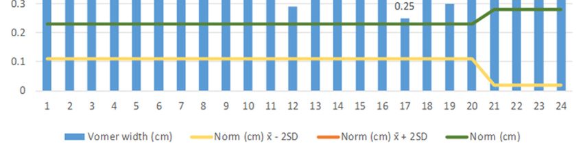
Medicina 2021, 57, 93                                                                                                  2 of 10

                        Table 1. Classification of congenital malformations of the nose according to Losee et al. (2004) [3].

                                                                 Half nose nasal aplasia, missing nose
                                                                 (arhinia), absence of some nasal structures
                           Type I–Hypoplasia and atrophy (62%) (often part of craniofacial malformations),
                                                                 stenosis, apertura piriformis, choanal
                                                                 atresia, and stenosis
                                                                 Proboscis lateralis, doubling of the nose and
                          Type II–Hyperplasia and duplicity (1%)
                                                                 doubling of the nostril
                                   Type III–Clefts (16%)         Clefts of the nose
                                                                 Encephaloceles, gliomas, dermoid cysts,
                                 Type IV–Neoplasia (20%)
                                                                 fistulas, and vascular anomalies

                           So far, there is no uniform theory on the cause of choanal atresia development.
                        Hengerer and Strome (1982) [6] defined four embryological theories:
                        (1) persistence of the buccopharyngeal membrane forming the bottom of the stomodea,
                        (2) persistence of the Hochstetter nasobuccal membrane and failure of its perforation in
                            the seventh week of intrauterine life,
                        (3) abnormal occurrence of mesoderm forming adhesion in the choanal area,
                        (4) abnormal direction of mesoderm migration (nasoseptal elements), and its excessive
                            growth due to exogenous and local factors.
                             The ectopic mesenchymal tissue excess in the central part of the face forms a
                        tangential atretic plate that extends from the vomer wall to the lateral wall of the nose.
                        According to this theory, the excessive growth of nasoseptal elements leads to stenosis or
                        bone atresia. Membranous atresia probably arises due to incomplete recanalization of the
                        epithelial plug in the 13th to 20th week of gestation. Other theories of atresia include the
                        Pasquini (2003) [7] and Lalani theory (2004) [8]:
                        (5) persistence of epithelial cells in the nasal cavity that proliferate between the sixth and
                            eighth week of gestation,
                        (6) excessive growth of lamina horizontalis and lamina perpendicularis ossis palatini.
                              The theory of persistence of the nasobuccal membrane (nasal sacs’ bottom) is the
                        most often mentioned in the literature. Nevertheless, it seems unlikely, as the nasobuccal
                        membrane forms near the outer nostrils, and after its perforation, primitive choanae start
                        to form. However, the atresia of choanae probably occurs only in the later stages of
                        development of a definitive secondary choanae. Due to the presence of associated
                        anatomical abnormalities of the nasal cavity and choanal area, which usually result in a
                        narrowing throughout half of the nasal cavity on the atresia side, the most probable cause
                        is a generalized disorder of mesenchymal cell migration that also affects the development
                        of the base of the skull [9].
                              Present diagnostic methods (endoscopy and CT imaging) have remarkably pushed
                        our knowledge about anatomical features in each patient further. It makes the diagnosis
                        of choanal atresia clear (type, thickness of the atretic segment). Typical changes in the
                        nasal cavity on the atretic side include the thickening of the posterior part of the vomer,
                        the thickening of the medial plate of the processus pterygoideus and the medialization of
                        the lateral nasal wall on the level of the choana [2]. The aim of our study was to find out
                        if the CT imaging evaluation with respect to the parameters followed in patients with
                        choanal atresia will help us determine the optimal surgical treatment.

                        2. Materials and Methods
                              We have evaluated the nasal cavity CT imaging of 24 pediatric patients diagnosed
                        with ICD-10: Q30.0 choanal atresia examined at the Department of Pediatric
                        Otorhinolaryngology of the Medical Faculty of the Comenius University and the National
                        Institute of Children’s Diseases in Bratislava (Slovakia). We have recorded the sex of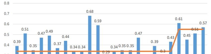
Medicina 2021, 57, 93 3 of 10
patients, the presence of syndromes, their age at the time of the nasal cavity CT
examination, and the types of atresia (congenital–acquired, unilateral–bilateral). In the
case of the unilateral atresia, we have examined which half of the nasal cavity was
affected. We determined the character of the atretic plate (membranous, bone-
membranous, and bone types) and their locations (unilateral, bilateral) [4]. Using the
method of Slovis et al. (1985) [10], a CT section paralleling the posterior hard palate at the
level of the pterygoid plates was obtained in all children (Figure 1). The sections were 0.5
to 1 mm thick. The TomoCon PACS system was used for CT scanning. The measurements
were obtained by one examiner with Tomocon Viewer application measuring tools.
Figure 1. The measurement plane: CT section paralleling the posterior hard palate at the level of
the pterygoid plates.
We measured the width of the vomer (V) at the posterior end, and the width of the
air space of the choanae or membranous atresia based on the measurement of the
dimension from the lateral wall of the nasal cavity to the edge of the vomer (LWNC-V) of
its posterior end (Figure 2). The obtained results were compared with the standards of
these parameters according to the Slovis et al. (1985) [10] (Table 2) and statistically
processed using the IBM SPSS Statistics for Macintosh, Version 27.0. Armonk, NY: IBM
Corp.
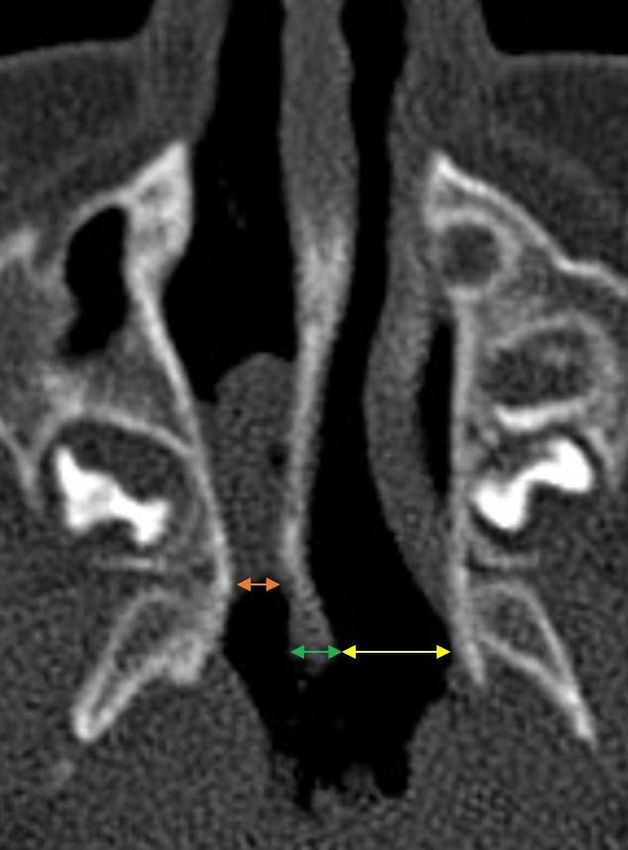
Figure 2. The measurement method: the lateral wall of the nasal cavity to the edge of the vomer
(LWNC-V) of the membranous atresia (orange arrow), the width of the vomer (green arrow) and
the LWNC-V of the choanal air space (yellow arrow).Medicina 2021, 57, 93 4 of 10
In patients with bone or bone-membranous atresia, the width of LWNC-V is non-
measurable.
Table 2. The standard dimensions of the choanae and vomer by patients’ age categories according
to Slovis et al. (1985) [10].
Measurement Age (Years) Mean (cm) ±2 Standard Deviations (cm)
Choanal air space Birth 0.67 0.34–1.01
>0–2 0.70 0.37–1.03
>2–4 0.75 0.42–1.09
>4–6 0.80 0.47–1.14
>6–8 0.86 0.53–1.19
>8–10 0.91 0.58–1.25
>10–12 0.97 0.63–1.30
>12–14 1.02 0.69–1.35
>14–16 1.07 0.74–1.41
>16–18 1.13 0.79–1.46
>18–20 1.18 0.85–1.51
Vomer width 0–8 0.23 0.11–0.34
>8–20 0.28 0.02–0.55
3. Results
In a group of 24 (100%) patients with a basic diagnosis made according to ICD-10
Q30.0 Choanal atresia (Table 3), there were 11 (46%) male and 13 (54%) female patients.
The age at the time of CT imaging was as follows: neonatal in 11 (45.7%) cases; 7 (30%)
children were from 1 month to 2 years old; 1 child between 2 and 4 years of age (4%); 1
child aged from 2 to 4 years (4%); 1 child between 14–16 years (4%); another 2 (8.3%)
patients were 16–18 years old; 1 patient in 18–20 years age group (4%). The associated
syndromes were present in 11 children (46%), with 7 patients (29%) having CHARGE
syndrome, the remaining four patients having Vacterl syndrome, Rubinstein–Taybi
syndrome, Treacher-Collins syndrome, and Down syndrome. Two families, where we
assumed the presence of CHARGE syndrome, refused to take genetic tests. All identified
cases of atresia were congenital and fully developed. During the CT examinations of the
nasal cavity, we found that 13 (54%) of cases represented a bone-membranous type of
atresia, in 8 cases (33%) it was a membranous type of atresia and in 3 patients (13%) it was
a bone type of atresia. Thirteen (54%) patients were unilaterally affected, and 11 (46%)
patients bilaterally. Of the 13 (100%) patients with unilateral choanal atresia, 8 were left-
sided (62%) and 5 right-sided (38%).
Table 3. The characteristics of the group (n = 24) taking into account the sex, age at the time of the nasal cavity CT
examination, presence of the syndrome, type of atresia according to its composition, and, in the case of unilateral atresia,
the lateral location.
LWNC-V on the LWNC-V on the
Vomer
Number Sex Age Type of Choanal Atresia Side of Soft Unaffected Side Syndrome
Width
Atresia (cm) (cm)
1 F Nb/2 d M–bilat. 0.39 0.29/0.29 - CHARGE
2 F Nb/2 d B–bilat. 0.51 - -
3 M Nb/3 d BM–unilat./left 0.35 - 0.49
4 M Nb/4 d BM–bilat. 0.47 - - Treacher-Collins
5 M Nb/4 d BM–bilat. 0.49 - - Vacterl
6 M Nb/6 d BM–unilat./right 0.37 - 0.32 Rubinstein–Taybi
7 F Nb/7 d BM–bilat. 0.44 - -
8 F Nb/7 d BM–bilat. 0.34 - - CHARGE
9 M Nb/10 d M–bilat. 0.34 0.27/0.31 - CHARGEMedicina 2021, 57, 93 5 of 10
10 F Nb/11 d B–bilat. 0.68 - - Down
11 M Nb/19 d BM–bilat. 0.59 - -
12 M 1.5 m M–unilat./left 0.29 0.49 0.75
13 F 2m M–unilat./left 0.34 0.36 0.59
14 M 2.5 m M–unilat./left 0.35 0.38 0.74 CHARGE
15 F 12 m M–unilat./right 0.35 0.39 0.73 CHARGE
16 F 14 m BM–unilat./left 0.47 - 0.62
17 F 20 m BM–unilat./left 0.25 - 0.89
18 M 24 m B–unilat./left 0.39 - 0.62
19 F 3y BM–unilat./right 0.30 - 0.74
20 F 4.5 y BM–unilat./right 0.43 - 0.81
21 M 15 y BM–bilat. 0.61 - - CHARGE
22 F 16 y M–unilat./right 0.45 0.54 0.9
23 M 16 y BM–bilat. 0.51 - - CHARGE
24 F 19 y BM–unilat./left 0.57 - 0.71
F = female, M = male, Nb = newborn, d = days, m = months, y = years, B = bony, M = membranous, BM = bony-membranous,
bilat. = bilateral, unilat. = unilateral, LWNC-V = lateral wall of nasal cavity-to-vomer measurement.
To evaluate the vomer width measurement results, we compared the individually
measured vomer width with the mean ± 2 SD in accordance with Slovis et al. (1985) [10]
(Figure 3). We found, in the age group of 0–8 years with 20 patients (patients 1–20), that
the average vomer width ± 2 SD in the whole group was 0.407 (0.305–0.51) cm,
significantly exceeding the norm. In the group of 4 patients aged >8–20 (patients 21–24),
two had unilateral choanal atresia and two bilateral choanal atresia. It can be seen from
the diagram that all patients have a vomer width ± 2 SD at the upper limit. In this group,
we also found that the average vomer width ± 2 SD is greater than the mean of 0.535
(0.474–0.59) cm.
Figure 3. The results of the comparison of individual vomer width in the group of patients with
choanal atresia (n = 24) with the norm ± 2 SD according to Slovis et al. (1985) [10]. Key: yellow
colour–norm-2 SD, green colour–norm, orange colour–norm + 2 SD in patients, blue colour–an
individual value of each patient.
We continued measuring the LWNC-V width (lateral wall nasal cavity–vomer) (1) on
the side of the membranous atresia at the end of the nasal cavity (Figure 4), and (2) on the
side of a unilaterally developed choana at the same level (Figure 5). The LWNC-V width
in patients with membranous choanal atresia (n = 8) aged 0 to 2 years of age was 0.363 ±
0.291–0.435, which is significantly less than the standard. The LWNC-V width in patientsMedicina 2021, 57, 93 6 of 10
with unilaterally developed choana was 0.639 ± 0.482–0.796, which is close to the norm
and does not exceed its upper limit.
1.2
1
0.8
0.6 0.49
0.36 0.38 0.39
0.4 0.29 0.27
0.2
0
1 2 4 5 6 7
LWNC-V on the side of the membranous atresia (cm)
Norm (cm) x̄ - 2SD
Norm (cm) x̄ + 2SD
Norm (cm)
Figure 4. Comparison of the norm ± 2 SD for the choana size according to Slovis et al. (1985) [10]
and LWNC-V width in patients with membranous choanal atresia (n = 7) aged 0–2 years. Key:
yellow color–norm-2 SD, green color–norm, orange color–norm + 2 SD in patients, blue color–an
individual value of each patient.
1.2
1 0.89
0.8 0.75 0.74 0.73
0.59 0.62 0.62
0.6 0.49
0.4 0.32
0.2
0
1 2 3 4 5 6 7 8 9
LWNC-V on the unaffected side (cm)
Norm (cm) x̄ - 2SD
Norm (cm) x̄ + 2SD
Norm (cm)
Figure 5. Comparison of the norm ± 2 SD for the choana size according to Slovis et al. (1985) [10].
LWNC-V of the developed choana in unilateral membranous choanal atresia in the observed
group (n = 9) of 0–2 years age. Key: yellow color–norm-2 SD, green color–norm, orange color–
norm + 2 SD in patients, blue color–an individual value of each patient.
Based on the Pearson correlation test between the vomer width and the age within
the observed group of patients with the choanal atresia (n = 24), we found a positiveMedicina 2021, 57, 93 7 of 10
correlation between the vomer width and the age. There is the same linear correlation as
in the normal population, i.e., in children with a standard finding in the nasal cavity. The
older the patient, the wider the vomer, even in patients with choanal atresia (r = 0.442, p =
0.031, 95% CI 0.108–0.760, SE 0.165). Besides, based on the Pearson correlation test,
between the vomer width and the presence of the unilateral or bilateral choanal atresia in
the group of 0–2 aged children (n = 20), we found that in the bilateral choanal atresia, the
vomer is on average wider than in the unilateral atresia (r = −0.578, p = 0.012, 95% CI 0.274–
0.807, SE 0.136).
4. Discussion
Anderhuber (2010) [11] states that the rupture of the membrana oronasalis occurs in
the fifth/sixth embryonic week when the primary choana is formed. Subsequently, a
secondary palatum is created from the tissue behind the foramen incisivum, definitively
separating the nasal and oral cavities, and creating the final shape of the choana. For this
reason, the most common theory for the development of choanal atresia is the notion of
membrana oronasalis persistence. The evaluated CT images of patients with choanal
atresia confirm that it is the case of a luminescence disorder of various degrees in the area
of the presumed choana with regard to the time of the disorder’s onset—from prominent
bone plate to bone-membrane plate to membranous plug, between the fifth and twentieth
week of embryo development, according to the available literature [12,13].
The causes of choanal atresia are still not clear. Retinoic acid deficiency is discussed
as it affects the ontogenesis and homeostasis of many tissues. It causes the overexpression
of FGF-8, which could lead to the persistence of the nasal valve. Other authors consider
the possible effect of thioamides used during pregnancy. Kurosaka (2018) focuses on the
role of genes involved in 50% in the development of choanal atresia. They are usually part
of the signaling pathways that control cellular activity and are essentially involved in the
normal or abnormal development of the neural plate, for example via the fibroblast
growth factor (FGF). Syndromes characterized by aberrant development of the cranial
part of the neural plate are referred to as neurocristopathies; these include CHARGE
syndrome, Treacher-Collins and DiGeorge syndrome, and others. Another group includes
syndromes caused by FGF mutations, such as Crouzon and Pfeiffer syndromes.
Furthermore, there are syndromes characterized by accelerated bone growth, for example
in craniodiaphyseal dysplasia or Marshall syndrome, then syndromes typical for middle
facial disorders, such as Fraser syndrome, and finally syndromes caused by mutations in
the SHH (sonic hedgehod) signaling pathway, such as Pallister–Hall syndrome [14].
Hengerer et al. (2008) [6] underline the importance of the correct identification of
obstruction locations in the nasal cavity, as they may represent limits to the success of
surgical treatment. They draw attention to the “2–1” rule, which relates to the
predominance of unilateral over bilateral choanae atresia, the predominant female sex,
and the more frequent right-sided occurrence of unilateral atresia. We did not identify
such a typical representation in the study group—unilateral atresia was 54%, the
representation of the female sex was also 54%, and on the contrary, left-sided atresia
predominated (62%) in the cases of unilateral atresia, but our results are limited by the
small count in the group. According to the literature, choanal atresia is at approximately
50% associated with the presence of syndromes, which corresponds to our result of 46%.
The most frequent is CHARGE syndrome; Lofty and Al-Noury (2011) [15] state that 75%
of 9 patients had CHARGE syndrome in their group. Burrow et al. (2009) [16] identified a
syndrome in 51% of 129 patients, predominantly CHARGE syndrome. These findings
correlate with our findings. In our group of 11 patients, 7 children (29%) had the
syndrome, and the two families, where presence had been expected, refused to undergo
a genetic examination. In his review of choanal atresia, Kwong (2015) [17] refuted the
historical fact that 90% of cases are bone atresia, and only 10% are membranous type.
Based on a retrospective evaluation of CT images and histological examination of the
findings in 63 patients, he determined that the bone atresia was present in only 29% ofMedicina 2021, 57, 93 8 of 10
patients and the others had a mixed syndrome form of atresia, which was present in 71%
cases. In our group of patients, the majority had bone-membranous atresia as well (n = 13,
54%).
Ginat and Robson (2015) [18] state that in children under 2 years of age, the width of
the postero-inferior part of the vomer is ≤2.3 mm and the diameter of the choana is ≥3.7
mm, while its width increases by 0.09 mm/year. They compared the nasal cavity CT
examinations of 11 patients with choanal atresia with a control group of 66 patients with
normal findings in the nasal cavity. In our work, we followed the standards set out by
Slovis et al. (1985) [10]. For neonates, they found that the space between the lateral wall of
the nasal cavity and the vomer was 0.67 mm, increasing by 0.27 mm each year until the
age of 20. The average vomer thickness was 2.3 mm in patients under 8 years of age and
2.8 mm in patients aged from 8 to 20 years. The thickness of bone atresia varied from 1 to
12 mm, depending on bone changes in the area of the lamina pterygoidea medialis. In our
group, we found in all examined parameters that the width of the vomer, LWNC-V, and
the width of the developed choana in unilateral choanal atresia are on average below the
standards set for a given age by Slovis et al. (1985) [10]. Aslan (2009) [19] examined 17
different parameters on HRCT nasal images in 9 children with bilateral choanal atresia,
compared to the control group of 104 pediatric patients. They found that in the presence
of atresia, the nasal cavity is narrowed to the full extent. They did not find statistically
significant differences in the length of the nasal septum, or in the area of the nasopharynx.
Lofty and Al-Noury (2011) [15] based on CT examinations of the nasal cavity
compared to the group of 9 pediatric patients, showed its great informative value in the
diagnosis of choanal atresia, as well as in the detection of associated maxillo-facial
anomalies, including stenosis of the apertura piriformis, which is a typical manifestation
of CHARGE syndrome. When evaluating CT images of the nasal cavity, we confirmed the
presence of typical anatomical changes accompanying choanal atresia, such as the
thickening of the posterior part of the vomer, medialization of the lateral nose wall at the
level of the medial plate processus pterygoideus and narrower dimensions of the choana
with the membranous atresia, as well as on the unaffected side in unilateral atresia. The
same changes are reported by several authors [6,9].
Members of IPOG (International Pediatric Otolaryngology Group) from eight
countries (Australia, Canada, France, Ireland, Italy, Portugal, United Kingdom and
United States) have agreed on the importance of HRCT nasal cavity examination as the
gold standard in the diagnosis and treatment of choanal atresia and evaluated the
experience of 22 tertiary centers in the consensual document of 2019. Regarding treatment,
89.3% of participants prefer an endoscopic approach to the treatment of choanal atresia;
the transpalatal approach is used in exceptional cases [20]. It also follows from our results
that in the case of separate transnasal curettage for the purpose of fenestration of atretic
choanae, the scope of intervention is insufficient, due to the above-mentioned changes. It
does not allow the removal of excess bone in the area of the choanal atresia to a sufficient
extent and leaves an enlarged back part of the vomer in place. Currently, we can achieve
an adequate extent of the fenestration by utilizing a microendoscopic technique with the
use of a drill or a bone shaver. This will remove the excess bone in the area of the atretic
plate and any membranous part of the choana atresia. A laser should be used to prevent
bleeding. Subsequently, the obtained space is enlarged using a posterior septostomy,
removing the dorsal excessively thickened part of the vomer. Rajan and Tunkel (2018) [2]
underline the importance of covering the wound areas in the neochoana with mucosal
grafts using fibrin glue.
The safety of surgery can be increased by the IGNS (Image Guided Navigation
Surgery). In the case study by Ji et al. (2020) [5], they report successful use of the IGNS in
the surgical endoscopic treatment of an unrecognized complex bilateral choanal atresia,
associated with significant deviation of the nasal septum, and with the Tessier 3 facial
cleft.Medicina 2021, 57, 93 9 of 10
5. Conclusions
Contrary to the recommendation by Slovis et al. (1985) [10] to indicate a transpalatal
approach for the bilateral bone atresia, and endoscopically address only unilateral choanal
atresia, based on the current modern endoscopic options, we have evidently moved to a
transnasal microendoscopic approach [12,20,21]. The consistent fenestration of the atretic
choana is combined with the posterior nasal septotomy. The results of our study strongly
contributed to the justification of this procedure; it allows the creation of a spacious
neochoana for both halves of the nasal cavity without the need for long-term stenting. The
transpalatal approach is indicated only for very special and complicated cases.
Author Contributions: Conceptualization I.Š. and J.B.; methodology and investigation, I.Š. and J.B.;
software and validation, J.B., I.Š. and I.V.; statistical and formal analysis I.V.; writing—original draft
preparation I.Š.; writing—review and editing, J.B. and I.V. All authors have read and agreed to the
published version of the manuscript.
Funding: This research received no external funding.
Institutional Review Board Statement: The study was conducted according to the guidelines of the
Declaration of Helsinki, and approved by the Ethics Committee of National Institute of Children’s
Diseases in Bratislava, Slovakia—protocol code: EK271-21.
Informed Consent Statement: Not applicable.
Data Availability Statement: All the data are available in this article (Table 3).
Conflicts of Interest: The authors declare no conflict of interest.
References
1. Ramsden, J.D.; Campisi, P.; Forte, V. Choanal atresia and choanal stenosis. Otolaryngol. Clin. North. Am. 2009, 42, 339–352,
doi:10.1016/j.otc.2009.01.001.
2. Rajan, R.; Tunkel, D.E. Choanal atresia and other neonatal nasal anomalies. Clin. Perinatol. 2018, 45, 751–767,
doi:10.1016/j.clp.2018.07.011.
3. Losee, J.E.; Kirschner, R.E.; Whitaker, L.A.; Bartlett, S.P. Congenital nasal anomalies: A classification scheme. Plast. Reconstr.
Surg. 2004, 113, 676–689, doi:10.1097/01.PRS.0000101540.32533.EC.
4. Brown, O.E.; Pownell, P.; Manning, S.C. Choanal atresia: A new anatomic classification and clinical management applications.
Laryngoscope 1996, 106, 97–101, doi:10.1097/00005537-199601000-00019.
5. Ji, Y.S.; Kyu-Sup, Ch.; Yong, C.B.; Seong, H.B. Image-guided navigation surgery for bilateral choanal atresia with a Tessier
number 3 facial cleft in an adult. Arch. Craniofacial. Surg. 2020, 21, 64–68, doi:10.7181/acfs.2019.00661.
6. Hengerer, A.S.; Strome, M. Choanal atresia: A new embryologic theory and its influence on surgical management. Laryngoscope
1982, 92, 913–921.
7. Pasquini, E.; Sciarretta, V.; Saggese, D.; Cantaroni, C.; Macrì, G.; Farneti, G. Endoscopic treatment of congenital choanal atresia.
Int. J. Pediat. Otorhinolaryngol. 2003, 67, 271–276.
8. Lalani, S.R.; Safiullah, A.M.; Molinari, L.M.; Fernbach, S.D.; Martin, D.M.; Belmont, J.W. SEMA3E mutation in a patient with
CHARGE syndrome. J. Med. Genet. 2004, 41, 94.
9. Smith, J.K.; Castillo, M.; Mukherji, S.; Buenting, J.; Drake, A. Imaging of Nasopharyngeal Atresia. Am. J. Neuroradiol. 1995, 16,
1936–1938.
10. Slovis, T.L.; Renfro, B.; Kuhns, L.R.; Belenky, W.; Spoylar, J. Choanal atresia: Precise CT evaluation. Radiology 1985, 155, 345–
348, doi:10.1148/radiology.155.2.3983384.
11. Anderhuber, W. Malformationen der Nase und der Nasennebenhöhlen. In Pädiatrische HNO-Heilkunde; Götte, K.; Nicolai, T.;
Eds.; Urban and Fischer: Műnchen, Germany, 2010; pp. 253–259.
12. Rontal, M.; Todd, D.O.; Anon, J.B.; Zinreich, S.J. Embryology and anatomy of the sinuses. In Bluestone and Stool’s Pediatric
Otolaryngology, 5th ed.; Bluestone, C.D., Ed.; PMPH-USA: Shelton, CT, USA, 2014; pp. 913–925.
13. Otteson, T.D. Congenital malformations of the nose and paranasal sinuses. In Bluestone and Stool´s Pediatric Otolaryngology, 5th
ed.; Bluestone, C.D., Ed.; PMPH-USA: Shelton, CT, USA, 2014; pp. 1029–1032.
14. Kurosaka, H. Choanal atresia and stenosis: Development and diseases of the nasal cavity. Wiley Interdiscip. Rev. Dev. Biol. 2018,
e336, doi:10.1002/wdev.336.
15. Lofty Al-Noury, K. Role of Multislice Computed Tomography and Local Contrast in the Diagnosis and Characterization of
Choanal Atresia. Int. J. Pediatr. 2011, 2011, 280763, doi:10.1155/2011/280763.
16. Burrow, T.A.; Saal, H.M.; De Alarcon, A.; Martin, L.J.; Cotton, R.T.; Hopkin, R.J. Characterization of congenital anomalies in
individuals with choanal atresia. Arch. Otolaryngol. Head. Neck. Surg. 2009, 135, 543–547, doi:10.1001/archoto.2009.53.
17. Kwong, K.M. Current updates on choanal atresia. Front. Pediatr. 2015, 3–52, doi:10.3389/fped.2015.00052.Medicina 2021, 57, 93 10 of 10
18. Ginat, D.T.; Robson, C.D. Diagnostic imaging features of congenital nose and nasal cavity lesions. Clin. Neuroradiol. 2015, 25, 3–
11, doi:10.1007/s00062-014-0323-5.
19. Aslan, S.; Yilmazer, C.; Yildirim, T.; Akkuzu, B.; Yilmaz, I. Comparison of nasal region dimensions in bilateral choanal atresia
patients and normal controls: A computed tomographic analysis with clinical implications. Int. J. Pediatr. Otorhinolaryngol. 2009,
73, 329–335, doi:10.1016/j.ijporl.2008.10.029.
20. Moreddu, E.; Rizzi, M.; Adil, E.; Balakrishnan, K.; Chan, K.; Cheng, A.; Daniel, S.J.; de Alarcon, A.; Hart, C.; Hartnick, C.; et al.
International pediatric otolaryngology group (IPOG) Consensus recommendations: Diagnosis, pre-operative, operative and
post-operative pediatric choanal atresia care. Int. J. Pediatr. Otorhinolaryngol. 2019, 123, 151–155, doi:10.1016/j.ijporl.2019.05.010.
21. LaCour, J.B.; Patel, M.R.; Zdanski, C. Image-guided endoscopic and microdebrider assisted repair of choanal atresia in a
neonate. Int. J. Pediatr. Otorhinolaryngol. 2009, 4, 21–24, doi:10.1016/j.pedex.2008.05.002.You can also read


