Diagnostic Accuracy of Trichoscopy in Trichotillomania: A Systematic Review
←
→
Page content transcription
If your browser does not render page correctly, please read the page content below
1/6
REVIEW ARTICLE
Diagnostic Accuracy of Trichoscopy in Trichotillomania:
ActaDV
A Systematic Review
Agnieszka KACZOROWSKA1, Lidia RUDNICKA1, Catherine M. STEFANATO2, Anna WAŚKIEL-BURNAT1, Olga WARSZAWIK-
HENDZEL1, Małgorzata OLSZEWSKA1 and Adriana RAKOWSKA1
1
Department of Dermatology, Medical University of Warsaw, Poland and 2Department of Dermatopathology, St John’s Institute of Dermatology,
King’s College London, London, UK
Acta Dermato-Venereologica
Trichotillomania is formally classified as a mental
health disorder, but it is commonly diagnosed by derma-
SIGNIFICANCE
tologists. The aim of this systematic review is to assess Trichotillomania is a hair loss condition, which is classified
the diagnostic value of trichoscopy in diagnosing tricho- as a mental health disorder. The affected person pulls out
tillomania. The analysis identified the 7 most specific their own hair until areas of alopecia are visible on the
trichoscopic features in trichotillomania. These features scalp. The patient (often a child) is usually not likely to ad-
had the following prevalence and specificity: trichop mit that they are pulling their own hair. This review deter-
tilosis (57.5%; 73/127 and 97.5%, respectively), v- mined 7 hair-related features that can help the dermatolo-
sign (50.4%; 63/125 and 99%), hook hairs (43.1%; gist identify hair pulling and protect the patient from being
28/65 and 100%), flame hairs (37.1%; 52/140 and misdiagnosed and mistakenly treated as having alopecia
96.5%), coiled hairs (36.8%; 46/125 and 99.6%), areata (which is hair loss with similar appearance). The
tulip hairs (36.4%; 28/77 and 89.6%), and hair powder hair abnormalities are visible when using a special non-
(35.6%; 42/118 and 97.9%). The 2 most common, invasive magnifying technique, called trichoscopy.
but least specific, features were broken hairs and
black dots. In conclusion, trichoscopy is a reliable new
diagnostic method for hair loss caused by hair pulling. psychiatric criteria. In a significant proportion of patients
Trichoscopy should be included as a standard proce- only criterion (i) is fulfilled (3). Clinically, trichotilloma-
dure in the differential diagnosis of trichotillomania in nia presents as a patchy alopecia (4, 5). The pull-test is
clinical practice. negative (3, 5). The scalp is the most common location
ActaDV
(72.8%) (4). The vertex area is commonly affected with
Key words: trichotillomania; hair-pulling disorder; trichoscopy;
dermoscopy; dermatoscopy. a so-called tonsure or “Friar Tuck” pattern of baldness (3,
5). Other common locations of trichotillomania include
Accepted Jun 28, 2021; Epub ahead of print Jun 29, 2021
eyebrows (56.4%), eyelashes (52.6%), and pubic area
Acta Derm Venereol 2021; 101: XX–XX. (50.7%) (4). Beard is rarely affected (4.3%) (4). Diag-
Corr: Lidia Rudnicka, Department of Dermatology, Medical University of
nosis of trichotillomania can be challenging, especially
Warsaw, Koszykowa 82A, PL-02-008 Warsaw, Poland. E-mail: lidia.rud- when patients deny or are unaware of hair pulling or do
nicka@dermatolodzy.com.pl
not meet all the psychiatric criteria.
Advances in dermatology and venereology
Trichoscopy is widely used to diagnose patients with
T richotillomania (hair-pulling disorder) is characteri-
zed by a recurring habit of pulling out of one’s hair,
resulting in hair loss (1). Diagnosis of trichotillomania
hair and scalp disorders (3, 6). Several studies were
performed to identify the typical trichoscopic features
of trichotillomania. However, no large-scale analysis of
is usually based on clinical examination. According to the specificity and sensitivity of trichoscopic findings
the Diagnostic and Statistical Manual of Mental Disor- in trichotillomania has been performed to date. The aim
ders 5 (DSM-5) criteria trichotillomania belongs to the of this study was to evaluate the role of trichoscopy in
obsessive-compulsive and related disorders group. The diagnosing trichotillomania by identifying characteristic
DSM-5 diagnostic criteria for trichotillomania are: (i) trichoscopic features of the disease and assessing the
recurrent pulling out of one’s hair, resulting in hair loss; sensitivity and specificity of each trichoscopic finding.
(ii) repeated attempts to decrease or stop hair pulling;
(iii) the hair pulling causes clinically significant distress
or impairment in social, occupational, or other important MATERIALS AND METHODS
areas of functioning; (iv) the hair pulling or hair loss is not A literature review was performed by searching 3 databases: Pub-
attributable to another medical condition (e.g. a derma- Med, Scopus and EBSCO. The search terms “trichotillomania”
tological condition); and (v) the hair pulling is not better and “hair pulling disorder” combined with “trichoscopy”, “der-
moscopy”, “dermatoscopy”, “videodermoscopy” or “videoder-
explained by the symptoms of another mental disorder
matoscopy” were used. Moreover, references of all relevant articles
(e.g. attempts to improve a perceived defect or flaw in were checked for further publications. Original studies and case
appearance in body dysmorphic disorder) (2). Patients series published in English were eligible for quantitative analysis.
visiting dermatologists’ offices often do not fulfil these Trichoscopic features that were reported in at least 10 patients with
This is an open access article under the CC BY-NC license. www.medicaljournals.se/acta doi: 10.2340/00015555-3859
Society for Publication of Acta Dermato-Venereologica Acta Derm Venereol 2021; 101: advxxxxx2/6 A. Kaczorowska et al.
trichotillomania in studies with a control group were included in
the quantitative analysis. If the study was conducted using both
ActaDV
polarized and non-polarized trichoscopy, the data from polarized
mode was used in this analysis. Studies with incomplete epide-
miological data, such as unknown number of patients or frequency
of trichoscopic findings, case reports, animal studies, reviews or
book chapters were excluded. Articles considering the frequency
of trichoscopic features based on the number of patches instead
of the number of patients were also excluded (Fig. 1). The review
presents the frequency of the most characteristic trichoscopic
features of trichotillomania. Their sensitivity, specificity, positive
predictive value (PPV), and negative predictive value (NPV) were
Acta Dermato-Venereologica
calculated. The review also considers trichoscopic findings that
are commonly observed, but are not disease-specific. This article
is based on previously conducted studies and does not contain any
studies with human participants or animals performed by any of
the authors. Prevalence ranges and prevalence mean values are
based on all studies in which particular trichoscopic features were
evaluated. Sensitivity, specificity, PPV, NPV are based only on
original studies that included patients with trichotillomania and
other hair diseases. Calculation methods are shown below:
Mean value= Number of patients with trichotillomania (TTM) and given feature
Total number of patients with TTM
Sensitivity= Number of patients with TTM and given feature
Number of patients with TTM and given feature + Number of patients
with TTM without given feature
Specificity= Number of patients without TTM and without given feature
Number of patients without TTM and without given feature + Number
of patients without TTM with given feature
PPV= Number of patients with TTM and given feature Fig. 1. Preferred Reporting Items for Systematic Reviews and
Number of patients with TTM and given feature + Number of patients Meta-Analyses (PRISMA) flowchart. *Excluded records: reviews, book
without TTM with given feature
chapters, articles in languages other than English, case reports, articles
NPV= Number of patients without TTM and without given feature concerning animals, articles concerning different diseases. **Excluded
Number of patients without TTM and without given feature + Number articles: 1 article was based on a number of patches instead of patients,
ActaDV
of patients with TTM without given feature
3 articles did not present the frequency of trichoscopic findings, 1 article
contained an unclear data considering trichoscopic findings, 1 article
described trichotemnomania instead of trichotillomania. ***10 original
RESULTS studies and 3 case series.
The review found 13 articles (10 original studies and
3 case series), which were selected and included in the or more hair shafts of a single follicular unit are pulled
simultaneously and break at the same length above the
quantitative analysis, as shown on the Preferred Re-
scalp’s surface. Normal, regrowing terminal hairs after
porting Items for Systematic Reviews and Meta-Analyses
shaving may look similar, but they cover the whole field
(PRISMA) flow diagram in Fig 1. Overall, 203 patients
of view, whereas the v-sign in trichotillomania is sur-
Advances in dermatology and venereology
with trichotillomania were included in the systematic
rounded by long terminal hairs (5). The v-sign is another
review. The results of the quantitative analysis are shown
feature that is characteristic of trichotillomania, with an
in Table I and described below.
estimated specificity of 99%. The frequency of the v-sign
in trichotillomania varied between 20% and 70% (mean
Trichoscopic findings in trichotillomania 50.4% (63/125)) (5, 9, 11, 13–16).
Trichoptilosis. Trichoptilosis presents as
longitudinal splitting of fractured distal hair Table I. Trichoscopic features of trichotillomania
shafts. It is a common finding in healthy, long Positive Negative
hairs that have not been cut for a while, but in predictive predictive
Trichoscopic Prevalence Prevalence Sensitivity Specificity value value
trichotillomania it affects short hairs instead. feature Ranges, % Mean, % % % % %
It can occasionally be detected in different Hook hairs 11.1−56.8 43.1 43.5 100.0 100.0 93.6
types of alopecia, including traction alopecia, Coiled hairs 4.3−100.0 36.8 29.0 99.6 90.0 92.4
alopecia areata, tinea capitis, and primary V-sign
Hair powder
20.0–70.0
10.0−88.9
50.4
35.6
48.9
30.6
99.0
97.9
86.3
68.4
93.5
90.3
cicatricial alopecias (3, 7). The incidence rate Trichoptilosis 34.1−100.0 57.5 50.5 97.5 67.1 95.2
of trichoptilosis varied between 34.1% and Flame hairs 25.0−100.0 37.1 36.2 96.5 52.1 93.5
Tulip hairs 10.0−47.7 36.4 47.7 89.6 38.2 92.7
100% (mean 57.5% (73/127)) of patients with Broken hairs 80.0−100.0 99.5 99.3 62.9 18.7 99.9
trichotillomania (5, 8–13). Black dots 27.3−100.0 66.7 65.1 61.7 12.7 95.4
V-sign. This term was introduced by Rudnicka The prevalence ranges and mean are based on studies in which given feature was evaluated
(5, 8–17, 20, 22). The sensitivity, specificity, PPV and NPV were calculated based on original
et al. in 2012 (3). A v-sign is formed when 2 articles (5, 8–10, 14, 16, 17, 20).
www.medicaljournals.se/actaTrichoscopy in diagnosis of trichotillomania 3/6
Hook hairs. Hook hairs or question mark hairs are par- induced alopecia, dissecting cellulitis, tinea capitis, and
tially coiled hairs (5). They are the most specific finding traction alopecia (18, 23–25). According to Rakowska
ActaDV
of trichotillomania, with the frequency of 11.1–56.8% et al. (5) black dots found in trichotillomania and tinea
(mean 43.1% (28/65)) (5, 14, 15). Hook hairs were also capitis are variable in diameter and shape (round, oval,
reported in traction alopecia (3). irregular), whereas those in alopecia areata tend to be
Flame hairs. Flame hairs result from traumatic pulling similar in size and shape.
anagen hairs. They are observed as hair residues that Yellow dots with black peppering. Yellow dots with black
are semi-transparent, wavy and cone-shaped. Flame peppering (containing a black dot, mostly in the central
hairs were first described by Rakowska et al. (5) in 2014 part), were first described in trichotillomania by Inui
and suggested as a specific sign for trichotillomania. et al. (21) in 2008. Isolated cases were also reported in
Acta Dermato-Venereologica
In analysed studies they were detected in patients with alopecia areata (5, 9). As they were present only in 11%
trichotillomania with the incidence rate between 25% (8/73) of patients with trichotillomania, they were not
and 100% (mean 37.1% (52/140)) (5, 9, 11, 13–15, included in the quantitative analysis. In contrast, yellow
17). However, flame hairs were also described in acute dots without black peppering are mainly associated with
chemo- and radiotherapy-induced alopecias, alopecia alopecia areata, but can also be found in androgenetic
areata, traction alopecia, and central centrifugal cicatri- alopecia, chronic cutaneous lupus erythematosus, and
cial alopecia (17, 18). dissecting cellulitis (26). In trichotillomania, they were
Coiled hairs. Coiled hairs are broken telogen/catagen described as very sparse and irregularly distributed,
hairs that curl back (19). They are similar to pigtail hairs, unlike most cases of alopecia areata (5).
but can be distinguished by their irregular appearance Burnt match-stick sign. The sign was first reported by
and frequently frayed ends (5). Coiled hairs are highly Malakar et al. (22) in 2017, as a dark bulbar proximal tip
specific of trichotillomania (99.6%), with the incidence with a linear stem of variable length. As there were only
rate varying between 4.3% and 100% (mean 36.8% 4 cases described in the literature (3 in trichotillomania
(46/125)) (5, 8, 9, 11–14). They can also be observed in and 1 in traction alopecia (14, 22)) this feature was not
traction alopecia (3). included in the evaluation of specificity and sensitivity.
Tulip hairs. Tulip hairs were first named by Rakowska Haemorrhages. The first description of haemorrhages in
et al. (5) in 2014. They are described as short hairs with trichoscopy of trichotillomania was published in a case
ActaDV
darker, tulip flower shaped ends, which form due to report by Peralta & Morais (27) in 2012. They show as
diagonal hair shaft fracture (5). The frequency of tulip a red, perifollicular dots and are caused by traumatic
hairs in trichotillomania varied between 10% and 47.7% hair pulling (11). In the study conducted by Elmas et
(mean 36.4% (28/77)) (5, 11, 13, 15). They can be found al. (13), hemorrhages were detected in 40% (8/20) of
in alopecia areata and traction alopecia (3, 5). the patients with trichotillomania. Moreover, follicular
Hair powder. Hair powder (black powder/sprinkled micro-haemorrhages were described as a unique sign
hairs) is another term introduced by Rakowska et al. for detecting coexisted trichotillomania in patients with
(5) in 2014. It develops as a result of total hair shaft da- alopecia areata, since these conditions often coexist (11,
mage, when only a sprinkled hair residue is visible (5). 28, 29).
Advances in dermatology and venereology
The incidence rate of hair powder in trichotillomania Mace sign. This sign was first described in 3 cases by
was estimated between 10% and 88.9% (mean 35.6% Malakar et al. (15) in 2016, and suggested as an exclu-
(42/118)) (5, 9, 11, 13–15). Recently, hair powder was sive sign of trichotillomania. The mace sign presents as
described in traction alopecia and alopecia areata (9, broken terminal hairs, which are uniform in diameter and
14). pigmentation with a bulging distal end that resembles a
Broken hairs. Broken hairs were detected in almost every mace. In the study performed by Elmas et al. (13) the
case of trichotillomania (mean 99.5%; 182/183), but they mace sign was reported in 45% (9/20) of patients with
have a low calculated PPV (18.7%) (5, 8–12, 14–16, trichotillomania.
20–22). They are common in other alopecias, especially Branched hairs. The term branched hairs was introduced
traction alopecia, alopecia areata, and tinea capitis. In in 2020 by Elmas et al. (13). Branched hairs are described
trichotillomania and traction alopecia hairs are broken as hair shafts with obliquely detached pieces. They form
at different levels above the scalp, which can be useful as a result of pushing hair shafts linearly from the distal
in making the diagnosis (3, 5, 9, 10, 13, 14). to the proximal end. They were reported in 40% (8/20)
Black dots. Black dots were detected in 27.3–100% of of patients with trichotillomania (13).
patients with trichotillomania (mean 66.7% (122/183)) Concentric hairs. Concentric hairs are another novel
with a low calculated PPV (12.7%) (5, 8–16, 20–22). finding, described by Elmas et al. (13). Concentric hairs
Black dots are pigmented residues of hairs broken or are broken hairs presenting as a central black dot sur-
destroyed at the scalp level (3). They are not specific rounded with a black circle. They are probably caused
and can be recognized in alopecia areata, chemotherapy- by attenuation of hair cortex associated with repetitive
Acta Derm Venereol 20214/6 A. Kaczorowska et al.
trauma. They were described in 35% (7/20) of patients DISCUSSION
with trichotillomania (13).
ActaDV
This systematic review was conducted to systematize cur-
Angulated hairs. Angulated hairs is a term introduced rent knowledge about the trichoscopic pattern of tricho-
in 2017 by Khunkhet et al. (9). They are described as tillomania. This analysis revealed the 7 most characteristic
fractured hairs forming a sharp angle along the hair trichoscopic findings: trichoptilosis (present in 57.5% of
shaft, regardless of the number of fracture sites. The term patients), v-sign (50.4%), hook hairs (43.1%), flame hairs
covers zigzag hairs, checkmark hairs, and trichorrhexis (37.1%), coiled hairs (36.8%), tulip hairs (36.4%), and
nodosa (9). In 2020, Elmas et al. (13) reported this sign hair powder (35.6%) (Fig. 2). Hook hairs, coiled hairs
in 40% (8/20) of patients with trichotillomania. and v-sign have the highest calculated PPV (100%, 90%
Exclamation mark hairs. Exclamation mark hairs are and 86.3%, respectively). Thus, their presence is highly
Acta Dermato-Venereologica
rare in trichotillomania. They are described as hairs with indicative of trichotillomania. Although, in the current
thin proximal ends and thicker distal ends. Exclamation analysis, hook hairs were present only in patients with
mark hairs are a hallmark feature of alopecia areata (30); trichotillomania, they were also described in patients with
however, they can also be present in trichotillomania, traction alopecia. Thus, hook hairs should not be consi-
tinea capitis, chemotherapy-induced alopecia, anagen dered as a pathognomonic sign of trichotillomania (3).
effluvium, and severe intoxication (31). According to The most common trichoscopic findings in trichotillo-
Rudnicka et al. (3) there are slight differences between mania were broken hairs and black dots. However, these
exclamation mark hairs in these 2 diseases; in trichotillo- features are not specific for trichotillomania (3).
mania they tend to have a flat distal end and a pigmented A relatively common, but not highly specific, feature of
proximal end, whereas in alopecia areata they usually trichotillomania is tulip hairs. The comparison between
have an uneven, ragged, distal end and a hypopigmented the tulip hairs (5) and the subsequently described mace
proximal end. Exclamation mark hairs were observed sign (15) is not clear. The mace sign is presented as a
in 33.3% (23/69) of patients with trichotillomania, with bulging at the distal end of a broken hair, with a rough
sensitivity 80.8% and PPV of only 11.4%. hair shaft that is uniform in pigmentation and darker than
surrounding hairs. On the contrary, tulip hair is described
Other trichoscopic findings as a black mark at tip of the hair with light-coloured hair
shaft and dark distal end. These features may indicate
Other reported trichoscopic features of trichotillomania
ActaDV
that the mace sign is a variant of tulip hairs. The slight
were not characteristic or common. They included: differences may result from using trichoscopy with dif-
upright re-growing hairs, vellus hairs, and yellow dots. ferent magnifications.
Based on the results of this analysis it may be sug-
Locations other than scalp gested that trichoscopy should play a major role in esta-
Trichotillomania may affect any site of the body (32). blishing the diagnosis of trichotillomania. The available
According to available case reports, trichoscopic features clinical criteria for trichotillomania were established
of eyebrows and scalp trichotillomania are similar, and on the basis of analysing groups of patients who are in
include: irregularly broken hairs, black dots, coiled hairs, psychiatric care. Many patients, in particular children and
Advances in dermatology and venereology
hook hairs, trichoptilosis, v-sign, flame hairs, tulip hairs, adolescents, do not all meet the criteria for trichotilloma-
yellow dots, and haemorrhages (33, 34). Trichoscopy of nia as described in the DSM-5. Many patients fulfil only
eyelashes trichotillomania presents irregularly broken criterion (i) of DSM-5 (recurrent pulling out of one’s
hairs, black dots, and flame hairs (35). In beard tricho- hair, resulting in hair loss), with no attempts to decrease
tillomania, a new trichoscopic finding called the ”pluck or stop hair pulling and no psychiatric comorbidities.
out sign” has recently been described in the literature. Furthermore, many patients, in particular children, do
It consists of round haemorrhages around hair shafts not admit recurrent pulling out of their hair, making the
and, according to the authors, can be very suggestive of correct diagnosis very challenging. The first perceived
trichotillomania (4, 36). symptom of the disease is loss of hair; hence dermato-
logists are usually the first doctors to deal with patients
suspected of trichotillomania.
Monitoring of treatment
Trichoscopy is likely to be a helpful tool in screening
In patients with trichotillomania, a significant increase in patients with hair loss who are suspected of trichotillo-
capillary density, the presence of i-hairs and hairs in the mania (3). A rapid in-office diagnosis may also accelerate
growth phase were reported in the 6-month follow-up. treatment, which could be important for the patients, who
I-hair has been a suggested marker of the trichotilloma- tend to have significant functional impairment and low
nia remission. It presumably is a modified black dot, quality of life (1). Moreover, trichoscopy could avoid the
developing into a normal hair as the condition improves need for semi-invasive diagnostic methods, such as scalp
(37, 38). biopsy. In doubtful cases, trichoscopy-guided biopsy may
www.medicaljournals.se/actaTrichoscopy in diagnosis of trichotillomania 5/6
ActaDV
Acta Dermato-Venereologica
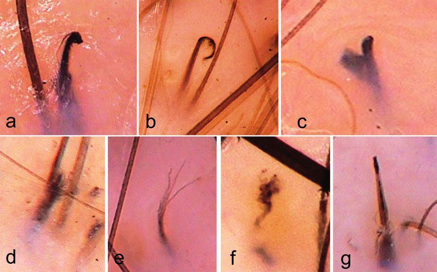
Fig. 2. The 7 most characteristic trichoscopic features of trichotillomania (×70). (a) Hook hairs: partly coiled hairs with a hook-like appearance. (b)
Coiled hairs: irregularly coiled hairs with a jagged end. (c) V-sign: created when 2 or more hairs emerging from 1 follicular unit are pulled simultaneously
and break at the same length above scalp surface. (d) Hair powder: residue of totally damaged hair shafts. (e) Trichoptilosis: longitudinal splitting of
the distal end of hair shaft (f) Flame hairs: hair residues, semi-transparent, wavy, and cone-shaped, resembling a fire flame. (g) Tulip hairs: short hairs
with a tulip leaf-like hyperpigmentation at the distal end.
ActaDV
be performed (39). The results of this systematic analysis efficacy, such as an increase in capillary density, presence
may serve as indication as to which common trichoscopic of i-hairs and hairs in the growth phase.
features of trichotillomania should be considered when This study has some limitations. The number of studies
performing a trichoscopy-guided biopsy. related to trichoscopy of trichotillomania is limited, and
Other trichoscopic features: yellow dots with black some did not include control groups. Various topics have
peppering (3), burnt match stick sign (14, 22), haemorr- not yet been assessed in clinical studies, including the
hages (11), mace sign (15), branched hairs (13), concen- role of trichoscopy in locations other than the scalp and in
tric hairs (13), and angulated hairs (9) were observed in monitoring treatment. There are only single reports about
Advances in dermatology and venereology
trichotillomania. These features were rare in the global those issues and more data is required for a full analysis.
analysis of all published data. Consequently, they were In conclusion, this systematic review found that the
not included in the quantitative analysis. Some of these 7 most specific trichoscopic findings in trichotillomania
features may have potential in future differential diagno- are: hook hairs, coiled hairs, v-sign, hair powder, flame
sis of hair loss, but further studies are needed to evaluate hairs, tulip hairs and trichoptilosis. The 2 most common
their significance. trichoscopic findings are broken hairs and black dots.
This review also collected information about trichos- Diagnosis of trichotillomania is simplified by the fact
copic findings of trichotillomania in locations other than that, usually, several different features mentioned above
the scalp. These findings were similar to features found are present in a single patient, creating a so-called chaotic
in the scalp and included irregularly broken hairs, black trichoscopic pattern. Thus, trichoscopy is a reliable new
dots, coiled hairs, hook hairs, trichoptilosis, v-sign, tool for identifying alopecia associated with hair pulling,
flame hairs, tulip hairs, yellow dots, and various areas and it should be included as a standard procedure in
of haemorrhages. Güleç et al. (33) described the v-sign diagnosing trichotillomania.
in eyebrow trichotillomania. However, eyebrow hairs
grow in one-hair follicular units. Therefore, we suggest
that the v-sign, observed in the eyebrows, was probably REFERENCES
2 hair shafts emerging from 2 different hair follicles, 1. Jafferany M, Patel A. Trichopsychodermatology: the psychia-
arranged in a way that imitated the v-sign. tric and psychosocial aspects of hair disorders. Dermatol
Ther 2020; 33: e13168.
Finally, the current study analysed trichoscopic fea- 2. American Psychiatric Association: Diagnostic and Statistical
tures that may be helpful in the monitoring treatment Manual of Mental Disorders, Fifth Edition. Arlington, VA:
Acta Derm Venereol 20216/6 A. Kaczorowska et al.
American Psychiatric Association, 2013: p. 251–257. 22. Malakar S, Mukherjee SS. Burnt matchstick sign – a new
3. Rudnicka L, Olszewska M, Rakowska A. Atlas of trichoscopy: trichoscopic finding in trichotillomania. Int J Trichology
ActaDV
dermoscopy in hair and scalp disease. London: Springer, 2017; 9: 44–46.
2012: p. 257–275. 23. Polat M. Evaluation of clinical signs and early and late trichos-
4. Woods DW, Flessner CA, Franklin ME, Keuthen NJ, Goodwin copy findings in traction alopecia patients with Fitzpatrick skin
RD, Stein DJ, et al. The Trichotillomania Impact Project (TIP): type II and III: a single-center, clinical study. Int J Dermatol
exploring phenomenology, functional impairment, and tre- 2017; 56: 850–855.
atment utilization. J Clin Psychiatry 2006; 67: 1877–1888. 24. Kowalska-Oledzka E, Slowinska M, Rakowska A, Czuwara
5. Rakowska A, Slowinska M, Olszewska M, Rudnicka L. New J, Sicinska J, Olszewska M, et al. ‘Black dots’ seen under
trichoscopy findings in trichotillomania: flame hairs, v-sign, trichoscopy are not specific for alopecia areata. Clin Exp
hook hairs, hair powder, tulip hairs. Acta Derm Venereol Dermatol 2012; 37: 615–619.
2014; 94: 303–306. 25. Rudnicka L, Olszewska M, Rakowska A, Slowinska M. Trichos-
6. Rudnicka L, Olszewska M, Rakowska A, Kowalska-Oledzka copy update 2011. J Dermatol Case Rep 2011; 5: 82–88.
E, Slowinska M. Trichoscopy: a new method for diagnosing 26. Lima CDS, Lemes LR, Melo DF. Yellow dots in trichoscopy:
Acta Dermato-Venereologica
hair loss. J Drugs Dermatol 2008; 7: 651–654. relevance, clinical significance and peculiarities. An Bras
7. Malakar S. Trichoscopy: a text and atlas. New Delhi: Jaypee Dermatol 2017; 92: 724–726.
Brothers Medical Publishers (P) Ltd, 2017: p. 73. 27. Peralta L, Morais P. Photoletter to the editor: The Friar Tuck
8. Moneib HA, El-Shiemy SMH, Saudi WM, El-Fangary MM, Nabil sign in trichotillomania. J Dermatol Case Rep 2012; 6: 63–64.
T, Mohy SM. Hair loss among a group of Egyptian children: 28. Ise M, Amagai M, Ohyama M. Follicular microhemorrhage:
a clinical and dermoscopic study. J Egypt Women’s Dermatol a unique dermoscopic sign for the detection of coexisting
Soc 2017; 14: 9–24. trichotillomania in alopecia areata. J Dermatol 2014; 41:
9. Khunkhet S, Vachiramon V, Suchonwanit P. Trichoscopic clues 518–520.
for diagnosis of alopecia areata and trichotillomania in Asians. 29. Brzezinski P, Cywinska E, Chiriac A. Report of a rare case
Int J Dermatol 2017; 56: 161–165. of alopecia areata coexisting with trichotillomania. Int J
10. Park J, Kim JI, Kim HU, Yun SK, Kim SJ. Trichoscopic findings Trichology 2016; 8: 32–34.
of hair loss in Koreans. Ann Dermatol 2015; 27: 539–550. 30. Waskiel A, Rakowska A, Sikora M, Olszewska M, Rudnicka
11. Ankad BS, Naidu MV, Beergouder SL, Sujana L. Trichoscopy L. Trichoscopy of alopecia areata: An update. J Dermatol
in trichotillomania: a useful diagnostic tool. Int J Trichology 2018; 45: 692–700.
2014; 6: 160–163. 31. Rudnicka L, Olszewska M, Waskiel A, Rakowska A. Trichosco-
12. Thakur BK, Verma S, Raphael V, Khonglah Y. Extensive ton- py in hair shaft disorders. Dermatol Clin 2018; 36: 421–430.
sure pattern trichotillomania-trichoscopy and histopathology 32. Grant JE, Dougherty DD, Chamberlain SR. Prevalence, gender
aid to the diagnosis. Int J Trichology 2013; 5: 196–198. correlates, and co-morbidity of trichotillomania. Psychiatr
13. Elmas Ö F, Metin MS. Trichoscopic findings of trichotilloma- Res 2020; 288: 112948.
nia: new observations. Postepy Dermatol Alergol 2020; 37: 33. Güleç AT. Trichoscopic features of eyebrow trichotillomania:
340–345. it looks similar to scalp trichotillomania. Dermatol Pract
14. Said M, El-Sayed SK, Elkhouly NDE. Trichoscopic evaluation Concept 2020; 10: e2020040.
of frontal hairline recession in Egyptian female patients. J 34. Adil M, Amin SS, Mohtashim M, Agrawal D. Concomitant
ActaDV
Cosmet Dermatol 2020; 19: 2706–2716. trichotillomania, trichotemnomania and skin picking disor-
15. Malakar S, Mukherjee SS. ‘Mace sign’ – a definitive sign of der in a woman. Ind J Dermatol Venereol Leprol 2020; 86:
trichotillomania? Our Dermatology Online/Nasza Dermato- 286–289.
logia Online 2017; 8: 491–492. 35. Slawinska M, Opalska A, Mehrholz D, Sobjanek M, Nowicki R,
16. Nikam VV, Mehta HH. A nonrandomized study of trichos- Baranska-Rybak W. Videodermoscopy supports the diagnosis
copy patterns using nonpolarized (contact) and polarized of eyelash trichotillomania. J Eur Acad Dermatol Venereol
(noncontact) dermatoscopy in hair and shaft disorders. Int 2017; 31: e477–e478.
J Trichology 2014; 6: 54–62. 36. Cutrone M, Grimalt R. The dermoscopic “pluck out sign”
17. Miteva M, Tosti A. Flame hair. Skin Appendage Disord 2015; for beard trichotillomania. Skin Appendage Disord 2018;
1: 105–109. 4: 15–17.
18. Rossi A, Caterina Fortuna M, Caro G, Cardone M, Garelli V, 37. Malakar S, Mehta PR. “i hair”: A prognostic marker in alo-
Advances in dermatology and venereology
Grassi S, et al. Monitoring chemotherapy-induced alopecia pecia areata & trichotillomania. Indian J Dermatol 2017;
with trichoscopy. J Cosmet Dermatol 2019; 18: 575–580. 62: 658–660.
19. Miteva M, Tosti A. Hair and scalp dermatoscopy. J Am Acad 38. Pinto ACVD, de Brito FF, Cavalcante MLLL, de Andrade TCPC,
Dermatol 2012; 67: 1040–1048. da Silva GV, Martelli ACC. Trichotillomania: a case report
20. Shim WH, Jwa SW, Song M, Kim HS, Ko HC, Kim BS, et al. with clinical and dermatoscopic differential diagnosis with
Dermoscopic approach to a small round to oval hairless patch alopecia areata. Anais Brasileiros de Dermatologia 2017;
on the scalp. Ann Dermatol 2014; 26: 214–220. 92: 118–120.
21. Inui S, Nakajima T, Nakagawa K, Itami S. Clinical significance 39. Miteva M, Tosti A. Dermoscopy guided scalp biopsy in ci-
of dermoscopy in alopecia areata: analysis of 300 cases. Int catricial alopecia. J Eur Acad Dermatol Venereol 2013; 27:
J Dermatol 2008; 47: 688–693. 1299–1303.
www.medicaljournals.se/actaYou can also read


