Imaging Modalities for Rotator Cuff and Labro-Ligamentous Complex of Shoulder Joint Evalution
←
→
Page content transcription
If your browser does not render page correctly, please read the page content below
Annals of R.S.C.B., ISSN:1583-6258, Vol. 25, Issue 2, 2021, Pages. 2028 - 2037
Received 20 January 2021; Accepted 08 February 2021.

                   Imaging Modalities for Rotator Cuff and Labro- Ligamentous Complex of
                                           Shoulder Joint Evalution
                                                                      Sahil Arora1
               1
                Department of Radiodiagnosis, Sri Lakshmi Narayana Institute of Medical Sciences Affiliated to Bharath Institute
                                       of Higher Education and Research, Chennai, Tamil Nadu, India

                                                                       ABSTRACT
                    To compare the positive and negative predictive value of USG and MRI in diagnosis of rotator cuff injuries. To
                    determine the diagnostic accuracy of ultra-sonography (US) compared with magnetic resonance imaging (MRI).
                    To compare the sensitivity and specificity of USG and MRI findings in diagnosis of rotator cuff injuries. To
                    compare the observed ultrasound and magnetic resonance imaging findings with the available arthroscopic or
                    operative findings. Demographic assessment of type of rotator cuff injuries..

                    Keywords:
                    Axial skeleton, shoulder, ultrasonography, arthritis and bone tumors

                                                                 1.        Introduction

              Structurally and functionally complex of freely moveable areas in the human body dueas a
              connecting link of the upper limb with the axial skeleton, the shoulder joint plays an imperative
              role in most daily activities, allowing us to position our hands in space. [1] Further, the joint acts
              as a small fulcrum for a long lever arm, predisposing the rotator cuff to injury, especially from
              the rapid acceleration and decelerations inherent to most sports and even some activities of daily
              living. [2] The large humeral head as compared with the glenoid fossa grants shoulder joint
              mobility at the expense of stability. Due to its greater mobility & unstable configuration it is
              frequently involved in dislocation & injury. Shoulder anatomy and biomechanics, particularly
              those of the rotator cuff, endow the gleno humeral joint dynamic and static stability throughout a
              substantial range of motion. The rotator cuff is composed of supraspinatus, infraspinatus, teres
              minor and subscapularis musculotendinous complexes. Because of the rotator cuff crucial role,
              rotator cuff pathology may lead to considerable limitations in daily routine work and
              leisure/sporting activities. Shoulder pain is the most common musculoskeletal complain after
              neck and low back pain and can be associated with impairment and marked disabilities. [3-5] As
              many as 20% of people experience shoulder pain at some stage in life. The first step for a
              clinician is to determine whether shoulder pain originates from the shoulder region itself or from
              the cervical region. Several aspects of a patient’s history like coexistent neck pain, pain that
              radiates distally below the elbow, paresthesia, bilateral shoulder pain etc. can suggest cervical
              pathology. [6] Differential diagnosis of painful shoulder comprise impingement syndrome,
              rotator cuff tears, glenohumeral joint instability, capsulitis , acromioclavicular joint pathologies,
              etc. [6] Shoulder impingement syndrome is the most common disorder among shoulder disorders
              resulting in functional loss and disability in patients it affects. In patients older than 40 years, the
              main cause of shoulder pain and / or functional deficit is adhesive capsulitis impingement and or
              rotator cuff disease. [7,8] The symptoms accompanying shoulder pathologies vary according to
              the specific site and thus provide important indication of the type of pathology. However, clinical
              history and physical and examination are rarely sufficient for making a precise diagnosis,
              radiological examination plays a determining role in diagnosing the various conditions involving
              shoulder joint. Conventional radiographsare the first line approach to the shoulder, and often they
              are sufficient for evaluating any traumatic conditions of the joint. It can detect most fractures,

             http://annalsofrscb.ro                                                                                         2028
Annals of R.S.C.B., ISSN:1583-6258, Vol. 25, Issue 2, 2021, Pages. 2028 - 2037
Received 20 January 2021; Accepted 08 February 2021.
dislocations, calcific tendinitis and other skeletal causes of pain like arthritis and bone tumors.
[9,10]
Most of patients the severity of injury acute or chronic, can be determined from a targeted
history, focused physical examination & diagnostic imaging. Misdiagnosis or mismanagement of
damage to supporting structures of the shoulder may lead to development of degenerative joint
disease, chronic shoulder pain range of motion and/or loss of shoulder function. [11]
Ultrasonography is a good screening modality for detecting rotator cuff tears but it is operator
dependent and is less sensitive for detection of labral pathologies. USG has the advantage of
being a rapid and accurate method of diagnosing rotator cuff bursal abnormalities, including
dynamic signs of impingement, calcific deposits, and irregularity of greater tuberosity are other
common findings that are clearly identified on USG. [12] It is very difficult to diagnose the
extent of lesion clinically in case of shoulder joint and the decision regarding the line of
management to be followed, that is conservative or surgical depends mainly on the extent of
lesion. It has been reported that sonography is less accurate for diagnosing partial- thickness tears
compared with full thickness tears. [13]There is difficulty distinguishing an extensive partial-
thickness tear from a full-thickness tear on sonography. The error occurs because of the
substantial loss of cuff substance and compressibility of the few remaining fibers with transducer.
An extensive partial-thickness tear involving greater than 50% of the cuff substance can mimic a
full-thickness tear by virtue of its compressibility. [14,15]
2. Materials And Methods
This was a comparative cross sectional study which was conducted at the Department of Radio-
diagnosis, Sri Lakshmi Narayana Institute of Medical Sciences, Puducherry. The study was
approved by the institutional ethics committee, and patients gave written informed consent. A
record was maintained in the Department of Radio-diagnosis, Sri Lakshmi Narayana Institute of
Medical Sciences, Puducherry.
This study included 62 patients presented to the Radiology Department of Sri Lakshmi Narayana
Institute of Medical Sciences with clinical history of shoulder pain for evaluating the rotator cuff
integrity through the period from 1st Nov 2016 to 31st October 2018from Department of
Orthopedics. All patients were taken for high resolution ultrasonography and conventional
shoulder MRI in 1.5 Tesla Siemens MR Scanner. The patients enrolled had undergone shoulder
arthroscopy and had confirmed rotator cuff tears.
Inclusion Criteria
Adult patients of either sex with any of the following: History of injury to shoulder. History of
recurrent dislocation of shoulder. Suspected ligamentous, labral, glenohumeral, rotator-cuff or
musculotendinous injuries on clinical examination
Exclusion criteria
Patients who have contraindications to an MR evaluation- patients with pacemakers,
claustrophobia, metallic implants. Past history of any operative intervention in the shoulder joint.
Ultrasonography was done using a high frequency linear transducer on ULTRASONIX Expert
Ultrasound machine with6-10 MHz linear-array transducer using musculoskeletal settings with
the following technique. X-ray findings suggestive of labro-ligamentous & rotator-cuff or
musculotendinous pathologies of shoulder joint and Magnetic Resonance Imaging, [MRI] was
performed using Siemens 1.5 Tesla MR system using flex coil. Examinations were done with the
http://annalsofrscb.ro 2029Annals of R.S.C.B., ISSN:1583-6258, Vol. 25, Issue 2, 2021, Pages. 2028 - 2037
Received 20 January 2021; Accepted 08 February 2021.
shoulder in external rotation as this anatomic position optimally orients the supraspinatus tendon
parallel and perpendicular to the oblique coronal and oblique sagittal.
Patient was seated on a chair in front of ultrasound machine and arm fully exposed from neck to
elbow with free mobility of the arm. The probe is held between the thumb, index and middle
fingers with the little finger extended to rest on the patient. Sequential examination of the
muscles was done. The long head of the biceps is used as the landmark in the rotator cuff
examination by US. Then scanning of the subscapularis followed by the supraspinatus then the
infraspinatusand teres minor muscles were performed.
Statistical Analysis
All the rotator cuff muscles were determined with Sonography and MRI for full-thickness and
partial tearing. Comparison between two methods was done by McNemar Chi-Square test.
Descriptive statistics (mean, standard deviation, median, range) were provided where appropriate.
The empirical distribution of age was reported with mean, standard deviation (SD) and range,
with absolute and relative frequencies in case of categorical variables. Agreement of the scores
measured with different methods was evaluated using Kappa statistic and McNemar-test
(Bowker-test in case of more than two categories). In all statistical tests, an effect was considered
to be statistically significant if the p-value was 0.05 or less. To evaluate the sensitivity and
specificity of the USG for detection of any supraspinatus, infraspinatus or subscapularis tendon
damage we pooled full-thickness-tears and partial-thickness-tears to one entity. To compare the
classification of supraspinatus, infraspinatus and subscapularis tendons in US and MRI to
intraoperative findings we also pooled full-thickness and partial-thickness tears into one entity.
To calculate the sensitivity and specificity of USG for the detection of long biceps pathologies we
pooled tendovaginitis, dislocation and ruptures. All data analysis was performed using statistical
software (SPSS16.0 Software for windows).
3. Results And Discussion
Age And Sex Distribution Of Patients
Table 1: Age and Sex distribution of patients
Age Male Female Total
70 years 0 0 0
Total 40 22 62
There were 42 male and 20 female patients in the study. Age ranged from 19 to 68 years with the
mean age 35.38 years. 42 cases with clinical diagnosis of recurrent dislocation of shoulder were
taken and 20 cases with rotator cuff tear were taken. Out of 42 cases of recurrent dislocation of
shoulder 4 had concomitant rotator cuff tear which was not suspected clinically. The average age
http://annalsofrscb.ro 2030Annals of R.S.C.B., ISSN:1583-6258, Vol. 25, Issue 2, 2021, Pages. 2028 - 2037
Received 20 January 2021; Accepted 08 February 2021.
of patients with recurrent dislocation of shoulder was 30.61 yrs. The average age of patients with
rotator cuff tear of shoulder was 45.4 yrs. 70.9 % of patients (44/62) belonged to the 21-40 years
age group. There were 42 male and 20 female patients in the study. Age ranged from 19 to 68
years with the mean age 35.38 years. Total 62 cases were included in the study out of which 42
cases had clinical diagnosis of recurrent dislocation of shoulder and 20 cases had suspected
rotator cuff tear. Out of 42 cases of recurrent dislocation of shoulder 4 had concomitant rotator
cuff tear which was not suspected clinically. The average age of patients with recurrent
dislocation of shoulder was 30.61 years. [16-19] The average age of patients with rotator cuff tear
of shoulder was 45.4 yrs. 70.9% of patients (44/62) belonged to the 21-40 years’ age group. The
most common age group for patients with rotator cuff tear was 41-50 years and that for recurrent
dislocation of shoulder was 21-30 yrs. The minimum age of affected female was 19 years and
that for male was 21yrs and both had history of instability syndrome after trauma. The maximum
age of affected female was 63 years and that for male was 68 years, both having rotator cuff tear.
The prevalence of partial- or full- thickness tears increased markedly after 50 years of age. These
were present in over 50% of dominant shoulder in the seventh decade and in 80 % of subjects
over 80 years of age in a study conducted by Milgrom et al89 A high correlation between the
onset of rotator cuff tears increasing age has also been reported by Fehringer et al 90 and
Yamamoto et al 91in their studies which correlates with our findings. [20]
Males were more commonly affected than females overall. (20:11) For recurrent dislocation of
shoulder overall ratio was even more skewed towards males with male: female ratio of 5:2. The
ratio for rotator cuff tear was same for males and females (1:1) and two patients with recurrent
dislocation of shoulder also had rotator cuff tear. In a study conducted by Zacchilli MA et al, on
epidemiology of shoulder dislocation the male incidence rate was 34.90 per 100,000 person-years
with an incidence rate ratio of 2.64 relative to the female incidence rate. [21,22] It was found that
71.8% of the dislocations were in males. Stratified by decade, the maximum incidence rate (47.8)
occurred in those between the ages of twenty and twenty-nine years; A young age and male sex
are risk factors for shoulder dislocation which correlated well with our study.
Out of 62 patients included in the study, right side was more commonly involved as compared to
left. (Right: left ratio was 18:13) In case of recurrent dislocation of shoulder, the ratio was 4:3
and in case of rotator cuff tear it was 3:2.
Figure: 1 Chart-distribution of patient according to side of involvement
25
20
15
10
5
0
ROTATOR CUFF RECURRENT RCT+RDC
TEAR DISLOCATION
http://annalsofrscb.ro 2031Annals of R.S.C.B., ISSN:1583-6258, Vol. 25, Issue 2, 2021, Pages. 2028 - 2037
Received 20 January 2021; Accepted 08 February 2021.
Out of 62 cases, 42 patients had history of recurrent dislocation of shoulder. Out of 42 patients 38
patients had history of anterior instability, 2 cases had posterior instability, and two patients had
multi directional instability. Hill Sach’s was the most common lesion, found in 34 out of 42
cases. Labral tear was noted in 9 cases on imaging and 34 cases on surgery. Ligamentous injury
was found in 4 cases, with inferior glen humeral ligament being must commonly involve (4
cases), and middle glen humeral ligament was the next most commonly involved ligament. It was
torn from its humeral attachment in 2 cases. ALPSA was detected in 14 out of 42 cases with
instability syndrome. Perthes lesion was noted in 8 cases, with 4of them being bony Bankart and
4 being cartilaginous. Reverse Hill Sach’s was seen in 4 cases. Reverse Bankert’s lesion was
noted in 2 cases.
Spectrum Of Injured Structures
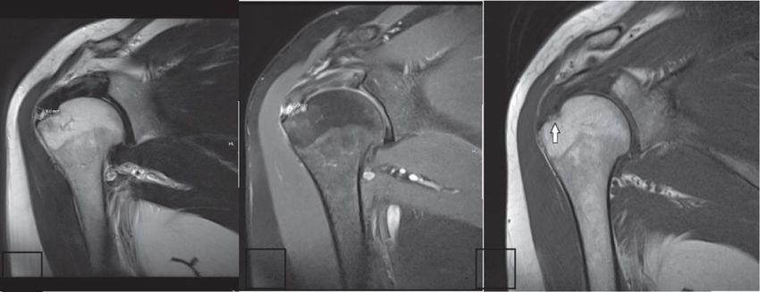
Figure: 2 Shows The Recurrent Dislocation Of Shoulder
MRI coronal T2 (a), PD with fat suppression (b) and T1 (c) WI revealed a full thickness tear of
the supraspinatus tendon near its humeral attachment with fluid signal seen in the gapping area
which measures about 6 mm (comparable to the US).
Figure: 3 Findings By Mri And Surgery In Patients With Recurrent Dislocation Of Shoulder, Pd-
Tse-Weighted Mri.
http://annalsofrscb.ro 2032Annals of R.S.C.B., ISSN:1583-6258, Vol. 25, Issue 2, 2021, Pages. 2028 - 2037
Received 20 January 2021; Accepted 08 February 2021.
Coronal PD fat sat (a) and STIR (b) images for the shoulder show abnormal hyperintense signal
(arrowheads) at the distal part of the supraspinatus tendon that was proved to be tendenosis Out
of total 62 patients had clinical suspect ion of rotator cuff tear, 24 patients had rotator cuff tear,
out of them 8 patients had full thickness tear and 16 had partial thickness tear. [23] Supraspinatus
was the most commonly involved muscle and was involved in all the 24 cases. Out of 24 cases,
full thickness tear was seen in 4 cases and partial thickness tear in 16 cases. Infraspinatus was the
second most commonly involved muscle, showing tear in 6 cases. 2 of the cases showed full
thickness tear and rest of them had partial thickness tear. Subscapularis tear was not observed in
any of the cases. (most likely due to less number of cases). [24-27]In patients withAnterior
dislocation was the most common type of dislocation seen in 38/42 cases. One patient had multi
directional instability and one patient had posterior dislocation.
Figure 4: Full-Thickness Supraspinatus Tear. Coronal Pd-Tse-Weighted Mri
plane (a) and transverse US image (b). The arrowspoint out the rupture, hyperintense in the MRI
and hypoechoic in the US. A ¼ Acromion; H ¼ Humerus;SSP = Supraspinatus tendon.
XRAY
An AP view of the shoulder was performed in all the cases. Assessment for signs of rotator cuff
pathology like degenerative changes (cystic and sclerotic) at greater tubercle (GT) or
undersurface of acromion or lesser tuberosity was done in patients with rotator cuff pathology (20
patients). Acromion-clavicular joint degeneration was also assessed. [13,27]
In patients of instability syndromes X ray was assessed for evidence of Hill Sachs lesion or Bony
Bankart’s lesion.
Table 2: X-ray findings in patients with rotator cuff pathology
XRAY CHANGES N %
GT IRREGULARITY 8/62 12.9
LT IRREGULARITY 2/62 3.22
http://annalsofrscb.ro 2033Annals of R.S.C.B., ISSN:1583-6258, Vol. 25, Issue 2, 2021, Pages. 2028 - 2037
Received 20 January 2021; Accepted 08 February 2021.
AC JOINT DEGENRATION 4/62 6.45
Greater tubercle irregularity was found to be fairly common (40%) in patients presenting with
symptoms of rotator cuff pathology.
Acromial Alignment On Mri:
Most of the cases had parallel alignment of acromion. Out of 42 cases of recurrent dislocation of
shoulder, 10 had inferolateral tilt and none had low lying acromion.
Table 3: Acromial Alignment On MRI
ACROMIAL ALIGAMENT NO. OF PATIENTS PRECENTAGE
Parallel 40 64.5%
Inferolateral 16 25.8%
Low lying 6 9.67%
Out of 20 cases with rotator cuff tear, 8 had parallel alignment of acromion, 6 had inferolateral
tilt and 6 had low lying acromion suggesting a possible co relation between the type of acromion
and rotator cuff tear.
Figure 5: Partial-Thickness Subscapularis Tear. Axial Pd-Tse-Weighted Mri
plane (a) and transverse US image (b). The arrowsshow the focal defect with a thinned-out
tendon. Co ¼ Coracoid; H ¼ Humerus, ISP ¼ Infraspinatus tendon, S ¼ Scapula;SSC ¼
Subscapularis tendon.
Acromio Humeral Distance & Rotator Cuff Tear:
7.05 mm was the average acromion-humeral distance in patients rotator cuff pathology, which
was lower than the overall average of 7.5mm which in concordance with the study by Saupeet al
who found that a >7mm acromion- humeral distance in their study was associated with 90% of
the rotator cuff tears. Our study showed that type I and type II acromion (90.3%) is the most
http://annalsofrscb.ro 2034Annals of R.S.C.B., ISSN:1583-6258, Vol. 25, Issue 2, 2021, Pages. 2028 - 2037
Received 20 January 2021; Accepted 08 February 2021.
common which is consistent with the findings in general population. [28] And also that Type II
and III are more likely to be associated with tears and impingement. 16/42 patients (25.8%)
showed inferolateral alignment of acromion with respect to clavicle Inferolateral alignment
showed higher incidence of rotator cuff tear. In a study on 91 patients with rotator cuff tears
Hiranoet al also found that there was little correlation between acromion type and presence of
tear. However, they reported a significantly higher number of type III acromion (39%) in patients
with rotator cuff tear. In our study we found that overall type ii and iii acromion (50%) were
associated with rotator cuff pathology more frequently (6/12 patients with rotator cuff tear). [29]
4. Conclusion
MRI is very sensitive for detection of full thickness tears of rotator cuff but it lacks sensitivity in
case of partial thickness tears of rotator cuff especially the articular surface partial thickness
tears.The detection of partial thickness tear is important because partial thickness tear of the
anterior supraspinatus fiber increases the strain upon the remaining supraspinatus fibers and
intact infraspinatus tendon, leading to tear propagation and potentially impacting the decision to
operate sooner as opposed to waiting. Ultrasound and MRI are comparable in both sensitivity and
specificity. Since USG is less expensive and more easily available, it could be considered as the
screening method when rotator cuff integrity is the main question, and when well-trained
radiologists and high resolution equipment are available.
Funding: No funding sources
Ethical approval: The study was approved by the Institutional Ethics Committee
Conflict Of Interest
The authors declare no conflict of interest.
Acknowledgments
The encouragement and support from Bharath University, Chennai is gratefully acknowledged.
For provided the laboratory facilities to carry out the research work.
References
[1] Scott J. Mc Mongale Emily N Vinson. MRI of the shoulder: Rotator cuff. Applied
Radiology. 2012;10(1):20-28.
[2] Van der Heijden GJMG. Shoulder disorders: a state-of-the-art review. Best Pract Res
Clin Rheumatology. 1999;13(2):287-309.
[3] Pierre F. Desmeules F. Shouldering the Pain: Practical tools for evaluating and
treating a painful Shoulder, The Canadian Journal of CME. 2003:119-121
[4] Neer CS. Impingement Lesion, Clinical orthopedics related research.
Shoulderdoc.1983;17(3):70-77.
[5] Teefey SA. Hasan SA. Ultrasonogrphy of the rotator cuff. A comparison of
Ultrasonography and arthroscopic findings in one hundred consecutive cases. J Bone
Joint Surg. 2000; 82:498-504.
http://annalsofrscb.ro 2035Annals of R.S.C.B., ISSN:1583-6258, Vol. 25, Issue 2, 2021, Pages. 2028 - 2037
Received 20 January 2021; Accepted 08 February 2021.
[6] Zlatkin MB. MRI of shoulder. 2nd Ed. Philadelphia; Lippincott Williams &
Wilkins;2003.
[7] Van Holsbeeck MT. Kolowich PA. Eyler WR. et al. Ultrasound depiction of partial
thickness tears of the rotator cuff. Radiology. 1995; 197:443-446
[8] Seeger LL. Gold RH. Basett LW. et al. Shoulder impingement syndrome: MR findings
in 53 shoulders. AJR. 1988;150(2): 343-347.
[9] Iannotti JP. Rotator cuff disorders, evaluation and treatment. In: American Academy
of orthopedic surgeons.1991; 21:41-46
[10] Berquist TH. MRI of musculoskeletal system.2012;6:578-685.
[11] Beltron J. Bercardino J et al. MR arthrography of the shoulder: variants and pitfalls.
Radiographics. 1997; 17:1403-1412
[12] Farley TE, Neuman CH et al. Full thickness tears of the rotator cuff of the shoulder:
Diagnosis with MR imaging. AJR. 1992; 158:347-351.
[13] De Jesus JO, Parker L, Frangos AJ et al. Accuracy of MRI, MR arthrography, and
ultrasound in the diagnosis of rotator cuff tears: a meta- analysis. AJR AM J
Roentenol. 2009; 192:1701-1707.
[14] C.L. Miller et al. Limited sensitivity of ultrasound for the detection of rotator cuff
tears. Skeletan radiol. 1989; 18:179-183.
[15] Whitley Vick C. et al. Rotator cuff tears: Diagnosis with Sonography. AJR 1990;
154:121-123.
[16] Magee T. Williams D. Mani N. Shoulder MR Arthrtography : which patient group
benefits most? AJR Am J Roentgenol. 2004; 183 (4): 969-74.
[17] Rowan KR. Andrews G. Spielman A. Leith J. Leith J. Foster BB. Shoulder
arthrography in patients younger than 40 years of age: frequency of rotator cuff tear
versus labroligamentous pathology. Australas radiol. 2007;51(3):257-9.
[18] Fehringer EV. Sun J. Van Oeveren LS. et al. Full-thickness rotator cuff tear prevalence
and correlation with function and co-morbidities in patients sixty-five years and older.
J Shoulder Elbow Surg. 2008; 17:881-885.
[19] Yamamoto A. Takagishi K. Osawa T. Yanagawa T. Nakajima D. Shitara H. Kobayashi
T. Prevalence and risk factors of a rotator cuff tear in the general population. J
Shoulder Elbow Surg. 2010;19(1):116-20.
[20] Hirano M. Ide J. Takagi K. Acromial shapes and extension of rotator cuff tear:
magnetic resonance imaging evaluation. J Shoulder Elbow Surg. 2002;11(6):576-8.
[21] Arslan G. Apaydin A. Kabaliglu A. Sindel T. Luleci E. Sonographically detected
subacromial/ subdeltoid bursal effutions and biceps tendon sheath fluid: reliable signs
of rotator cuff tears? J Clin Ultrasound.1999; 27(6):335-40.
[22] Theodoropoulos JS. Andreisek G. Harvey EJ. Wolin P. Magnetic resonance imaging
and magnetic resonance arthrography of the shoulder: Dependence on the level of
training of the performing radiologist for diagnostic accuracy. Skeletal Radiol. 2010;
39:661-667.
http://annalsofrscb.ro 2036Annals of R.S.C.B., ISSN:1583-6258, Vol. 25, Issue 2, 2021, Pages. 2028 - 2037
Received 20 January 2021; Accepted 08 February 2021.
[23] Garneau RA. Renfrew DL. Moore TE. EI-Khoury GY. Nepola JV. Lemke JH. Glenoid
labrum: evaluation with MR imaging. Radiology 1991;179:519-522
[24] Toyoda H. Ito Y. Nakao Y. Koike T. Takaoka K. Evaluation of rotator cuff tears with
magnetic resonance arthrography. Clin Orthop Relat Res. 2005 Oct; 439:109-15.
[25] Jacobson SR. Speer KP. DH et al. Reliability of radiographic assessment of acromial
morphology. J shoulder elbow surgery 1995;4:449-453
[26] Bonutti PM. Norfay JF. Friedman RJ. Reenz BM. Kinematic MRI of the shoulder.
JCAT.1993;17:666-669
[27] Waldt S. Burkart A. Imhoff AB. Bruegel M. Rummeny EJ. Woertler K. Anterior
shoulder instability: Accuracy of MR Arthrography in the classification of anterior
labroligamentous injuries. Radiol. 2005; 237:578-83
[28] Wakdt S. Burkat A. Lange P. Imhoff AB. Rummeny EJ. Woertler K. Diagnostic
performance of MR arthrography in the assessment of superior labral anteroposterior
lesions of the shoulder. AJR Am J Roentgenol. 2004;182:1271-78
[29] Rowan KR. Keogh C. Andrews G. Cheong Y. Forster BB. Essentials of shoulder MR
arthrography: A practical guide for the general radiologist. Clin Radiol. 200;59:327-34
http://annalsofrscb.ro 2037You can also read


