Examination of endobronchial ultrasound guided transbronchial needle aspiration using a puncture needle with a side trap - Nature
←
→
Page content transcription
If your browser does not render page correctly, please read the page content below

www.nature.com/scientificreports
OPEN Examination of endobronchial
ultrasound‑guided transbronchial
needle aspiration using a puncture
needle with a side trap
Kazuhito Miyazaki1*, Yuya Hirasawa1,4, Masaharu Aga1,4, Naoto Aiko1,4,
Yusuke Hamakawa1,4, Yuri Taniguchi1,4, Yuki Misumi1,4, Yoko Agemi1,4, Tsuneo Shimokawa1,4,
Hiroyuki Hayashi2,4, Katsuhiko Naoki3,4 & Hiroaki Okamoto1,4
Endobronchial ultrasound-guided transbronchial needle aspiration (EBUS-TBNA) is useful for
diagnosing hilar and mediastinal lymph node enlargement; however, specimens obtained are often
small and inadequate for pathologic diagnosis. In June 2017, EchoTip ProCore, a puncture needle
with a side trap, was launched in Japan. In this single-center prospective interventional study, 57
patients with lymph nodes, intrapulmonary tumor or pleural mass were diagnosed using EBUS-TBNA
with EchoTip ProCore between June 2017 and February 2020. EBUS-TBNA was performed for 57
patients and 53 patients had sufficient specimen for histologic diagnosis. The following pathologic
subtypes were diagnosed: non-small cell lung cancer, 22; small cell lung cancer, 8; cancer of unknown
primary, 2; neuroendocrine tumor (G2) recurrence, 1; lymphoma, 2; metastatic renal cell carcinoma,
3; thymoma recurrence, 1; sarcoidosis, 4; tuberculosis, 1; and non-malignancy, 9. In addition, the
cytology showed Class V in 31 out of 57 cases (54.4%). In total, a definitive pathological diagnosis
was obtained in 50 out of 57 cases (87.7%). The only complication was pneumonia caused by BAL
simultaneously combined with EBUS-TBNA in one patient. Among 13 patients with inadequate
specimens or without malignancy, only one patient was subsequently diagnosed with malignancy, and
the median follow-up period was 300 days. EBUS-TBNA using EchoTip ProCore can obtain a sufficient
specimen size for pathologic diagnosis.
Endobronchial ultrasound-guided transbronchial needle aspiration (EBUS-TBNA) is a useful diagnostic modal-
ity for thoracic lymphadenopathy, including primary lung cancer, malignant lymphoma, tuberculosis, and
sarcoidosis1–3. Tissue and cytological specimens can be obtained using EBUS-TBNA. Notably, tissue samples
obtained using this technique can undergo tests for epidermal growth factor receptor (EGFR) mutations and
immunohistochemical screening for anaplastic lymphoma kinase (ALK) rearrangement in patients with non-
small cell lung cancer (NSCLC). However, the specimens obtained are often small and inadequate for pathologic
diagnosis. Specifically, only 66% (19/29) of specimens obtained using EBUS-TBNA at our hospital between 2015
and 2016 were adequate for histological diagnosis.
In June 2017, EchoTip ProCore Endobronchial HD Ultrasound Needle (EchoTip ProCore; Cook Medical,
Bloomington, Indiana, United States), a puncture needle with a side trap that had been previously released for
gastrointestinal endoscopes, was launched in Japan. EchoTip ProCore needles are available in different sizes,
including 22- and 25-gauge needles. They are designed with a core trap proximal to the needle tip that receives
the sample during fine-needle aspiration (FNA) through the needle tip (Fig. 1). Therefore, EchoTip ProCore
is considered to obtain core biopsy for histologic evaluation rather than only cytological material, as in other
needles4. This study aimed to investigate the diagnostic utility of EBUS-TBNA with EchoTip ProCore for medi-
astinal lymphadenopathy.
1
Department of Respiratory Medicine, Yokohama Municipal Citizen’s Hospital, Yokohama, Kanagawa,
Japan. 2Department of Pathology, Yokohama Municipal Citizen’s Hospital, Yokohama, Kanagawa,
Japan. 3Department of Respiratory Medicine, Kitasato University School of Medicine, Sagamihara, Kanagawa,
Japan. 4These authors contributed equally: Yuya Hirasawa, Masaharu Aga, Naoto Aiko, Yusuke Hamakawa,
Yuri Taniguchi, Yuki Misumi, Yoko Agemi, Tsuneo Shimokawa, Hiroyuki Hayashi, Katsuhiko Naoki and Hiroaki
Okamoto. *email: ka07-miyazaki@city.yokohama.jp
Scientific Reports | (2021) 11:9789 | https://doi.org/10.1038/s41598-021-89244-x 1
Vol.:(0123456789)
www.nature.com/scientificreports/
Figure 1. The slow-pull technique using EchoTip ProCore. (A) is where the slow pull method is performed in
our hospital. B is an ultrasound image of EchoTip ProCore.
Materials and methods
Patients selection. This single-center prospective interventional study was conducted at the Department
of Respiratory Medicine, Yokohama Municipal Citizen’s Hospital (Kanagawa, Japan). We included patients with
radiological features of mediastinal or hilar lymphadenopathy who underwent EBUS-TBNA between June 2017
and February 2020. Chest computed tomography (CT) was performed before EBUS-TBNA, which revealed at
least one enlarged mediastinal or hilar lymphadenopathy of > 10 mm in all patients except for two patients with
a tumor around the trachea. Finally, we included 57 patients in this study.
EBUS‑TBNA. All enrolled patients underwent EBUS-TBNA using a convex endobronchial ultrasound
bronchoscope probe (BF-UC260FW; Olympus, Tokyo, Japan) under fentanyl-induced sedation (0.025-0.05 mg/
body) and midazolam (1–3 mg/body). A 22- and 25-gauge EchoTip ProCore Endobronchial HD Ultrasound
Needle was used. The 25-gauge needle was employed if contrast-enhanced CT revealed blood vessel enrich-
ment in a lymph node or if there was a high suspicion of hypervascular tumor metastasis, including renal cell
carcinoma. We performed 2–4 punctures using the traditional vacuum syringe method and slow-pull technique.
The specimens obtained with each needle pass were extracted to the dish by pushing the stylet and flushing the
syringe with saline solution and air. Since rapid on-site cytological examination (ROSE) was not available at
our hospital, the puncture was repeated up to 4 times when the volume of the collected sample was considered
insufficient macroscopically. With regard to sample processing, each tissue from one puncture was placed in
an embedding cassette and fixed in 10% neutral buffered formalin solution. A paraffin block of the sample was
then prepared and histologically assessed. The remaining liquid components and small tissue fragments were
collected with a dropper and placed in a spitz tube, a spitz tube was centrifuged at 2,000 rpm for 5 min and the
sediment was subjected to liquid-based cytology (LBC) using CytoRich Red Preservative (Becton Dickinson,
Franklin Lakes, New Jersey, United States) and assessed by cytologic diagnosis. At our hospital, clinical labora-
tory technicians (some of whom are cytotechnologists) process samples in the bronchoscope room, and the
tissue was fixed with formalin immediately after all punctures were completed. Cytological samples were taken
back to the laboratory with spitz tubes and sample processing was started immediately.
Slow‑pull technique. Originally, the slow-pull technique involved slow stylet retraction with needle fan-
ning in the mass. Ten to twenty to-and-fro movements were made with minimal negative pressure provided
by slow pulling of the stylet. The length of stylet retraction was about 1 m and the time of stylet retraction was
from 40 to 60 s5–7. However, to obtain larger specimens, we used a modified version of the slow-pull technique.
In short, although the stylet was slowly pulled out in 1 m, 10–30 to-and-fro movements with keeping negative
pressure were repeated in every 5–10 cm up to the end.
IRB approval. The study protocol was approved by the independent ethics committee of the institutional
review board of Yokohama Municipal Citizen’s Hospital (No. 20-11-11). Informed consent was obtained in the
form of opt-out on the web-site. Those who rejected were excluded. Topical subheadings are allowed. Authors
must ensure that their Methods section includes adequate experimental and characterization data necessary for
others in the field to reproduce their work.
Statement of human rights. All procedures performed in studies involving human participants were in
accordance with the ethical standards of the institutional and/or national research committee and with the 1964
Helsinki declaration and its later amendments or comparable ethical standards.
Results
Patients characteristics. Table 1 presents the baseline characteristics. This study enrolled 57 patients
who underwent EBUS-TBNA using EchoTip ProCore between June 2017 and February 2020. We used 22- and
25-gauge EchoTip ProCore for 51 and 6 patients, respectively. The median age was 71 (range: 18–89) years, and
44 (77%) patients were male. The stations of the punctured lymph nodes were as follows: 1 for #2R; 20 for #4R; 3
for #4L; 26 for #7; 4 for #11 s; 2 for #11i; and 2 for #11L. In all patients, the lymph nodes were in contact with the
trachea or bronchial wall. However, none of the tumor was visible in the central airway lumen when broncho-
scope was performed. Additionally, a right upper lobe tumor and pleural mass were punctured in two separate
Scientific Reports | (2021) 11:9789 | https://doi.org/10.1038/s41598-021-89244-x 2
Vol:.(1234567890)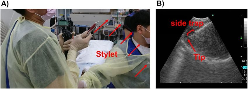
www.nature.com/scientificreports/
N = 57 N (%)
Age, yr (Range) 71 (18–89)
Sex
Male 44 77.2
Female 13 22.8
Punctured lymph nodes
#2R 1 1.7
#4R 20 33.3
#4L 3 5.3
#7 26 43.3
#11s 4 6.7
#11i 2 3.3
#11L 2 3.3
Right upper lobe mass 1 1.7
Pleural mass 1 1.7
Median maximum diameter of lymph nodes
mm (Range) 27 (14–70)
Maximum diameter of right upper lobe mass, mm 21
Maximum diameter of pleural mass, mm 25
EchoTip ProCore
22G 51 89.5
25G 6 10.5
Table 1. Patients characteristics. Baseline demographic and clinical characteristics of 57 patients who
underwent EBUS-TBNA with EchoTip ProCore between June 2017 and February 2020.
N (%)
NSCLC 22 38.6
SCLC 8 14.0
RCC (metastasis) 3 5.3
CUP (Sq) 2 3.5
NET(G2) recurrence 1 1.8
lymphoma 2 3.5
thymoma recurrence 1 1.8
sarcoidosis 4 7.0
necrotic tissue 1 1.8
tuberculosis 1 1.8
non-malignancy 9 15.8
cyst 1 1.8
inadequate 2 3.5
Class V 31 54.4
Class IV 0 0
Class III 8 14.0
Class II 4 7.0
Class I 14 24.6
Table 2. Pathological diagnosis. Pathological findings of tissue or cytological specimen obtained using EBUS-
TBNA. NSCLC non-small cell lung cancer, SCLC small cell lung cancer, RCCrenal cell cancer, CUP cancer
of unknown primary, NET neuroendocrine tumor, Sq squamous cell carcinoma, Class I Absence of atypical
or abnormal cells, Class II Atypical cytology but no evidence of malignancy, Class III Cytology suggestive of,
but not conclusive for malignancy, Class IV Cytology strongly suggestive of malignancy, Class V Cytology
conclusive for malignancy.
Scientific Reports | (2021) 11:9789 | https://doi.org/10.1038/s41598-021-89244-x 3
Vol.:(0123456789)www.nature.com/scientificreports/
Figure 2. Tissue samples obtained using EchoTip ProCore. (A) presents a tissue sample obtained using a
22-gauge EchoTip ProCore where the #11i lymph node was punctured. (B) presents a tissue sample obtained
using a 22-gauge EchoTip ProCore where the #4R lymph node was punctured in other cases. As shown here,
this needle can obtain very large tissue samples.
patients (Table 2). Among 57 patients, 60 lymph nodes were punctured, with two lymph nodes being punctured
in three patients. The median maximum diameter of the lymph nodes was 27 (range: 14–70) mm. Maximum
diameter of right upper lobe mass was 21 mm, and pleural mass was 25 mm, respectively.
EBUS‑TBNA. Figure 1 illustrates the slow-pull technique using EchoTip ProCore. As shown in Fig. 1(A), the
puncture is repeated with gradual pulling out of the stylet. Figure 1B presents an ultrasound image of EchoTip
ProCore. There is a side trap on the central tip side that shaves off the tissue. Figure 2 shows the tissue samples
obtained using EchoTip ProCore. Figure 2A presents a tissue sample obtained using a 22-gauge EchoTip Pro-
Core where the #11i lymph node was punctured. Hematoxylin–eosin staining showing large, strongly atypical
cells, as well as positive TTF-1 staining, led to a histological diagnosis of metastatic lung adenocarcinoma. Sub-
sequently, PD-L1 immunohistochemistry (IHC) revealed that 95% of the cells were positive; however, the cell
number had been reduced by re-thinning (Fig. 3). Figure 2B presents details of the other patients. There was
Scientific Reports | (2021) 11:9789 | https://doi.org/10.1038/s41598-021-89244-x 4
Vol:.(1234567890)www.nature.com/scientificreports/
Figure 3. Pathological examination of one case of adenocarcinoma. (A) Hematoxylin–eosin staining. (B)
Thyroid transcription factor-1 (TTF-1) staining using TTF-1 antibody. C) PD-L1 staining using PD-L1 IHC
22C3 PharmDx assay.
PD-L1 (TPS) N (%)
50% > 8 42.0
1–49% 5 26.0
1% < 6 32.0
Table 3. PD-L1 status of NSCLC. PD-L1 status non-small cell lung cancer was examined through IHC using
the PD-L1 IHC 22C3 PharmDx assay. TPS tumor proportion score.
lymph node enlargement of the right supraclavicular fossa and mediastinum; subsequently, EBUS-TNBA with
EchoTip ProCore was performed to differentiate among lymphoma, lymphoproliferative disease, and mediasti-
nal lung cancer. A 22-gauge EchoTip ProCore was used for #4R lymph node puncture. The lymph node was soft,
and the tissue was obtained using the slow-pull technique, which yielded a large tissue with a size > 10 cm, as
shown in Fig. 2B. Pathological examination revealed an epithelioid granuloma; moreover, the interferon-gamma
release assay for tuberculosis was positive; thus, the patient was diagnosed as having active tuberculosis.
As for the complications of EBUS-TBNA using EchoTip ProCore, none of the patients had massive bleed-
ing, mediastinitis, or pneumothorax. Only one case of suspected sarcoidosis with bronchoalveolar lavage (BAL)
developed pneumonia after bronchoscopy, probably owing to BAL, which was positive for the pneumococcal
urinary antigen and was relieved by oral levofloxacin treatment.
Pathological diagnosis. Among 57 patients, one had a cyst, one had necrotic tissue, and two had inad-
equate specimens; however, the remaining 53 (93%) patients had sufficient specimen for histologic diagnosis
using EBUS-TBNA (Fig. 3). The following pathologic subtypes were diagnosed: NSCLC, 22; small cell lung can-
cer (SCLC), 8; cancer of unknown primary (squamous cell carcinoma), 2; neuroendocrine tumor (G2) with thy-
mus origin recurrence, 1; lymphoma, 2; metastatic renal cell carcinoma, 3; thymoma recurrence, 1; sarcoidosis,
4; tuberculosis, 1; and non-malignancy, 9. Moreover, cytology revealed that 68% (39/57) of patients were class III
or higher. In total, a definitive pathological diagnosis was obtained in 50 out of 57 cases (87.7%). A patient with
necrosis on histological examination was diagnosed as having SCLC based on cytological examination. Two
patients lacked a definitive diagnosis based on histological examination; however, cytological examination was
indicative of class III, and a lymphoma was detected on surgical lymph node biopsy. One of the 10 patients with
class II according to cytological examination was diagnosed as Hodgkin’s lymphoma based on surgical lymph
node biopsy. Among the remaining patients, 8 were followed up, with 1 being excluded after being requested
to be examined in another hospital. The median observation period for the 8 patients was 300 (range 223–665)
days. However, for one patient who underwent lung cancer surgery, CT and positron emission tomography did
not reveal changes in the punctured lymph nodes; however, brain recurrence occurred.
Histological diagnosis revealed lung cancer in 97% (30/31) of patients, and EBUS-TBNA with EchoTip
ProCore was used for diagnosis in all patients, including cytological diagnosis.
PD‑L1 immunohistochemistry. The PD-L1 status was measured based on IHC using the PD-L1 IHC
22C3 PharmDx assay (Agilent Technologies, Santa Clara, California, United states). Among 22 NSCLC patients,
IHC of PD-L1 using the 22C3 antibody was possible in 19 patients except for 1 patient each where EBUS-TBNA
was performed to differentiate between lung and breast cancer recurrence for staging purposes and to search for
T790M mutation in EGFR-positive NSCLC, respectively. The PD-L1 tumor protein scores (TPSs) were ≥ 50%,
1–49%, and < 1% in 8 (42%), 5 (26%), and 6 (32%) patients, respectively (Table 3).
Scientific Reports | (2021) 11:9789 | https://doi.org/10.1038/s41598-021-89244-x 5
Vol.:(0123456789)www.nature.com/scientificreports/
N = 17 N (%)
EGFR
Wild type 12 70.6
Mutation 4 23.5
Not examined 1 5.9
ALK
Wild type 15 88.2
Rearrangement 0 0
Not examined 2 11.8
ROS1
Wild type 9 52.9
Rearrangement 0 0
Not determined but examined 5 29.4
Not examined 3 17.6
Table 4. Driver mutation status of non-Sq-NSCLC. EGFR mutation, ALK rearrangement, ROS1
rearrangement status of non squamous non-small cell lung cancer was examined.
Driver mutation test. At the time of study initiation, EGFR, ALK and PD-L1 examinations were approved
in Japan. Thereafter ROS1 and comprehensive genome sequencing (Oncomine test) were sequentially approved
between the study period. Therefore, the frequency of each biomarker test was different among the patients.
Among 22 NSCLC patients, 17 cases were adenocarcinoma, 4 cases were squamous cell carcinoma (Sq), 1
case was non-small cell lung carcinoma, not otherwise specified (NSCLC, NOS). In 18 cases of non-Sq-NSCLC,
gene testing was performed except for one case of staging. EGFR mutations were wild/mutation/not examined:
12/4/1, ALK rearrangement were wild/ rearrangement/not examined: 15/0/2, and ROS1 rearrangement were
wild/rearrangement/not determined but examined/not examined: 9/0/5/3 cases (Table 4). Of the above results,
Oncomine Dx. Target Test (Thermo Fisher Scientific, San Jose, CA, United States) was used in only one case.
Discussion
Endoscopic ultrasound-FNA using EchoTip ProCore was primarily employed in gastroenterology (especially
in pancreatic cancer) and rarely in the respiratory field8,9. To our knowledge, this is the first report regarding the
large-scale utility of EBUS-TBNA with EchoTip ProCore. Bronchoscopic examination is widely used for initial
diagnosis. Specifically, EBUS-TBNA is considered the first choice for mediastinal lymphadenopathy given that
it has an almost similar diagnosis rate as mediastinoscopy and is minimally i nvasive10.
Treatment of advanced lung cancer has dramatically changed since the discovery of the EGFR gene mutation
in the 2000s and various driver gene mutations, including ALK rearrangement11–13. Furthermore, nivolumab,
which is a programmed death-1 antibody, was approved in December 2015 in Japan; subsequently, multiple
immune checkpoint inhibitors have been a pproved14–17. The PD-L1 expression rate in tissues is an important
predictor of the effect of immune checkpoint inhibitors14,15. Therefore, EBUS-TBNA with mediastinal lymphad-
enopathy has been a useful procedure for lung cancer. There was an increasing need for a reliable method to
obtain tissue samples.
From 2015 to 2016, EBUS-TBNA was performed in 29 cases at our hospital; however, tissue diagnosis was
possible in 19 cases (66%), and the diagnosis was confirmed using tissues in 10 cases (34.5%) and including
cytology 13 cases (44.8%). In contrast, in the present study, tissue samples were available for 54 (94.7%) of 57
EBUS-TBNA cases using EchoTip ProCore, and a definitive diagnosis was obtained for 50 (87.7%) cases. A
definite diagnosis was made for 30/31 (97%) lung cancer cases; a diagnosis was obtained for each of the 31 cases
including cytological diagnosis. In the literatures, the diagnostic rate of EBUS-TBNA using a conventional
puncture needle was reported to be about 51% for both 21-gauge and 22-gauge for mediastinal and hilar lymph
nodes18. Other reports showed the diagnostic rate using conventional EBUS-TBNA methods were in the range
of 61 to 80%19,20. Although the patient characteristics between our study and other reports were different, the
diagnostic rate of current study seems to be superior than that of conventional EBUS-TBNA methods.
Although there was a difference between the number of cases that underwent EBUS-TBNA before and after
the introduction of EchoTip ProCore (13.5 and 20.7 cases/year, respectively), there was no significant increase
or decrease in the number of bronchoscopies in our hospital, suggesting an improvement in the tissue diagnosis
rate after the introduction of EchoTip ProCore. Therefore, the introduction of EchoTip ProCore contributed to
more accurate diagnoses. Three of 31 lung cancer cases were difficult to diagnose because of peripheral lesions
that could not be pathologically diagnosed, and EBUS-TBNA was successfully used for tissue diagnosis. From
this evidence, the diagnostic rate of EchoTip ProCore is superior to that of EBUS-TBNA using a standard needle.
Regarding safety, only one of 57 patients who had punctured a cyst received prophylactic antimicrobials after
EBUS-TBNA. None of the patients developed mediastinitis, and only one case of suspected sarcoidosis with BAL
developed pneumonia after bronchoscopy, probably owing to BAL, which was positive for the pneumococcal
urinary antigen and was relieved by oral levofloxacin treatment. No other complications were observed. The
incidence of complications after EBUS-TBNA has been reported to be 0.15 to 0.46%21,22, and EchoTip ProCore
was safe as it did not increase the incidence of complications. Furthermore, several studies have reported that
Scientific Reports | (2021) 11:9789 | https://doi.org/10.1038/s41598-021-89244-x 6
Vol:.(1234567890)www.nature.com/scientificreports/
samples obtained using EBUS-TBNA as a cell block can allow PD-L1 staining23,24. The present study shows that
EBUS-TBNA with EchoTip ProCore can consistently yield large tissues suitable for direct immunostaining of
PD-L1 (Fig. 3). Of course, tests for EGFR, ALK, and ROS1 were also available. Additionally, in one patient,
Oncomine Dx. Target Test, which is the first FDA-approved next-generation sequencing-based companion
diagnostic method for diagnosing EGFR mutation, ALK rearrangement, ROS1 rearrangement, and BRAF V600E
mutation in patients with NSCLC, was performed without complications using our methods. Specifically, EBUS-
TBNA with EchoTip ProCore can collect appropriate samples for next-generation sequencing examinations.
Among 22 patients with NSCLC who could yield histological specimens, the PD-L1 TPS was successfully exam-
ined in 19 patients except for one patient for staging reasons, one for differentiation between breast and lung
cancer recurrence, and one for T790M search purposes. This high success rate could contribute to the selection
of more appropriate individual therapy for patients with NSCLC.
This study had some limitations. First, it was difficult to puncture small lymph nodes with EchoTip ProCore
given its side core. Second, this was a small uncontrolled study, a single-center study and did not perform com-
parisons with other needles. However, as shown in Fig. 2, this needle allowed us to consistently obtain large tissue
samples. Finally, it was unclear whether the superior diagnostic rate was attributed to the slow-pull technique or
EchoTip ProCore. Although examples are not shown, we attempted EBUS-TBNA in a small number of patients
at our institution with the slow-pull technique using the 22-gauge Vizishot2 needle (Olympus, Tokyo, Japan).
However, tissue specimen collection was not as stable as with EchoTip ProCore. This indicates that the slow-pull
technique using EchoTip ProCore may be more important.
In conclusion, our method allowed pathological diagnosis of 54 of 57 patients using histology, cytology, or
both. The PD-L1 TPS was successfully examined in all 19 patients with NSCLC who could yield histological
specimens and required examination. Additionally, none of the patients had complications. Among 13 patients
with inadequate specimens or without malignancy, only one patient was subsequently diagnosed as having malig-
nancy with a median follow-up period of 300 days. Therefore, EBUS-TBNA using a puncture needle with a side
trap should be considered for patients with hilar and mediastinal lymphadenopathy in routine clinical practice.
Received: 16 February 2021; Accepted: 22 April 2021
References
1. Okamoto, H. et al. Endobronchial ultrasonography for mediastinal and hilar lymph node metastases of lung cancer. Chest 121,
1498–1506 (2002).
2. Yasufuku, K. et al. Real-time endobronchial ultrasound-guided transbronchial needle aspiration of mediastinal and hilar lymph
nodes. Chest 126, 122–128 (2004).
3. Medford, A. R., Bennett, J. A., Free, C. M. & Agrawal, S. Endobronchial ultrasound-guided transbronchial needle aspiration
(EBUS-TBNA): applications in chest disease. Respirology 15, 71–79 (2010).
4. Dincer, H. E., Andrade, R., Zamora, F. & Podgaetz, E. A new needle on the block: EchoTip ProCore endobronchial ultrasound
needle. Med. Dev. (Auckl). 9, 467–473 (2016).
5. Nakai, Y. et al. Slow pull versus suction in endoscopic ultrasound-guided fine-needle aspiration of pancreatic solid masses. Dig.
Dis. Sci. 59, 1578–1585 (2014).
6. Paik, W. H. et al. Prospective evaluation of new 22 gauge endoscopic ultrasound core needle using capillary sampling with stylet
slow-pull technique for intra-abdominal solid masses. J. Clin. Gastroenterol. 49, 199–205 (2015).
7. Lee, J. M. et al. Slow-pull using a fanning technique is more useful than the standard suction technique in EUS-guided fine needle
aspiration in pancreatic masses. Gut Liver. 12, 360–366 (2018).
8. Inoue, T. et al. Assessment of factors affecting the usefulness and diagnostic yield of core biopsy needles with a side hole in endo-
scopic ultrasound-guided fine-needle aspiration. Gut Liver 10, 51–57 (2016).
9. Bang, J. Y. & Varadarajulu, S. Procore and flexible 19 gauge needle can replace trucut biopsy needle?. Clin. Endosc. 46, 503–505
(2013).
10. Yasufuku, K. et al. A prospective controlled trial of endobronchial ultrasound-guided transbronchial needle aspiration compared
with mediastinoscopy for mediastinal lymph node staging of lung cancer. J. Thorac. Cardiovasc. Surg. 142, 1393–1400 (2011).
11. Mitsudomi, T. et al. Gefitinib versus cisplatin plus docetaxel in patients with non-small-cell lung cancer harbouring mutations of
the epidermal growth factor receptor (WJTOG3405): an open label, randomised phase 3 trial. Lancet Oncol. 11, 121–128 (2010).
12. Solomon, B. J. et al. First-line crizotinib versus chemotherapy in ALK-positive lung cancer. N. Engl. J. Med. 371, 2167–2177 (2014).
13. Hida, T. et al. Alectinib versus crizotinib in patients with ALK-positive non-small-cell lung cancer (J-ALEX): an open-label,
randomised phase 3 trial. Lancet 390, 29–39 (2017).
14. Reck, M. et al. Pembrolizumab versus chemotherapy for PD-L1-positive non-small-cell lung cancer. N. Engl. J. Med. 375, 1823–1833
(2016).
15. Mok, T. S. K. et al. Pembrolizumab versus chemotherapy for previously untreated, PD-L1-expressing, locally advanced or metastatic
non-small-cell lung cancer (KEYNOTE-042): a randomised, open-label, controlled, phase 3 trial. Lancet 393, 1819–1830 (2019).
16. Brahmer, J. et al. Nivolumab versus docetaxel in advanced squamous-cell non-small-cell lung cancer. N. Engl. J. Med. 373, 123–135
(2015).
17. Borghaei, H. et al. Nivolumab versus docetaxel in advanced nonsquamous non-small-cell lung cancer. N. Engl. J. Med. 373,
1627–1639 (2015).
18. Yarmus, L. B. et al. Comparison of 21-gauge and 22-gauge aspiration needle in endobronchial ultrasound-guided transbronchial
needle aspiration: results of the American College of Chest Physicians Quality Improvement Registry, Education, and Evaluation
Registry. Chest 143, 1036–1043 (2013).
19. Herth, F., Becker, H. D. & Ernst, A. Conventional vs endobronchial ultrasound-guided transbronchial needle aspiration: a rand-
omized trial. Chest 125, 322–325 (2004).
20. Dhooria, S. et al. Diagnostic yield and complications of EBUS-TBNA performed under bronchoscopist-directed conscious seda-
tion: single center experience of 1004 subjects. J. Bronchol. Interv. Pulmonol. 24, 7–14 (2017).
21. Gu, P. et al. Endobronchial ultrasound-guided transbronchial needle aspiration for staging of lung cancer: a systematic review and
meta-analysis. Eur. J. Cancer 45, 1389–1396 (2009).
22. Asano, F. et al. Deaths and complications associated with respiratory endoscopy: a survey by the Japan Society for Respiratory
Endoscopy in 2010. Respirology 17, 478–485 (2012).
Scientific Reports | (2021) 11:9789 | https://doi.org/10.1038/s41598-021-89244-x 7
Vol.:(0123456789)www.nature.com/scientificreports/
23. Sapalidis, K. et al. EBUS-TNBA 22G samples: comparison of PD-L1 expression between DAKO and BIOCARE®. J. Cancer 10,
4739–4746 (2019).
24. Heymann, J. J. et al. PD-L1 expression in non-small cell lung carcinoma: comparison among cytology, small biopsy, and surgical
resection specimens. Cancer Cytopathol. 125, 896–907 (2017).
Author contributions
K.M., H.O., are responsible for the study concept and design. All authors contributed to the acquisition, analysis,
or interpretation of data. All authors contributed to the drafting of the manuscript. All authors contributed to the
intellectual content. The authors meet criteria for authorship as recommended by the International Committee
of Medical Journal Editors Author contributions.
Funding
This research did not receive any specific grant from funding agencies in the public, commercial, or not-forprofit
sectors.
Competing interests
The authors declare no competing interests.
Additional information
Correspondence and requests for materials should be addressed to K.M.
Reprints and permissions information is available at www.nature.com/reprints.
Publisher’s note Springer Nature remains neutral with regard to jurisdictional claims in published maps and
institutional affiliations.
Open Access This article is licensed under a Creative Commons Attribution 4.0 International
License, which permits use, sharing, adaptation, distribution and reproduction in any medium or
format, as long as you give appropriate credit to the original author(s) and the source, provide a link to the
Creative Commons licence, and indicate if changes were made. The images or other third party material in this
article are included in the article’s Creative Commons licence, unless indicated otherwise in a credit line to the
material. If material is not included in the article’s Creative Commons licence and your intended use is not
permitted by statutory regulation or exceeds the permitted use, you will need to obtain permission directly from
the copyright holder. To view a copy of this licence, visit http://creativecommons.org/licenses/by/4.0/.
© The Author(s) 2021
Scientific Reports | (2021) 11:9789 | https://doi.org/10.1038/s41598-021-89244-x 8
Vol:.(1234567890)You can also read