Assessment of Leptomeningeal Carcinomatosis Diagnosis, Management and Outcomes in Patients with Solid Tumors Over a Decade of Experience
←
→
Page content transcription
If your browser does not render page correctly, please read the page content below
Rinehardt et al. LMC: Diagnosis, Management and Outcomes

Original Article                                                                                                Eur J Breast Health 2021; 17(4): 371-377
                                                                                                              DOI: 10.4274/ejbh.galenos.2021.2021-4-10

Assessment of Leptomeningeal Carcinomatosis
Diagnosis, Management and Outcomes in Patients with
Solid Tumors Over a Decade of Experience
    Hannah Rinehardt1, Mahmoud Kassem2,3, Evan Morgan2,3, Marilly Palettas4, Julie A. Stephens4,
    Anupama Suresh2,3, Akansha Ganju2,3, Maryam Lustberg2,3, Robert Wesolowski2,3, Sagar Sardesai2,3,
    Daniel Stover2,3, Jeffrey Vandeusen2,3, Mathew Cherian2,3, Maria del Pilar Guillermo Prieto Eibl5, Abdul Miah2,
    Iyad Alnahhas5, Pierre Giglio5, Vinay K. Puduvalli5, Bhuvaneswari Ramaswamy2,3, Nicole Williams2,3,
    Anne M. Noonan2
1
  The Ohio State University College of Medicine, Columbus, OH, USA
2
  Division of Medical Oncology, Comprehensive Cancer Center, The Ohio State University Wexner Medical Center, Columbus, OH, USA
3
  Stefanie Spielman Comprehensive Breast Cancer, The Ohio State University, Columbus, OH, USA.
4
  Center for Biostatistics, Department of Biomedical Informatics, The Ohio State University College of Medicine Columbus, OH, USA
5
  Division of Neuro-oncology, The Ohio State University Wexner Medical Center, Columbus, OH, USA

    ABSTRACT
    Objective: Leptomeningeal carcinomatosis (LMC), a common complication of advanced malignancies, is associated with high morbidity and mortality,
    yet diagnosis and treatment decisions remain challenging. This study describes the diagnostic and treatment modalities for LMC and identifies factors
    associated with overall survival (OS).
    Materials and Methods: We performed a single-institution retrospective study (registration #: OSU2016C0053) of 153 patients diagnosed with LMC
    treated at The Ohio State University, Comprehensive Cancer Center, (OSUCCC)-James between January 1, 2010 and December 31, 2015.
    Results: Median age at diagnosis was 55.7 years, and 61% had Eastern Cooperative Oncology Group baseline performance status ≤1. Most common
    primary tumors were breast (43%), lung (26%), and cutaneous melanoma (10%). At presentation, most patients were stage III-IV (71%) with higher grade
    tumors (grade III: 46%). Metastases to bone (36%), brain (33%), and lung (12%) were the most common sites with a median of 0.5 years (range, 0-14.9
    years) between the diagnosis of first metastasis and of LMC. 153 (100%) patients had MRI evidence of LMC. Of the 67 (44%) who underwent lumbar
    puncture (LP), 33 (22%) had positive cerebrospinal fluid (CSF) cytology. Most patients received radiotherapy for LMC (60%) and chemotherapy (93%)
    for either the primary disease or LMC. 28 patients received intrathecal chemotherapy, 22 of whom had a primary diagnosis of breast cancer. 98% died with
    median OS of all patients was 1.9 months (95% CI: 1.3-2.5 months).
    Conclusion: Despite improved treatments and targeted therapies, outcomes of LMC remain extremely poor. Positive CSF cytology was associated with
    lower OS in patients who had cytology assessed and specifically in patients with breast cancer. CSF cytology serves as an important indicator for prognosis
    and helps aid in developing individualized therapeutic strategies for patients with LMC.
    Keywords: Leptomeningeal carcinomatosis, breast cancer, metastasis, cerebrospinal fluid, magnetic resonance imaging

    Cite this article as: Rinehardt H, Kassem M, Morgan E, Palettas M, Stephens JA, Suresh A, Ganju A, Lustberg M, Wesolowski R, Sardesai S, Stover D,
    Vandeusen J, Cherian M, del Pillar Guillermo M, Eibl P, Miah A, Alnahhas I, Giglio P, Puduvalli VK, Ramaswamy B, Williams N, Noonan AM. Assessment
    of Leptomeningeal Carcinomatosis Diagnosis, Management and Outcomes in Patients with Solid Tumors Over a Decade of Experience.
    Eur J Breast Health 2021; 17(4): 371-377.

    Key Points
    •     LMC most commonly presents with late-stage cancers with cancers of the breast, lung and melanoma being the most common primary cancers.
    •     Diagnosis of LMC may be challenging and imaging with MRI brain and spine was most frequently used in our study as an aid in diagnosis and in
          some cases as the primary tool for diagnosis.
    •     CSF cytology is the gold standard for diagnosis but is not always technically possible to obtain as demonstrated by only 67 of 153 patients in this review
          having CSF sampled.

Corresponding Author:                                                                                                                            Received: 27.04.2021
Anne M. Noonan, Anne.Noonan@osumc.edu                                                                                                            Accepted: 18.07.2021   371

©Copyright   2021 by the the Turkish Federation of Breast Diseases Societies / European Journal of Breast Health published by Galenos Publishing House.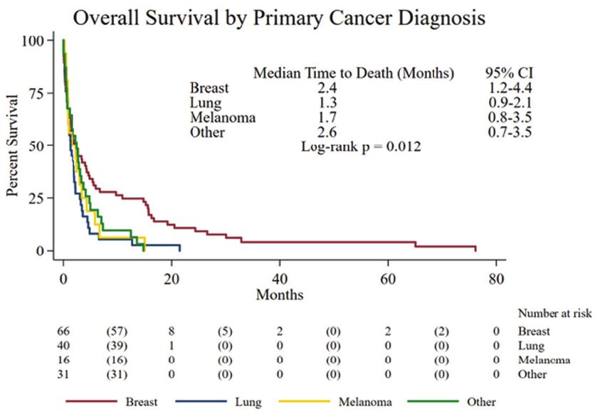
Eur J Breast Health 2021; 17(4): 371-377

        •     Prognosis was worse in patients with positive CSF cytology versus equivocal or negative cytology.
        •     Treatment of LMC either by intrathecal chemotherapy, radiation to the brain or spine, or systemic therapy was associated with an improvement in
              survival versus no treatment.

      Introduction                                                                     Materials and Methods
      Leptomeningeal carcinomatosis (LMC) is defined as metastatic                     Study design and data collection
      involvement of the leptomeninges, subarachnoid space and                         This study was an IRB-approved (registration #: OSU2016C0053)
      cerebrospinal fluid (CSF) (1). Malignant tumor cells spread and                  retrospective chart review of clinical and histopathologic data from
      disseminate to the subarachnoid space by hematogenous, perineural,               patients treated at The Ohio State University Comprehensive
      lymphatic, or perivascular mechanisms or by direct extension from                Cancer Center, (OSUCCC)-James that was initially approved on
      superficial brain metastases or bone metastases of the calvarium or              05/04/2016 between January 1st, 2010 and December 31st, 2015.
      spine (2-4). The incidence of LMC is increasing as patient survival              Eligible patients were identified by ICD-9 and ICD-10 codes
      improves with advances in the management of metastatic solid                     (198.4/ C79.32, C79.49, respectively) and included patients who
      tumors and as magnetic resonance imaging (MRI) becomes more                      were diagnosed with leptomeningeal carcinomatosis or unspecified
      widely utilized (5, 6). LMC occurs in approximately 4%–15% of                    meningeal disease, as well as patients who were diagnosed with a
      patients with malignant solid tumors, most commonly melanomas                    malignant solid tumor, who had undergone a procedure indicative
      and malignancies of the breast, lung, and gastrointestinal organs                of leptomeningeal carcinomatosis according to current procedural
      (7-11). Signs and symptoms of LMC include headaches, vomiting,                   terminology (CPT) codes. These procedures included insertion
      seizures, focal neurologic deficits, radicular neck and back pain,               of cerebrospinal fluid drainage device or catheter, LP, intrathecal
      cerebellar dysfunction, altered mental status, cauda equina syndrome,            infusion or injection of a therapeutic or prophylactic substance,
      dizziness, or syncope (12-14). The sensitivity and specificity of                injection or infusion of cancer chemotherapeutic substance with
      MRI in the diagnosis of LMC is difficult to estimate due to poor                 destruction of blood brain barrier, or MRI imaging of the brain or
      concordance with the gold standard diagnostic test of positive CSF               spinal cord. Patients without LMC, patients with LMC secondary
      cytology (15, 16). MRI with and without contrast is the initial and              to leukemia, lymphoma, or primary central nervous system
      often the sole diagnostic tool for LMC (17). Definitive diagnosis                malignancies, patients with incomplete clinical data and those
      of LMC depends on the presence of malignant cells in the CSF,                    treated at other institutions were excluded. Per EANO-ESMO
      but sensitivity is limited at about 50%–60% for the first lumbar                 Clinical Practice Guidelines, MRI is the gold standard imaging tool
      puncture (LP) (6, 18, 19). If the first CSF analysis is negative, a              for imaging suspected cases of LMC. Given the technical challenges
      second LP can increase sensitivity to 80%–85% (20). As a result of               of doing a lumbar puncture on some poor performance patients,
      low sensitivity and patient intolerance one or more LPs, a probable              we defined a case of LMC as having either positive CSF cytology or
      diagnosis of LMC is made when MRI findings are present in the                    MRI imaging indicative of LMC. Of 469 medical records reviewed,
      setting of systemic malignancy, even in the absence of positive CSF              153 patients were determined eligible.
      cytology (16).
                                                                                       Data for the eligible patients were initially obtained from The Ohio
      Once diagnosed with probable or definitive LMC, median survival time             State University Information Warehouse and uploaded into REDCap
      for patients is 2–6 months with treatment (21-25). Most treatment                (29). Data missing from the initial query were populated using manual
      recommendations are based on clinical experience or studies with a               review of each patient’s electronic medical record.
      low level of evidence due to a lack of prospective, randomized trials
      for patients with LMC (26). Intrathecal chemotherapy is the direct               Outcome measures
      instillation of chemotherapy into the subarachnoid space, making it a            The primary objectives of this study were to assess the overall
      promising treatment strategy. Intra-CSF pharmacotherapy should be                survival (OS) of patients with LMC at the OSU-CCC James, and
      reserved mainly for patients with a positive cytology on LP given that           to examine if primary tumor characteristics, diagnostic information,
      clearance of CSF cytology is used as one indicator for efficacy of this          management modalities (locoregional, systemic, or combined
      treatment (27). This is usually provided via an Ommaya reservoir after           therapy) and demographic factors were associated with OS. We
      adequate CSF flow is confirmed using 111Indium-DTPA flow study.                  performed a specific subgroup analysis to assess treatment strategies
                                                                                       and outcomes among LMC patients with primary breast cancer
      Whole Brain Radiotherapy (WBRT) with whole spine irradiation                     overall and each histologic subtype of breast cancer including
      can target the entire craniospinal axis and thus a larger area of disease        hormone receptor positivity.
      burden in LMC, however its use is limited by significant myelotoxicity
      (26). Focal external beam radiation to areas of bulky leptomeningeal             A change in treatment after LMC diagnosis was defined as a patient
      involvement of the spine causing CSF obstruction can be utilized                 receiving any of the following new treatments or changes in initial
      to relieve symptoms and allow for the administration of intrathecal              therapy: focal radiation therapy to brain metastases, bulky sites of
      administration (26, 28). The survival benefit of the various radiation           LMC burden or whole brain radiation therapy (WBRT); initiating IT
      therapy modalities in LMC is unclear.                                            chemotherapy, discontinuing previous systemic therapy, or initiating
                                                                                       new systemic therapy. If a patient did not undergo any of the previously
      We conducted a retrospective study to assess the diagnosis,                      mentioned changes, they were considered as having no new treatment,
      management and outcomes of leptomeningeal carcinomatosis at The                  even if continuing with any previous systemic therapy treatments or
372   Ohio State University.                                                           opting for supportive care alone.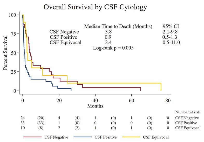
Rinehardt et al. LMC: Diagnosis, Management and Outcomes

Statistical analysis
                                                                           Table 1. Demographic Summary
Demographic and clinical characteristics were summarized using
descriptive analysis reported as medians and interquartile range for
continuous variables and frequencies and percents for categorical                                                     Total (n = 153)
variables. Overall survival (OS) was defined as the time from the date     Age at LMC
of diagnosis to date of death due to any cause or last known follow-up.                           Median [IQR]      55.7 years [48, 62.5]
                                                                           diagnosis
Patients were censored at the date last known to be alive. OS estimates
                                                                                                  White                   128 (84%)
were generated using Kaplan-Meier methods and compared using
                                                                           Race                   Black                   16 (10%)
log-rank tests. All data analyses were performed using SAS 9.4 (SAS
Institute Inc., Cary, NC) or Stata 14 (StataCorp LLC, College Station,                            Other                    9 (6%)
TX). For comparison of continuous data of one variable between two         Site of primary cancer diagnosis
groups, student’s t-test was used. A two-sided p-value of
Eur J Breast Health 2021; 17(4): 371-377
Table 1. Continued
Total (n = 153)
ECOG performance status
0 31 (20%)
1 62 (41%)
2 33 (22%)
3 17 (11%)
4 3 (2%)
Unknown 7 (5%)
Site of first metastasis
Bone 55 (36%)
Brain 51 (33%)
Lung 18 (12%)
Liver 8 (5%)
Spinal Cord 1 (1%)
Other 17 (11%)
None 2 (1%)
Missing 1 (1%)
LMC: Leptomeningeal carcinomatosis, IQR: Interquartile range, HER2:
Human epidermal growth factor receptor 2, CSF: cerebrospinal fluid, ECOG:
Eastern Cooperative Oncology Group, n: Number
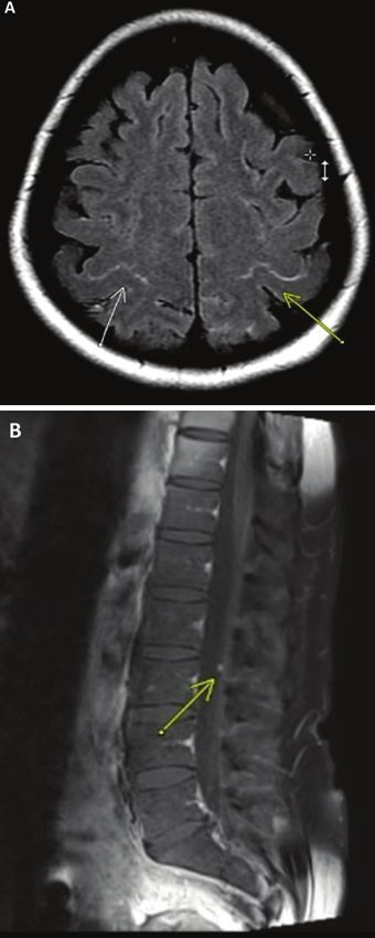
Figure 2. a) MRI brain with leptomeningal enhancement in the
parietal sulci b) A leptomeningal enhancing focus along a nerve root
in the lumbar spine.
Figure 1. Kaplan-Meier survial curves showing the overall survival
MRI: Magnetic resonance imaging
for patients with LMC secondary to breast cancer, lung cancer,
melanoma, and other tumors
equivocal (suspicious or atypical cells present) in 15%, and negative in systemic chemotherapy agent. The most common addition was
36%. Figure 3 shows an example of CSF cytology showing LMC from radiotherapy in 30 patients (42%). The most likely new agent was
a patient with poorly differentiated gastric carcinoma with signet ring the addition of capecitabine in six patients (8%). Twenty-eight
features. As depicted in Figure 4, the Kaplan-Meier curves revealed (18%) patients received intrathecal chemotherapy with 27 (96%)
differences in OS by CSF cytology: median OS for CSF negative receiving liposomal cytarabine and one (4%) receiving thiotepa. The
patients was 3.8 months (95% CI: 2.1, 9.8), for CSF equivocal was median OS for patients with no new treatment after LMC diagnosis
2.4 months (95% CI: 0.5, 11.0), and for CSF positive patients was 0.9 was 0.7 months (95% CI: 0.6, 1.2) and for those with a change
months (95% CI: 0.5, 1.3) (pRinehardt et al. LMC: Diagnosis, Management and Outcomes
characteristics). Thirty-seven breast cancer patients received Discussion:
radiotherapy for LMC (56%) and 64 received chemotherapy for
In patients with solid tumor malignancies, LMC is considered one of
either the primary disease or LMC (97%), with 22 patients (36%)
the most serious complications. We present a comprehensive overview
receiving intrathecal chemotherapy and 42 patients (64%) receiving
of diagnostic methods and treatments of patients with LMC associated
hormonal therapy.
with solid tumors over a 10-year period at our institution. LMC is
Of the 66 patients, there were 64 (97%) observed deaths; and the commonly associated with breast cancer, lung cancer, skin melanoma
along with various other cancers (5, 30). In our cohort, all patients
survival differed for patient based on their biomarker status. Median
underwent MRI of the brain and/or spine and 97% demonstrated
OS for all patients was 2.4 months (95% CI: 1.2–4.4). Median OS for
radiographic evidence of LMC. This high rate demonstrates that at
ER+/PR+/HER2- patients (n = 40, 61%) was 4.1 months (CI: 1.7,
our institution MRI is the preferred initial diagnostic modality prior
9.8), for triple negative breast cancer (TNBC) patients (n = 17, 26%)
to attempting high volume LP.
was 0.9 months (CI: 0.2, 1.9) and for HER2+ patients (n = 6, 9%)
was 0.7 months (CI: 0.0, 15.8). A significant difference in OS between The presence of malignant cells in the CSF versus equivocal or negative
subtypes based on hormone receptor status was found (p-0.002, log- cytology was associated with a significantly lower overall survival in our
rank test). OS was improved with new treatment after LMC diagnosis, cohort (0.9 months vs 3.8 months). This highlights the importance of
with median OS of 2.8 months (CI: 1.3, 5.7) in treated patients (n = repeating LP if CSF is initially negative as accurate CSF cytology is
57, 86%) compared to 1.2 months (CI: 0.03, 3.6) in untreated patients essential to further delineate an individual patient’s prognosis.
(n = 9, 14%) (p-0.026). The median OS in CSF negative patients was
Patients with LMC at our institution most commonly presented with
15.3 months (CI: 3.6, 30.1), 6.9 months in CSF equivocal patients
stage IV breast cancer, lung cancer, or melanoma with metastases to the
(CI: 1.5, 76.2), and 0.9 months in CSF positive patients (CI: 0.4, 2.0)
brain or bone. In the literature, the survival from the time of diagnosis
(p = 0.009, Log rank test).
of LMC is 4 to 6 weeks without treatment and 2 to 6 months with
therapy (5, 6, 22-25, 31). Our cohort included 153 patients with a
mixed population including patients who received treatment and some
who proceeded with comfort care or hospice alone following diagnosis
of LMC. The median OS of our cohort was 1.9 months (CI: 1.3, 2.5).
In our study, treatment of LMC either by intrathecal chemotherapy,
radiation to the brain or spine, or systemic therapy was associated with
an improvement in survival versus no treatment (Figure 4). The higher
CSF protein level present in patients with LMC demonstrates that
there is likely a blood-brain barrier disruption and resultant increased
levels of systemic chemotherapy delivered to the subarachnoid space
(32). Systemic chemotherapy is primarily based on the histology of the
primary tumor as in other forms of metastatic disease. Use of systemic
cytotoxic agents such as high-dose methotrexate can induce a response
in LMC from various solid tumors and improve survival outcomes,
however its use is limited due to systemic side effects, the potential
Figure 3. CSF cytology showing LMC from a patient with poorly for significant hematologic toxicity and the need for inpatient
differentiated gastric carcinoma with signet ring features administration (32). A significant limitation to the efficacy of systemic
CSF: Cerebrospinal fluid, LMC: Leptomeningeal carcinomatosis
chemotherapy in the treatment of LMC is resistance to therapy as
most patients developed disease progression despite multiple lines of
systemic chemo and/or hormonal therapy prior to development of
LMC.
Intrathecal methotrexate is a commonly utilized and relatively well-
tolerated agent associated with leukoencephalopathy (33). The
efficacy of intrathecal trastuzumab is currently unclear and is being
investigated for LMC from HER2-positive breast cancer given that
systemic trastuzumab appears to have poor penetration into the CSF
(26, 27). Liposomal cytarabine administered intrathecally has been
associated with complete cytological remission likely due to its unique
formulation which allows for persistence for up to 28 days in the CSF
(19). However, this agent is no longer available for clinical use due
to the manufacturer discontinuing production of this preparation;
the shorter acting version can still be utilized. The decision to use
intrathecal chemotherapy in the setting of LMC must be carefully
Figure 4. Kaplan-Meier survival curves showing the overall survival considered taking into account the extent and status of systemic
based on the CSF cytology disease, the patient’s functional status, and impact of the treatment
CSF: Cerebrospinal fluid and frequency of administration on the quality of life. 375Eur J Breast Health 2021; 17(4): 371-377
Breast cancer appears to be particularly responsive to therapy with References
overall survival of 7.5 months with therapy in the literature (34).
However, as evidenced in our cohort of breast cancer patients, TNBC 1. Nugent JL, Bunn PA Jr, Matthews MJ, Ihde DC, Cohen MH, Gazdar A,
et al. CNS metastases in small cell bronchogenic carcinoma: increasing
and HER2+ patients have a significantly worse prognosis as compared
frequency and changing pattern with lengthening survival. Cancer 1979;
to ER+/PR+/HER2- patients. Patients with a primary lung cancer
44: 1885-1893. (PMID: 227582) [CrossRef ]
or melanoma appear to be less responsive. In these patients, targeted
2. Boyle R, Thomas M, Adams JH. Diffuse involvement of the leptomeninges
therapy in the setting of certain actionable mutations (e.g osimertinib
by tumour--a clinical and pathological study of 63 cases. Postgrad Med J
in EGFR mutant NSCLC or BRAF inhibitor or checkpoint inhibitors 1980; 56: 149-158. (PMID: 7393804) [CrossRef ]
in melanoma) have shown preliminary evidence of activity against
3. Yung WA, Horten BC, Shapiro WR. Meningeal gliomatosis: a review of
LMC in these tumors (35). In this mixed cohort of patients with 12 cases. Ann Neurol 1980; 8: 605-608. (PMID: 6260012) [CrossRef ]
and without treatment, the median OS for primary breast cancer was
4. Price RA, Johnson WW. The central nervous system in childhood
2.4 months which was significantly longer than primary lung cancer leukemia. I. The arachnoid. Cancer 1973; 31: 520-533. (PMID:
(OS: 1.3 months) and primary melanoma (OS: 1.7 months). Despite 4511909) [CrossRef ]
treatment, prognosis remains poor and confirmation of diagnosis is 5. Groves MD. New strategies in the management of leptomeningeal
challenging. metastases. Arch Neurol 2010; 67: 305-312. (PMID: 20212228)
[CrossRef ]
Strengths and limitations
6. Freilich RJ, Krol G, DeAngelis LM. Neuroimaging and cerebrospinal
A strength of the study was the relatively large cohort size of fluid cytology in the diagnosis of leptomeningeal metastasis. Ann Neurol
153 patients given the relative rarity of LMC. We used not only 1995; 38: 51-57. (PMID: 7611725) [CrossRef ]
the ICD9 and ICD10 codes for carcinomatous meningitis or 7. Groves MD. Leptomeningeal disease. Neurosurg Clin N Am 2011; 22:
unspecified meningeal disease, but we also included patients who 67-78, vii. (PMID: 21109151) [CrossRef ]
were diagnosed with a malignant solid tumor, who had undergone 8. Yap HY, Yap BS, Tashima CK, DiStefano A, Blumenschein GR.
a procedure indicative of leptomeningeal carcinomatosis according Meningeal carcinomatosis in breast cancer. Cancer 1978; 42: 283-286.
to CPT codes. There are several limitations to our study including (PMID: 667799) [CrossRef ]
its retrospective nature, somewhat limited sample size for specific 9. Aroney RS, Dalley DN, Chan WK, Bell DR, Levi JA. Meningeal
treatment modalities, and the 5-year period of review during carcinomatosis in small cell carcinoma of the lung. Am J Med 1981; 71:
which time imaging techniques and treatment options changed 26-32. (PMID: 6264785) [CrossRef ]
significantly for many solid tumors. The range of treatments and 10. Bruna J, Simó M, Velasco R. Leptomeningeal metastases. Curr Treat
histologic diagnoses was too heterogeneous, and sample sizes were Options Neurol 2012; 14: 402-415. (PMID: 22736147) [CrossRef ]
too small to statistically assess the impact of specific drugs or 11. Kesari S, Batchelor TT. Leptomeningeal metastases. Neurol Clin 2003;
treatment modalities on specific cancer diagnoses. Future multi- 21: 25-66. (PMID: 12690644) [CrossRef ]
institution studies may reveal more information specific to LMC of 12. Grossman SA, Trump DL, Chen DC, Thompson G, Camargo EE.
difference histologies. Cerebrospinal fluid flow abnormalities in patients with neoplastic
meningitis. An evaluation using 111indium-DTPA ventriculography. Am
In conclusion, the risks and benefits of treatment in patients with J Med 1982; 73: 641-647. (PMID: 6814249) [CrossRef ]
LMC must be considered in detail on an individual basis. This study 13. Chamberlain MC, Corey-Bloom J. Leptomeningeal metastases:
may provide additional information for physicians to communicate 111indium-DTPA CSF flow studies. Neurology 1991; 41: 1765-1769.
prognostic information to patients based on an individual’s cancer (PMID: 1944906) [CrossRef ]
type, stage, grade, molecular status, and CSF cytology results. 14. Siegal T, Mildworf B, Stein D, Melamed E. Leptomeningeal metastases:
reduction in regional cerebral blood flow and cognitive impairment. Ann
Neurol 1985; 17: 100-102. (PMID: 3985577) [CrossRef ]
Ethics Committee Approval: This study was an IRB-approved (registration
15. Costa R, Kumthekar P. Management of central nervous system metastases
#: OSU2016C0053) retrospective chart review of clinical and histopathologic in breast cancer. Breast 2018: 942-690. e7. [CrossRef ]
data from patients treated at The Ohio State University Comprehensive Cancer
16. Le Rhun E, Weller M, Brandsma D, Van den Bent M, de Azambuja E,
Center, (OSUCCC)-James that was initially approved on 05/04/2016 between
Henriksson R, et al; EANO Executive Board and ESMO Guidelines
January 1st, 2010 and December 31st, 2015.
Committee. EANO-ESMO Clinical Practice Guidelines for diagnosis,
Informed Consent: Retrospective study. treatment and follow-up of patients with leptomeningeal metastasis
from solid tumours. Ann Oncol 2017;28(Suppl 4):iv84-iv99. (PMID:
Peer-review: Externally peer-reviewed. 28881917) [CrossRef ]
17. Yousem DM, Patrone PM, Grossman RI. Leptomeningeal metastases:
Author Contributions MR evaluation. J Comput Assist Tomogr 1990; 14: 255-261. (PMID:
Conception: H.R., M.K., E.M., A.S., A.G.; Design: H.R., M.K., E.M., A.S., 2312855) [CrossRef ]
A.G.; Data Collection and/or Processing: H.R., M.K., E.M., M.D.P.G.P.E., 18. Chamberlain MC. Leptomeningeal metastasis. Semin Neurol 2010; 30:
A.M., I.A.; Analysis and/or Interpretation: M.P., J.A.S., M.L., R.W., S.S., D.S., 236-244. (PMID: 20577930) [CrossRef ]
J.V., M.C., P.G., V.K.P., N.W., B.R., A.M.N.; Writing: H.R., M.K.; Critical 19. Bohn JP, Reinstadler V, Pall G, Stockhammer G, Steurer M, Oberacher
Review: M.L., R.W., S.S., D.S., J.V., M.C., P.G., V.K.P., N.W., B.R., A.M.N. H, et al. Cerebrospinal fluid drug concentrations and clinical outcome of
patients with neoplastic meningitis treated with liposomal cytarabine. Eur
Conflict of Interest: No conflict of interest was declared by the authors.
J Drug Metab Pharmacokinet 2019; 44: 845-851. (PMID: 31435852)
Financial Disclosure: The authors declare that this study received no financial [CrossRef ]
376 support.Rinehardt et al. LMC: Diagnosis, Management and Outcomes
20. Kaplan JG, DeSouza TG, Farkash A, Shafran B, Pack D, Rehman F, 28. Hermann B, Hültenschmidt B, Sautter-Bihl ML. Radiotherapy of the
et al. Leptomeningeal metastases: comparison of clinical features and neuroaxis for palliative treatment of leptomeningeal carcinomatosis.
laboratory data of solid tumors, lymphomas and leukemias. J Neurooncol Strahlenther Onkol 2001; 177: 195-199. (PMID: 11370554) [CrossRef ]
1990; 9: 225-229. (PMID: 2086737) [CrossRef ] 29. Harris PA, Taylor R, Thielke R, Payne J, Gonzalez N, Conde JG. Research
21. Chamberlain MC. Neoplastic meningitis. J Clin Oncol 2005; 23: 3605- electronic data capture (REDCap)--a metadata-driven methodology and
3613. Retraction in: J Clin Oncol 2010; 28: 4018. (PMID: 15908671) workflow process for providing translational research informatics support.
[CrossRef ] J Biomed Inform 2009; 42: 377-381. (PMID: 18929686) [CrossRef ]
22. Clarke JL, Perez HR, Jacks LM, Panageas KS, Deangelis LM. 30. Chamberlain MC. Leptomeningeal metastases: a review of evaluation
Leptomeningeal metastases in the MRI era. Neurology 2010; 74: 1449- and treatment. J Neurooncol 1998; 37: 271-284. (PMID: 9524085)
1454. (PMID: 20439847) [CrossRef ] [CrossRef ]
23. Herrlinger U, Förschler H, Küker W, Meyermann R, Bamberg M, 31. Mayer RJ, Berkowitz RS, Griffiths CT. Central nervous system
Dichgans J, et al. Leptomeningeal metastasis: survival and prognostic involvement by ovarian carcinoma: a complication of prolonged survivial
factors in 155 patients. J Neurol Sci 2004; 223: 167-178. (PMID: with metastatic disease. Cancer 1978; 41: 776-783. (PMID: 630551)
15337619) [CrossRef ] [CrossRef ]
24. Oechsle K, Lange-Brock V, Kruell A, Bokemeyer C, de Wit M. Prognostic 32. Scott BJ, Kesari S. Leptomeningeal metastases in breast cancer. Am J
factors and treatment options in patients with leptomeningeal metastases Cancer Res 2013; 3: 117-126. (PMID: 23593536) [CrossRef ]
of different primary tumors: a retrospective analysis. J Cancer Res Clin 33. Zairi F, Kotecki N, Rodrigues I, Baranzelli M-C, Andre C, Dubois F, et
Oncol 2010; 136: 1729-1735. (PMID: 20204406) [CrossRef ] al. Prospective follow-up of a cohort of 112 patients with leptomeningeal
25. Waki F, Ando M, Takashima A, Yonemori K, Nokihara H, Miyake M, et al. metastases of breast cancer recruited from 2007 to 2011: Prognostic
Prognostic factors and clinical outcomes in patients with leptomeningeal factors. Am Soc Clin Oncol 2012; 30(Suppl): 2070. [CrossRef ]
metastasis from solid tumors. J Neurooncol 2009; 93: 205-212. (PMID: 34. Chamberlain MC, Kormanik PR. Carcinomatous meningitis secondary
19043775) [CrossRef ] to breast cancer: predictors of response to combined modality therapy. J
26. Mack F, Baumert BG, Schäfer N, Hattingen E, Scheffler B, Herrlinger U, Neurooncol 1997; 35: 55-64. (PMID: 9266441) [CrossRef ]
et al. Therapy of leptomeningeal metastasis in solid tumors. Cancer Treat 35. Geukes Foppen MH, Brandsma D, Blank CU, van Thienen JV,
Rev 2016; 43: 83-91. (PMID: 26827696) [CrossRef ] Haanen JB, Boogerd W. Targeted treatment and immunotherapy in
27. Beauchesne P. Intrathecal chemotherapy for treatment of leptomeningeal leptomeningeal metastases from melanoma. Ann Oncol 2016; 27: 1138-
dissemination of metastatic tumours. Lancet Oncol 2010; 11: 871-879. 1142. (PMID: 26961150) [CrossRef ]
(PMID: 20598636)
377You can also read