Development and validation of a novel nomogram to predict aneurysm rupture in patients with multiple intracranial aneurysms: a multicentre ...

Page created by Clarence Erickson
 
CONTINUE READING
Development and validation of a novel nomogram to predict aneurysm rupture in patients with multiple intracranial aneurysms: a multicentre ...
Open access                                                                                                                    Original research

                                         Development and validation of a novel

                                                                                                                                                              Stroke Vasc Neurol: first published as 10.1136/svn-2020-000480 on 5 February 2021. Downloaded from http://svn.bmj.com/ on March 15, 2021 by guest. Protected by copyright.
                                         nomogram to predict aneurysm rupture
                                         in patients with multiple intracranial
                                         aneurysms: a multicentre
                                         retrospective study
                                         Xin Feng,1 Xin Tong ‍ ‍,2,3 Fei Peng,2,3 Hao Niu,2,3 Peng Qi,1 Jun Lu,1 Yang Zhao,4
                                         Weitao Jin,4 Zhongxue Wu,2 Yuanli Zhao,4 Aihua Liu ‍ ‍,2,3 Daming Wang1

To cite: Feng X, Tong X,                 ABSTRACT                                                           also found to be associated with unfavourable
Peng F, et al. Development and           Background and purpose Approximately 15%–45%                       outcomes after rupture.6 In addition, patient
validation of a novel nomogram           of patients with unruptured intracranial aneurysms have
to predict aneurysm rupture
                                                                                                            counselling in the elective setting is very
                                         multiple intracranial aneurysms (MIAs). Determining which          challenging because good models for specif-
in patients with multiple
intracranial aneurysms: a                one is most likely to rupture is extremely important for           ically predicting subarachnoid haemorrhage
multicentre retrospective study.         treatment decision making for MIAs patients. This study            (SAH) in these patients are lacking; instead,
Stroke & Vascular Neurology              aimed to develop and validate a nomogram to evaluate the
                                                                                                            information is extrapolated from varied data
2021;0. doi:10.1136/svn-2020-            per-­aneurysm rupture risk of MIAs patients.
000480                                                                                                      sets including patients with both single IA
                                         Methods A total of 1671 IAs from 700 patients with MIAs
                                         were randomly dichotomised into derivation and validation          and MIAs. Moreover, the treatment strategy
►► Additional material is
                                         sets. Multivariate logistic regression analysis was used           for patients with MIAs is typically complex
published online only. To view
please visit the journal online          to select predictors and construct a nomogram model for            and is driven by evaluation of rupture risks
(http://​dx.​doi.​org/​10.​1136/​svn-​   aneurysm rupture risk assessment in the derivation set.            of each IA (particularly when considering
2020-​000480).                           The discriminative accuracy, calibration performance and           endovascular management). Therefore, to
                                         clinical usefulness of this nomogram were assessed. We             prevent the catastrophic consequences of IA
XF and XT contributed equally.
                                         also developed a multivariate model for a subgroup of 158          rupture, it is clinically important to identify
AL and DW contributed equally.
                                         subarachnoid haemorrhage (SAH) patients and compared               and prophylactically treat the IA that is most
Received 3 July 2020                     its performance with the nomogram model.                           likely to rupture.
Revised 11 November 2020                 Results Multivariate analyses identified seven variables              Several studies characterised ruptured
Accepted 14 December 2020                that were significantly associated with IA rupture (history
                                                                                                            IAs in SAH patients with MIAs, and found
                                         of SAH, alcohol consumption, female sex, aspect ratio
                                                                                                            that the aspect ratio (AR), irregular shape,2
                                         >1.5, posterior circulation, irregular shape and bifurcation
                                         location). The clinical and morphological-­based MIAs
                                                                                                            size1 and size ratio (SR)4 could identify the
                                         (CMB-­MIAs) nomogram model showed good calibration                 ruptured IA. However, those studies only
                                         and discrimination (derivation set: area under the curve           included MIAs patients with SAH and not
© Author(s) (or their                                                                                       those without. Although those studies may
                                         (AUC)=0.740 validation set: AUC=0.772). Decision curve
employer(s)) 2021. Re-­use
permitted under CC BY-­NC. No            analysis demonstrated that the nomogram was clinically             aid in identifying responsible IAs in these
commercial re-­use. See rights           useful. Compared with the nomogram model, the AUC of               patients with SAH, they may not be adequately
and permissions. Published by            multivariate model developed from SAH patients had lower           helpful in treatment decision making of MIAs
BMJ.                                     value of 0.730.                                                    patients without SAH, that is, whether to
1
 Neurosurgery Department,                Conclusions This CMB-­MIAs nomogram for MIAs rupture               treat or manage conservatively and to iden-
Beijing Hospital, Beijing, Beijing,      risk is the first to be developed and validated in a large         tify the IA that should be treated. This may
China                                    multi-­institutional cohort. This nomogram could be used in
2
 Neurointervention Center,                                                                                  be attributed to the fact that decision making
                                         decision-­making and risk stratification in MIAs patients.
Beijing Neurosurgical Institute,                                                                            requires comprehensive consideration of the
Beijing, China                                                                                              characteristics of both patients and IAs.
3
 Neurointervention Center,               INTRODUCTION                                                          Significant demographic, clinical and
Beijing Tiantan Hospital, Beijing,
                                         Multiple intracranial aneurysms (MIAs) are                         morphological (CMB) differences are found
China
4
 Neurosurgery Department,                encountered in approximately 15%–45% of                            between patients with single IA and MIAs,3 6 7
Peking University International          patients with intracranial aneurysms (IAs).1–4                     indicating that the underlying pathogenesis
Hospital, Beijing, China                 The Japanese ‘small unruptured IA verifica-                        of MIAs may differ from that of single IA.8–10
                                         tion study’ found that patients with MIAs are                      Given the lack of analyses of large data
 Correspondence to
 Dr Aihua Liu;                           at a higher risk of IA growth and rupture.5                        samples, the true relevance of potential MIAs
​liuaihuadoctor@​163.c​ om               Compared with single IA cases, MIAs were                           predictors remains unknown, and the existing

                                                  Feng X, et al. Stroke & Vascular Neurology 2021;0. doi:10.1136/svn-2020-000480                         1
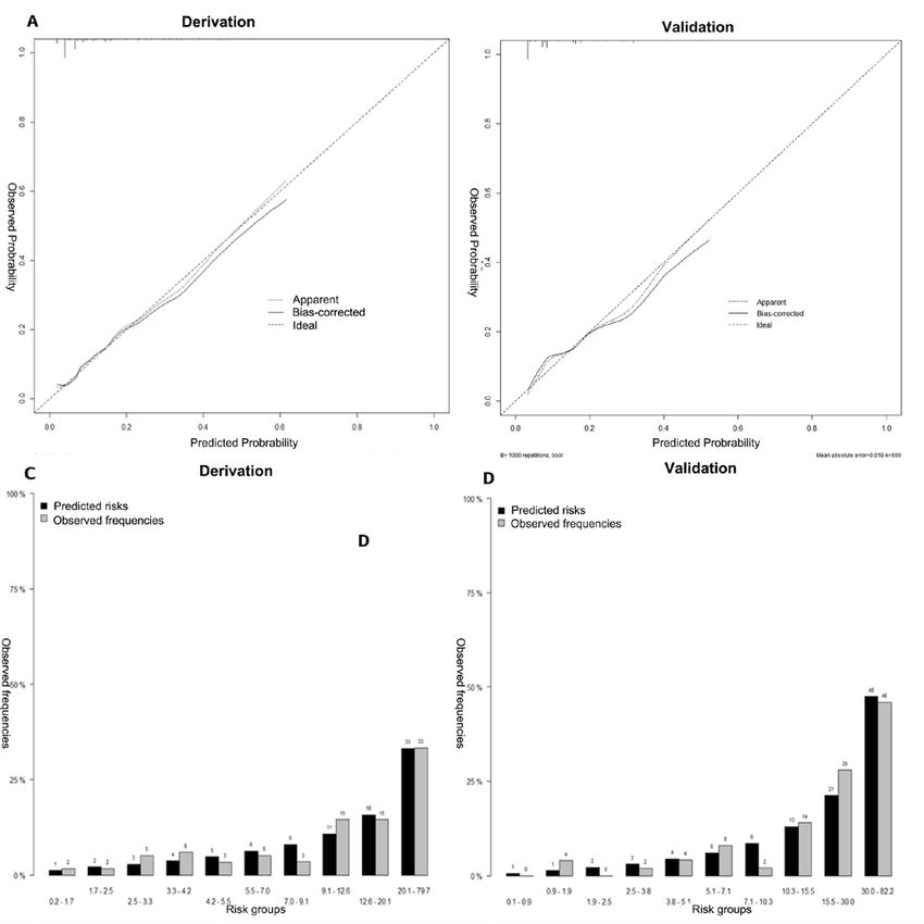
Development and validation of a novel nomogram to predict aneurysm rupture in patients with multiple intracranial aneurysms: a multicentre ...
Open access

                                                                                                                                                  Stroke Vasc Neurol: first published as 10.1136/svn-2020-000480 on 5 February 2021. Downloaded from http://svn.bmj.com/ on March 15, 2021 by guest. Protected by copyright.
scoring system is not suitable for these patients. One of       MIAs morphology
the most popular models for predicting the IA rupture           All patients in this study had digital subtraction angiog-
risk, the Population, Hypertension, Age, Size, Earlier          raphy (DSA) images and three-­dimensional (3D) recon-
SAH and Site (PHASES) score, was developed using 8382           structions. All DSA angiograms and 3D reconstructions
participants from six prospective cohort studies to predict     of the 1671 IAs were re-­evaluated and measured by two
patient’s risk of IA rupture.11 However, the PHASES score       researchers on a 0.1 mm scale; they were supervised by
only used the characteristics of the largest IA when eval-      two senior neurointerventionists with 15 years of expe-
uating MIAs patients, and data of other coexisting IA(s)        rience (online supplemental figure S2). The AR (dome
were ignored. In addition, previous studies showed that         height/neck width), SR (maximum IA height/average
one-­quarter of the largest IAs did not rupture in patients     of the parent diameter), and bottleneck factors (dome
with SAH and MIAs.1 2 Thus, it is necessary to develop          width/neck width) were calculated. IAs were categorised
a new method for assessing the rupture risk of each IA          as regular or irregular (with multiple lobes, daughter sacs
among MIAs patients to improve clinical decision making,        or other types of wall protrusions). The inflow angle was
especially in patients without SAH.                             defined as the angle from the parent artery into the IA,
   In this study, we aimed to identify the independent risk     and the outflow angle was defined as that at which the
factors for IA rupture in a large multi-­institutional cohort   IA flowed outward to the distal parent artery. The main
of MIAs patients. We also aimed to develop and validate a       branching angle was defined as the angle of the parent
nomogram model for rupture risk assessment of each IA,          artery (in case of a sidewall IA) or that between the parent
and evaluate its clinical usefulness in aiding clinical deci-   artery and the daughter branch most approaching 180°
sion making and improving ongoing treatment efforts.            (in case of a bifurcation IA).2 All the associated angles
                                                                were measured on a 1° scale (online supplemental figure
                                                                S2). For bifurcation IAs, the branching to parent ratio
                                                                was defined as the ratio of the sum of the diameters of
METHODS
                                                                the branch vessels to the diameter of the parent artery (in
Study population
                                                                case of a sidewall IA, the branching to parent ratio was set
We retrospectively obtained data regarding cerebro-
                                                                to 1). The neck to parent ratio was defined as the ratio of
vascular images and medical records from a consecu-
                                                                the neck width to the parent artery diameter.
tive series of patients with at least two saccular IAs from
three centres (Beijing Tiantan Hospital, Beijing Hospital
                                                                Statistical analyses
and Peking University International Hospital) in China
                                                                Continuous and categorical variables of patients’ baseline
between September 2015 and December 2018. Aneu-
                                                                characteristics have been presented as means±SD and
rysms were divided into ruptured and unruptured groups          percentages, respectively. The 1671 IAs were randomly
depending on whether they were ruptured at admission.           divided into two subsets with similar event rates, namely,
In patients with SAH, we only included those in whom            the derivation subset (1171/1671, 70%) and the vali-
the responsible IA could be identified; they included:          dation subset (500/1671, 30%). Mean imputation was
(1) patients who underwent treatment by clipping in             used with low missing data; 20 factors with complete data
whom the ruptured IA was confirmed through micro-               (gender, age, SAH history, number of IAs, hypertension,
scopic visual assessment and (2) patients who under-            diabetes, hypercholesterolaemia, heart diseases, history
went either endovascular or no treatment; in these cases,       of stroke, smoking, drinking, size, neck, AR, branching
we included only those with a definitive haemorrhage            to parent ratio, neck to parent ratio, SR, posterior circu-
pattern on computed tomographic images (localised to            lation (PC, basilar tip, basilar-­superior cerebellar artery,
one IA). Examples of definitive haemorrhage patterns are        vertebral artery-­ posterior inferior cerebellar artery, or
provided in online supplemental figure S1. Patients with        vertebrobasilar junction), irregular shape and bifur-
fusiform or dissecting IAs, other cerebrovascular diseases,     cation location) and three factors with less than 10%
IAs previously treated at other neurological centres, and       missing data (inflow angle (missing 31/1671, 1.9%),
incomplete clinical and imaging data were excluded.             outflow angle (missing 31/1671, 1.9%) and branching
                                                                angle (missing 35/1671, 2.1%)) were included in this
Clinical presentation                                           study. Both, complete case and imputed data set analyses
Data were collected regarding the following: age, sex, pres-    showed similar results.
ence of hypertension (yes/no; diagnosed by the general
practitioner before admission and requiring medical             Development and validation of the nomogram
treatment), smoking, both current and previous (previ-          Multivariate logistic regression analysis was performed
ously smoked regularly and quit at least 1 year before          for per-­aneurysm rupture risk in MIAs patients using a
admission), alcohol consumption (yes/no; current or             forward stepwise method that included all variables with
previous intake >5 drinks per day),12 presence of cardi-        a p
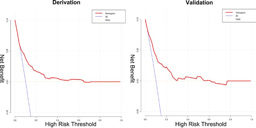
Development and validation of a novel nomogram to predict aneurysm rupture in patients with multiple intracranial aneurysms: a multicentre ...
Open access

                                                                                                                                                Stroke Vasc Neurol: first published as 10.1136/svn-2020-000480 on 5 February 2021. Downloaded from http://svn.bmj.com/ on March 15, 2021 by guest. Protected by copyright.
variables in the derivation set was evaluated by variation                      Multivariable logistic regression including all 1671 MIAs
inflation factors (VIF, with values
Open access

                                                                                                                                                           Stroke Vasc Neurol: first published as 10.1136/svn-2020-000480 on 5 February 2021. Downloaded from http://svn.bmj.com/ on March 15, 2021 by guest. Protected by copyright.
Table 1 Univariate analysis between unruptured and ruptured groups in the derivation and validation set
                                             Derivation cohort                                    Validation cohort
                                             (n=1171)                                             (n=500)
                                             Unruptured          Ruptured                         Unruptured             Ruptured
Characteristic                               N (%)               N (%)            P value         N (%)                  N (%)              P value
No of aneurysm                               1067 (91.1)         104 (8.9)                        446 (89.2)               54 (10.8)
Female sex                                    734 (68.8)          79 (76.0)         0.130         294 (65.9)               40 (74.1)          0.229
Age (years)
 1.5                                        196 (18.4)          41 (39.4)      1.2                 187 (17.5)          33 (31.7)      1.2                      325 (30.5)          41 (39.4)        0.060         138 (30.9)               14 (25.9)          0.449
SR >1.7                                        204 (19.1)          42 (40.4)
Open access

                                                                                                                                                     Stroke Vasc Neurol: first published as 10.1136/svn-2020-000480 on 5 February 2021. Downloaded from http://svn.bmj.com/ on March 15, 2021 by guest. Protected by copyright.
 Table 2 Multivariate analysis of all 1671 multiple aneurysms in 700 patients and 400 multiple aneurysms in 158 patients who
 presented with SAH
 Model 1*                    OR (95% CI)                    P value      VIF     Model 2*          OR (95% CI)                P value    VIF
 SAH history                 5.094 (2.848 to 8.929)         1.55            2.449 (1.302 to 4.641)     0.006      1.218
 Irregular shape             1.883 (1.225 to 2.897)          0.004       1.025 Irregular shape     1.886 (1.070 to 3.336)     0.028      1.120
 Alcohol use                 2.022 (1.016 to 3.920)           0.040      1.400 Neck >4 mm          1.994 (1.015 to 3.944)     0.046      1.307
 Female                      1.852 (1.053 to 3.395            0.038      1.419 PC                  2.522 (1.156 to 5.667)     0.022      1.007
 AR >1.5                     2.371 (1.507 to 3.698)
Open access

                                                                                                                                                                                                                                                                                                                                                                                                                                                                                         Stroke Vasc Neurol: first published as 10.1136/svn-2020-000480 on 5 February 2021. Downloaded from http://svn.bmj.com/ on March 15, 2021 by guest. Protected by copyright.
                                                                                                                                                                         0.730 (0.683 to 0.782)
                                                                                        Total group (400 IAs)

                                                                                                                                                                                                             0.3663
                                                                                                                                            0.202

                                                                                                                                                                                                                                    *Model 1 was developed by all 1671 multiple aneurysms in patients with or without SAH; model 2 was developed by 400 multiple aneurysms in 158 patients with SAH.
                                                                                                                                                                         0.717 (0.632 to 0.802)
                                                                                        Validation Group

                                                                                                                                                                                                                                                                                                                                                                                                       Figure 3 Decision curve analysis of CMB-­MIAs nomogram
                                                                                                                                                                                                                                                                                                                                                                                                       in derivation and validation cohort.
                                                                                                                                            0.207

                                                                                                                                                                                                             0.269

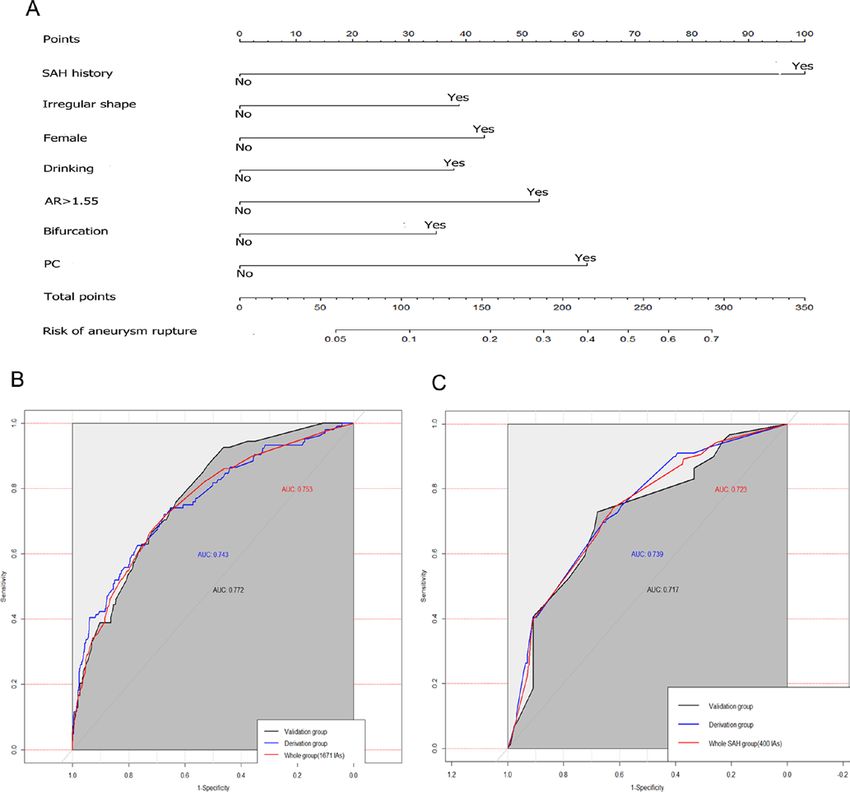
                                                                                                                                                                                                                                                                                                                                                                                                       study, we applied this score to evaluate the rupture risk
                                                                                                                                                                                                                                                                                                                                                                                                       of 1671 MIAs. The results showed that the AUC of their
                                                                                                                                                                         0.739 (0.677 to 0.801)

                                                                                                                                                                                                                                                                                                                                                                                                       model for the 1671 MIAs in this cohort was 0.577; this
                                                                                        Total group (1671 IAs) Derivation Group

                                                                                                                                                                                                                                                                                                                                                                                                       was worse than that of our model (AUC=0.753). This may
                                                                                                                                                                                                                                                                                                                                                                                                       be attributed to the fact that although the study included
                                                                                                                                                                                                                                                                                                                                                                                                       a large sample of IAs, only 13.7% of derivation data
                                                                             Model 2*

                                                                                                                                                                                                                                                                                                                                                                                                       and 12.7% of validation data were from MIAs patients;
                                                                                                                                                                                                                                                                                                                                                                                                       therefore, their model may be more suitable for single
                                                                                                                                            0.199

                                                                                                                                                                                                             0.893

                                                                                                                                                                                                                                                                                                                                                                                                       IA patients. In addition, this study may have selection
                                                                                                                                                                                                                                                                                                                                                                                                       bias. Some unruptured IAs with a high risk of rupture
                                                                                                                                                                         0.753 (0.712 to 0.794)

                                                                                                                                                                                                                                                                                                                                                                                                       were treated and not included in the follow-­up cohort.
                                                                                                                                                                                                                                                                                                                                                                                                       This necessitates the development of a prediction model
                                                                                                                                                                                                                                                                                                                                                                                                       specific for MIAs.
                                                                                                                                                                                                                                                                                                                                                                                                          To date, several studies have focused on identifying
                                                                                                                                                                                                                                                                                                                                                                                                       risk factors for MIAs rupture.1 2 4 8 23 However, the popu-
                                                                                                                                                                                                                                                                                                                                                                                                       lations were limited to MIAs patients with SAH, and the
                                                                                                                                            0.083

                                                                                                                                                                                                             0.983

                                                                                                                                                                                                                                                                                                                                                                                                       variables analysed were limited to IA morphological char-
                                                                                                                                                                                                                                    AUC, areas under curve; IA, intracranial aneurysm; SAH, subarachnoid haemorrhage.
      Evaluation of discrimination and calibration abilities of the models

                                                                                                                                                                                                                                                                                                                                                                                                       acteristics. These studies mainly intended to accurately
                                                                                                                                                                                                                                                                                                                                                                                                       identify ruptured IAs in SAH patients with MIAs. As we
                                                                                                                                                                         0.772 (0.686 to 0.858)

                                                                                                                                                                                                                                                                                                                                                                                                       pooled individual patient data from three larger centres
                                                                                        Validation Group

                                                                                                                                                                                                                                                                                                                                                                                                       in China, we were able to analyse clinical risk factors and
                                                                                                                                                                                                                                                                                                                                                                                                       morphological features of IAs in MIAs patients with or
                                                                                                                                                                                                                                                                                                                                                                                                       without SAH; we were also able to calculate the absolute
                                                                                                                                                                                                             0.4654

                                                                                                                                                                                                                                                                                                                                                                                                       risks of individual IA rupture. In addition, we developed
                                                                                                                                            0.085

                                                                                                                                                                                                                                                                                                                                                                                                       a multivariate model for SAH patients with MIAs (model
                                                                                                                                                                                                                                                                                                                                                                                                       2). In this model, the AUC in the derivation and vali-
                                                                                                                                                                                                                                                                                                                                                                                                       dation groups were 0.739 and 0.717, respectively; these
                                                                                                                                                                         0.743 (0.690 to 0.796)

                                                                                                                                                                                                                                                                                                                                                                                                       results were worse than those of the model including all
                                                                                        Derivation Group

                                                                                                                                                                                                                                                                                                                                                                                                       MIAs patients.
                                                                                                                                                                                                                                                                                                                                                                                                          Risk scores are more reliable if they include already
                                                                                                                                                                                                                                                                                                                                                                                                       well-­established risk factors for IA rupture.11 Most risk
                                                                             Model 1*

                                                                                                                                                                                                                                                                                                                                                                                                       factors included in our model are known to cause IA
                                                                                                                                                                                                                           0.9146
                                                                                                                                            0.073

                                                                                                                                                                                                                                                                                                                                                                                                       rupture; these included the following: history of SAH,11 24
                                                                                                                                                                                                  Calibration, goodness of fit

                                                                                                                                                                                                                                                                                                                                                                                                       female sex,25 alcohol consumption,26 27 bifurcation loca-
                                                                                                                                                                                                                                                                                                                                                                                                       tion,28 29 location of PC,19 AR30 and IA shape.18 19 DCA
                                                                                                                                  Overall performance

                                                                                                                                                                                                  P value of Hosmer-­

                                                                                                                                                                                                                                                                                                                                                                                                       evaluates whether a model is useful in clinical decisions
                                                                                                                                                                                                                                                                                                                                                                                                       and identifies the model leading to the best decision.31 In
                                                                                                                                                                                                  Lemeshow test
                                                                                                                                                        Discrimination

                                                                                                                                                                                                                                                                                                                                                                                                       the present study, when the threshold probabilities were
                                                                                                                                                                         AUC (95%)
                                                                                                                                  Brier score

                                                                                                                                                                                                                                                                                                                                                                                                       between 10% and 65%, DCA showed that the nomogram
      Table 3

                                                                                                                                                                                                                                                                                                                                                                                                       was more beneficial than either the treat-­all or treat-­none
                                                                                                                                                                                                                                                                                                                                                                                                       strategy (figure 3). These results indicate the clinical
                                                                                                                                                                                                                                                                                                                                                                                                       benefits of using the CMB-­MIAs nomogram.

6                                                                                                                                                                                                                                                                                                                                                                                                       Feng X, et al. Stroke & Vascular Neurology 2021;0. doi:10.1136/svn-2020-000480
Open access

   We found that IA size and number,12 and smoking

                                                                                                                                                                        Stroke Vasc Neurol: first published as 10.1136/svn-2020-000480 on 5 February 2021. Downloaded from http://svn.bmj.com/ on March 15, 2021 by guest. Protected by copyright.
                                                                              introduced bias, which may have affected our analysis.
status were not associated with IA rupture when other risk                    Prospective multicentre validation is needed to acquire
factors were considered. This does not imply that these                       high-­level evidence for further clinical application.
factors are not important for MIAs rupture in isolation;                      Second, although our nomogram exhibited favourable
it indicates that these factors were not significantly asso-                  discriminatory and calibration ability regarding predic-
ciated with IA rupture in addition to the seven predictors                    tive value and net benefit in the derivation and validation
used. Aneurysm size was ranked the most important risk                        cohort, it should be noted that our model was developed
factor; physicians widely accept that larger IAs are more                     using data from patients treated at Chinese institutions.
dangerous than smaller ones.32 However, a large cohort                        Thus, caution is needed when generalising our results
study of patients with IA SAH and MIAs2 revealed that                         to other countries and ethnicities. Third, our study did
the largest IA had not ruptured in 36 (29%) of 124 MIAs                       not include other factors that may be associated with
patients. Interestingly, for SAH patients with MIAs, IA size                  aneurysm rupture, such as blood pressure levels, family
was significantly associated with IA rupture; the risk of                     histories and imaging indicators such as haemodynamic
IA rupture increased with size in the range of 3–15 mm,                       analysis and vascular wall enhancement on MRI.33 Fourth,
(online supplemental figure S5A). This suggests that                          morphological measurements may change during and
IA size may help identify ruptured IAs in SAH patients;                       after rupture. A large-­  scale prospective cohort study
however, this is not necessarily applicable to all MIAs                       including unruptured MIAs is needed to investigate the
patients, and especially in those with IAs with irregular                     CMB risk factors of both unruptured IAs that eventu-
shapes (online supplemental figure S6). A previous study                      ally rupture and those that do not. Finally, we excluded
revealed that in MIAs patients, each IA is not associated                     patients with MIAs in whom the ruptured IA could not be
with an increased risk of rupture; however, these patients                    identified; this may have caused selection bias.
are subject to the cumulative risk from all individual IAs.19
Similarly, we found that the IA number is not associated
with an increased risk of rupture. In addition, compared                      CONCLUSIONS
with patients with three or more IAs, those with two IAs                      Using a large multicentre cohort, we first devised and
demonstrated more ruptures (online supplemental                               validated a nomogram, that is simple to use, for clinically
figure S5B). Regarding smoking status, we only had data                       evaluating the per-­aneurysm rupture risk of patients with
regarding the time of IA detection, and not for the inten-                    MIAs. Data on the seven independent factors included
sity and duration of smoking or passive smoking. The                          this nomogram are easy to obtain; these include: SAH
association between smoking and MIA rupture requires                          history, alcohol consumption, sex, AR >1.5, PC, irregular
further in-­depth research, with more detailed question-                      shape and bifurcation location. With favourable calibra-
naires or prospective studies. We found that hypertension                     tion and discrimination, this nomogram may be useful
was not a significant factor on univariate analysis in both,                  for decision making and risk stratification in patients with
derivation and validation cohorts. However, statistical                       MIAs.
differences were observed between ruptured and unrup-                         Acknowledgements We thank Yuesong Pan from the China National Clinical
tured groups in the entire cohort. This may be explained                      Research Center for Neurological Diseases, for analysis and interpretation of data.
by the fact that the results of randomisation caused a slight                 Contributors DW and AL conceived and designed the study. FP, HN, YZ and WJ
change in the distribution of this subgroup. However, we                      collected the data. PQ, JL, YZ and ZW were responsible for quality control. XF and
believe that this will not significantly impact the results                   XT conceived of the project, analysed the data and wrote the paper. All authors
of multivariate because factors with p
Open access

                                                                                                                                                               Stroke Vasc Neurol: first published as 10.1136/svn-2020-000480 on 5 February 2021. Downloaded from http://svn.bmj.com/ on March 15, 2021 by guest. Protected by copyright.
REFERENCES                                                                   17 Malhotra A, Wu X, Forman HP, et al. Management of unruptured
 1 Björkman J, Frösen J, Tähtinen O, et al. Irregular shape identifies          intracranial aneurysms in older adults: a cost-­effectiveness analysis.
   ruptured intracranial aneurysm in subarachnoid hemorrhage patients           Radiology 2019;291:411–7.
   with multiple aneurysms. Stroke 2017;48:1986–9.                           18 Lindgren AE, Koivisto T, Björkman J, et al. Irregular shape of
 2 Backes D, Vergouwen MDI, Velthuis BK, et al. Difference in                   intracranial aneurysm indicates rupture risk irrespective of size in a
   aneurysm characteristics between ruptured and unruptured                     population-­based cohort. Stroke 2016;47:1219–26.
   aneurysms in patients with multiple intracranial aneurysms. Stroke        19 UCAS Japan Investigators, Morita A, Kirino T, et al. The natural
   2014;45:1299–303.                                                            course of unruptured cerebral aneurysms in a Japanese cohort. N
 3 Jabbarli R, Dinger TF, Darkwah Oppong M, et al. Risk factors for             Engl J Med 2012;366:2474–82.
   and clinical consequences of multiple intracranial aneurysms: a           20 Mayer TE. The unruptured intracranial aneurysm treatment score: a
   systematic review and meta-­analysis. Stroke 2018;49:848–55.                 multidisciplinary consensus. Neurology 2016;86:792.2–3.
 4 Rajabzadeh-­Oghaz H, Wang J, Varble N, et al. Novel models for            21 Backes D, Rinkel GJE, Greving JP, et al. Elapss score for prediction
   identification of the ruptured aneurysm in patients with subarachnoid        of risk of growth of unruptured intracranial aneurysms. Neurology
   hemorrhage with multiple aneurysms. AJNR Am J Neuroradiol                    2017;88:1600–6.
   2019;40:1939–46.                                                          22 Tominari S, Morita A, Ishibashi T, et al. Prediction model for 3-­year
 5 Sonobe M, Yamazaki T, Yonekura M, et al. Small unruptured                    rupture risk of unruptured cerebral aneurysms in Japanese patients.
                                                                                Ann Neurol 2015;77:1050–9.
   intracranial aneurysm verification study: SUAVe study, Japan. Stroke
                                                                             23 Jagadeesan BD, Delgado Almandoz JE, Kadkhodayan Y, et al. Size
   2010;41:1969–77.
                                                                                and anatomic location of ruptured intracranial aneurysms in patients
 6 Kaminogo M, Yonekura M, Shibata S. Incidence and outcome of
                                                                                with single and multiple aneurysms: a retrospective study from a
   multiple intracranial aneurysms in a defined population. Stroke
                                                                                single center. J Neurointerv Surg 2014;6:169–74.
   2003;34:16–21.
                                                                             24 Burkhardt J-­K, Chua MH, Winkler EA, et al. Incidence, classification,
 7 Nurmonen HJ, Huttunen T, Huttunen J, et al. Polycystic kidney
                                                                                and treatment of angiographically occult intracranial aneurysms
   disease among 4,436 intracranial aneurysm patients from a defined
                                                                                found during microsurgical aneurysm clipping of known aneurysms.
   population. Neurology 2017;89:1852–9.
                                                                                J Neurosurg 2019;132:1–8.
 8 McDowell MM, Zhao Y, Kellner CP, et al. Demographic and clinical          25 Müller TB, Vik A, Romundstad PR, et al. Risk factors for unruptured
   predictors of multiple intracranial aneurysms in patients with               intracranial aneurysms and subarachnoid hemorrhage in a
   subarachnoid hemorrhage. J Neurosurg 2018;128:961–8.                         prospective population-­based study. Stroke 2019;50:2952–5.
 9 Takeshima Y, Kaku Y, Nishi T, et al. Multiple cerebral aneurysms          26 Sauvigny T, Mohme M, Grensemann J, et al. Rate and risk factors
   associated with neurofibromatosis type 1. J Stroke Cerebrovasc Dis           for a hyperactivity delirium in patients with aneurysmal subarachnoid
   2019;28:e83–91.                                                              haemorrhage. Neurosurg Rev 2019;42:481–8.
10 Backes D, Rinkel GJE, Laban KG. Patient- and Aneurysm-­Specific           27 Can A, Castro VM, Ozdemir YH, et al. Alcohol consumption
   risk factors for intracranial aneurysm growth: a systematic review and       and aneurysmal subarachnoid hemorrhage. Transl Stroke Res
   meta-­analysis. Stroke 2016;47:951–7.                                        2018;9:13–19.
11 Greving JP, Wermer MJH, Brown RD, et al. Development of                   28 Feng X, Qi P, Wang L, et al. Relationship between cerebrovascular
   the phases score for prediction of risk of rupture of intracranial           atherosclerotic stenosis and rupture risk of unruptured intracranial
   aneurysms: a pooled analysis of six prospective cohort studies.              aneurysm: a single-­center retrospective study. Clin Neurol Neurosurg
   Lancet Neurol 2014;13:59–66.                                                 2019;186:105543.
12 Jeon JS, Ahn JH, Huh W, et al. A retrospective analysis on the            29 Leemans EL, Cornelissen BMW, Rosalini G, et al. Impact of
   natural history of incidental small paraclinoid unruptured aneurysm. J       intracranial aneurysm morphology and rupture status on the particle
   Neurol Neurosurg Psychiatry 2014;85:289–94.                                  residence time. J Neuroimaging 2019;29:487–92.
13 Andic C, Aydemir F, Kardes O, et al. Single-­stage endovascular           30 You S-­H, Kong D-­S, Kim J-­S, et al. Characteristic features of
   treatment of multiple intracranial aneurysms with combined                   unruptured intracranial aneurysms: predictive risk factors for
   endovascular techniques: is it safe to treat all at once? J Neurointerv      aneurysm rupture. J Neurol Neurosurg Psychiatry 2010;81:479–84.
   Surg 2017;9:1069–74.                                                      31 Van Calster B, Wynants L, Verbeek JFM, et al. Reporting and
14 Huhtakangas J, Lehto H, Seppä K, et al. Long-­Term excess mortality          interpreting decision curve analysis: a guide for Investigators. Eur
   after aneurysmal subarachnoid hemorrhage: patients with multiple             Urol 2018;74:796–804.
   aneurysms at risk. Stroke 2015;46:1813–8.                                 32 Shojima M, Morita A, Nakatomi H, et al. Size is the most important
15 Dong Q-­L, Gao B-­L, Cheng Z-­R, et al. Comparison of surgical and           predictor of aneurysm rupture among multiple cerebral aneurysms:
   endovascular approaches in the management of multiple intracranial           post hoc subgroup analysis of unruptured cerebral aneurysm study
   aneurysms. Int J Surg 2016;32:129–35.                                        Japan. Neurosurgery 2018;82:864–9.
16 Johnston SC, Tiny L. Leaving tiny, unruptured intracranial                33 Khan MO, Toro Arana V, Rubbert C, et al. Association between
   aneurysms untreated: why is it so hard? JAMA Neurol                          aneurysm hemodynamics and wall enhancement on 3D vessel wall
   2018;75:13–14.                                                               MRI. J Neurosurg 2020:1–11.

8                                                                             Feng X, et al. Stroke & Vascular Neurology 2021;0. doi:10.1136/svn-2020-000480
You can also read