Differences in Peripheral Neuropathy in Xeroderma Pigmentosum Complementation Groups A and D
←
→
Page content transcription
If your browser does not render page correctly, please read the page content below
Differences in Peripheral Neuropathy in Xeroderma
Pigmentosum Complementation Groups A and D
Tanya Lehky ( lehkyt@ninds.nih.gov )
NINDS: National Institute of Neurological Disorders and Stroke https://orcid.org/0000-0001-6421-505X
Paul E. Sackstein
Genetics and Developmental Biology Research Unit: Genetique et biologie du developpement
Deborah Tamura
Laboratory of Cancer Biology and Genetics, NCI, NIH
Martha Quezado
Laboratory of Pathology, NCI, NIH
Tianxia Wu
Clinical Trials Unit, NINDS, NIH
Sikandar G Khan
Laboratory of Cancer Biology and Genetics, NCI, NIH
Nicholas J Patronas
Radiology and Imaging Sciences, CC, NIH
Edythe Wiggs
Clinical Trials Unit, NINDS, NIH
Carmen C Brewer
Otolaryngology Branch, NIDCD, NIH
John J DiGiovanna
Laboratory of Cancer Biology and Genetics, NCI, NIH
Kenneth H Kraemer
Laboratory of Cancer Biology and Genetics, NCI, NIH
Research article
Keywords: Xeroderma pigmentosum, peripheral neuropathy, neurodegeneration, sensorineural hearing loss, DNA repair
Posted Date: March 23rd, 2021
DOI: https://doi.org/10.21203/rs.3.rs-351476/v1
License: This work is licensed under a Creative Commons Attribution 4.0 International License. Read Full License
Page 1/15
Abstract
BACKGROUND

Xeroderma pigmentosum (XP) is a rare autosomal recessive genetic disorder with defective DNA nucleotide excision repair
and associated with a high frequency of skin cancer. Approximately 25% of patients develop progressive neurological
degeneration. Complementation groups XP-A and XP-D are most frequently associated with neurological disorders.

METHODS

This is a retrospective review of patients with XP who were evaluated at NIH from 1986 to 2015 and had nerve conduction
studies (NCS). Audiological, brain imaging, neuropsychological assessments were also performed on most of the
patients. Limited neuropathology of XP-A and XP-D patients were compared.

RESULTS

The 33 patients had NCS: XP-A (9 patients), XP-C (7 patients), XP-D (10 patients), XP-E (1 patient), XP-V (4 patients), and XP-
unknown (2 patients). Peripheral neuropathy was documented only in two complementation groups: 78% (7/9) of XP-
A patients had a sensorimotor neuropathy while 50% (5/10) of XP-D patients had a sensory neuropathy only. The severity
of the neuropathy correlated with hearing loss.

CONCLUSIONS

Despite similar brain imaging and audiological findings patients, XP-A and XP-D complementation groups differ in the type
of neuropathy, sensorimotor versus sensory alone. A few cases suggest that sensorineural hearing loss and absent deep
tendon reflexes may precede abnormal NCS in XP and therefore serve as valuable clinical indicators of XP patients that will
later develop peripheral neuropathy.

TRIAL REGISTRATION: Patients were evaluated under clinical protocols, NCT00001813 and NCT00046189, approved by the
NIH Institutional Review Boards.

Background
Xeroderma pigmentosum (XP) is a rare autosomal recessive disorder involving defective DNA nucleotide excision
repair (NER) that is characterized by extreme sensitivity to the damaging effects of ultraviolet radiation (UV) resulting in
a greater than 2,000-fold increase in the frequency of melanoma and non-melanoma skin cancers; 25% of XP patients
develop a progressive neurodegeneration.1 XP is classified into 8 different complementation groups, denoted as XP A-G
based on defects in DNA nucleotide excision repair (NER) genes and XP-Variant (XP-V) that has a defect in
the eta polymerase gene.2 Patients in complementation groups XP-A and XP-D have the highest frequency of neurological
complications along with the greatest photosensitivity or burning on minimal sun exposure .3-5 A four-decade longitudinal
study on the natural history of XP since 1971 showed that the leading cause of death in XP patients was skin cancer (34%,
n=10). However, neurological degeneration was also a major cause of death (31%, n=9).1 Brain atrophy, cerebellar and basal
ganglia degeneration leading to ataxia and severe cognitive decline have all been observed in XP-A and XP-D patients.6, 7 In
conjunction with CNS deterioration, these patients have been observed to become areflexic and develop an axonal
polyneuropathy as well as sensorineural hearing loss.8 Though visual loss occurs in XP, it is usually the result of direct UV-
exposure of the anterior components of the eye rather than specific optic nerve deterioration.9

The frequency of peripheral neuropathy and its association with CNS degeneration, in different XP complementation groups
remains unknown. In this study, we performed a retrospective review of nerve conduction studies of XP patients examined at
NIH to assess the frequency and type of peripheral neuropathy among patients in different XP complementation groups. We

                                                           Page 2/15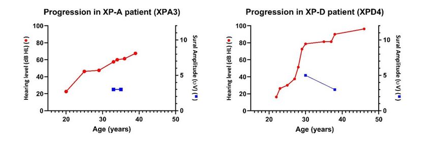
also examined MRI/CT, audiology, neuropsychological and clinical studies to determine if the peripheral neuropathy was
associated with other neurological findings.

Methods
Patients: Patients were evaluated under clinical protocols, NCT00001813 and NCT00046189, approved by the NIH
Institutional Review Boards. Written, informed consent and assent, was obtained from all patients. The patients underwent
multimodality assessment including clinical10, genetic, MRI, neuropsychological, laboratory and electrodiagnostic
evaluation. Records of deep tendon reflexes were available on most of the subjects evaluated. Absence of deep tendon
reflexes were considered to be a clinical manifestation of peripheral neuropathy. Available data was retrieved by
retrospective review of charts of patients with a clinically confirmed diagnosis of XP at the NIH from 1986-2018. For clarity
in this paper, complementation group A subjects were designated XPA1-9 and complementation group D were designated
XPD 1-10 but have also included the prior protocol designated subject numbers in the tables. The following subjects have
been previously reported in other articles, XPA3 (XP19BE)4, XPA5 (XP12BE)3, 4, 8, 11, XPA6 (XP360BE)12, XPD1 (XP29BE)4, 13,
14,
      and XPD4 (XP33BE)4.

Clinical Neurophysiology Studies: Nerve conduction studies were performed using standard methodology on a Nicolet
Viking Select equipment (Natus, Middleton, WI) for the studies performed after 2004 and were compared to department-
based normative values. All prior studies used earlier versions of the Nicolet Viking equipment. In most patients, the median
and sural sensory nerves and the median and fibular motor nerves were tested although other nerves were selected for
clinical indications. No needle EMG studies were performed. For consistency, the studies were reviewed by one neurologist
(TJL).

Audiological evaluations: Audiologic testing included pure-tone air-conduction (0.25–8 kHz) and bone-conduction (0.25–4
kHz) thresholds and speech audiometry using clinical audiometers in double-walled sound suites that met American
National Standards Institute criteria. Audiology results were presented as the 4F-PTA, a four frequency (0.5/1/2/4-kHz) pure-
tone average using the right ear results only. Classification of the hearing loss is: < 20 dBHL = normal hearing, >20-40 dBHL
= mild hearing loss, >40-70 dBHL = moderate hearing loss, >70-95 = severe hearing loss, > 95 profound hearing loss. For
consistency, the studies were reviewed by one audiologist (CCB).

Brain Imaging: MRI or CT imaging of brain was performed at NIH or reviewed from outside sources. Cerebral and cerebellar
atrophy were classified on a four-point scale: normal (0), mild (1), moderate (2), or severe (3) depending on the extent of
volume loss at each patient’s age. For consistency, the studies were reviewed and graded by one neuroradiologist (NP).

Neuropsychological studies: Wechsler scales appropriate for age were performed to yield a Full-Scale IQ. Those XP patients
unable to do the Wechsler test because of limited cognitive ability or very young age, were excluded. For the Full-Scale IQ, a
score of less than 80 was considered to be abnormal. For consistency, the studies were reviewed by one neuropsychologist
(EW).

Neuropathology: Pathology slides from autopsy were available for one XP-A patient (XPA5) and one XP-D patient (not
evaluated by NCS) for review. These autopsy reports had been previously published3, and available specimens of muscle
and nerve were reexamined. New H&E stained sections of peripheral nerve and muscle were prepared from formalin-fixed
paraffin embedded material. For consistency, the studies were reviewed by one neuropathologist (MQ).

Data Analysis: Demographic and clinical results are given as means ± standard deviations (SD) in tables and two-sample t-
test was used to evaluate the difference between XP complementation groups. For each of nerve conduction outcomes,
motor and sensory amplitudes, analysis of covariance (ANCOVA) was performed to examine the interaction between group
and hearing level. Pearson correlation analysis was used to evaluate the association between hearing level and each of

                                                          Page 3/15
nerve conduction outcomes. The correlation analysis was performed for XP-A and XP-D combined (interaction effect:
p>0.05) or separately depending on the interaction test.
Data Availability Policy: Anonymized data will be shared by request from any qualified investigator.
Results
Demographics: For the study, 106 patients had been evaluated for XP phenotype/genotype over 40 years (Figure 1).
Electrodiagnostic studies were performed on 54 of the patients. The majority of the electrodiagnostic studies were
performed between 2004-2015, though 7 patients had studies performed between 1986-1993. Twelve patients had XP-TTD
complex1 and were not included in the final analysis. Nine patients were excluded from the analysis because of
confounding comorbidities or incomplete studies. In the XP-C complementation group, six patients were excluded; one
patient had multiple sclerosis, one patient was treated with alpha interferon and had a preexisting neuropathy, one patient
was the product of a close consanguineous relationship and had atypical features of nonprogressive early hearing loss,
another patient was also the product of close consanguity and had fetal alcohol syndrome. Two patients lacked lower
extremity nerve conduction studies. In the XP-D complementation group, one patient was excluded for coexistence of limb
girdle muscular dystrophy. One patient with XP-E was excluded for a prior history of transverse myelitis, and one patient with
XP-G had a possible Cockayne syndrome presentation. Of the remaining 33 patients (Table 1), 9 (27%) patients were in
complementation group XP-A, 7 (21%) patients were in XP-C, 10 (30%) patients were in XP-D, 1 (3%) was in XP-E, 4 (12%)
were in XP-V, and 2 (6%) had an unknown mutation causing XP (Table 1).
Of the XP patients who underwent NCS, there were 14 males and 19 females and the mean age at initial NCS study was
20.7±13.6 years (range 3-54 years). The patients in XP-A, XP-C, and XP-D complementation groups were generally younger
than the XP-E and XP-V complementation groups. For the XP-A and XP-D complementation groups used in the analysis of
peripheral neuropathy characteristics, there was no significant difference in age (p=0.48) in the two groups (mean ±
standard deviation, range; XP-A -18.5±2.1, 3-24, XP-D - 16.7±9.0, 5-43). Six patients in the XP-A and XP-D complementation
groups had follow-up electrodiagnostic evaluations. Summary of patients with NCS, audiology, MRI or CT imaging and were
evaluated for deep tendon reflexes testing is in Table 1.
XP-A Findings:
Seven of the nine (78%) XP-A patients had axonal sensorimotor polyneuropathy on nerve conduction studies with the
findings summarized in Table 1,2. The 4F-PTA showed that six of these patients (XPA1-6) also had mild to severe
sensorineural hearing loss and MRI abnormalities. One subject (XPA-7) with neuropathy, a 3 year old, did not have audiology
testing. MRI findings was noted in the same subjects with neuropathy include cerebral atrophy, cerebellar atrophy and in 2
pediatric patients, hypomyelination was noted. Six XP-A patients had Full-Scale IQ testing and all were abnormal, regardless
of the presence or absence of neuropathy. XP-A patients who had neuropathy, expectedly, also had absent DTRs. However,
one XP-A patient (XPA-9) eventually lost her DTRs though her NCS remained normal.
XP-D findings:
In the XP-D patients, 5 patients of 10 studied (XPD1-5) or 50% had evidence for an axonal sensory neuropathy with the
findings summarized in Table 1,3. These patients (XPD1-5) also had mild to moderate sensorineural hearing loss and
abnormal MRIs of the brain. The MRI findings included varying degrees of cerebral and/or cerebellar atrophy. XP-D patients
without neuropathy had normal hearing and brain imaging, though one patient (XPD6) showed development of cerebral
atrophy between ages 9 and 14. Low Full-Scale IQs was observed in all patients with neuropathy and in two patients without
neuropathy. All XP-D patients with peripheral neuropathy had absent DTRs while only one patient without peripheral
neuropathy (XPD6) had absent DTRs. This was the same patient noted to be developing recent signs of brain atrophy in
brain imaging.
Page 4/15Other XP complementation group findings: In the XP-C patients, the NCS (7 patients), audiology (6 patients), and brain imaging studies (2 MRI and 2 CT) were all normal and only one patient (XPC1) had a low Full-scale IQ at age 5. Patients in the XP-E, XP-V and XP-U complementation groups had normal nerve conduction studies. These patients had normal hearing with the exception of one XP-E patient had a mild unilateral hearing loss and one XP-V patient developed mild high frequency hearing loss over 5 years. The XP-V had normal MRIs of the brain with the exception of a few punctate white matter lesions observed in one MRI (XPV4). Comparison of XP-A and XP-D Complementation Groups: Comparison of the nerve conduction studies between the XP-A and XP-D complementation groups, using a two-sample t- test, showed a significant difference in the fibular motor amplitudes (p = 0.0208) but not the sural sensory, median sensory and median motor amplitudes. There was no significant difference in the hearing loss, cortical atrophy on MRI imaging and Full-scale IQ between the two complementation groups. Association evaluations (XP-A and XP-D): For sural and median sensory amplitude (Figure 2a,b), ANCOVA showed that the interaction between XP complementation groups (XP-A vs. XP-D) and hearing loss was not significant (p = 0.635, Figure 2a), indicating that the relationship between SNC response and hearing was consistent across the two groups and reflecting deterioration together in sensory and hearing functions in both complementation groups. The difference in sural sensory amplitudes between XP-A and XP-D complementation group was not significant (p=0.0792) and the interactions for MRI severity and Full-Scale IQ were also not significant. Therefore, Pearson correlation analysis based on the combined XP-A and XP-D groups was used to examine the linear correlations between sural sensory amplitude and hearing loss, MRI severity, and Full-scale IQ. The analysis showed that the linear correlation was significant for hearing loss (r=-0.845, R2=0.714, p
lower extremity motor nerves were not available. Patient XPA8 (XP337BE) did not have overt signs of neurologic
deterioration and motor and sensory NCS at age 14 and 23 years have remained stable.

In the XP-D complementation group, the two patients, XPD1 (XP29BE) and XPD4 (XP33BE) with peripheral neuropathy (XPD-
1 and XPD-4) and neurological deterioration showed a minimal decline in the sensory responses with stable motor
responses (Figure 3b). Patient XPD6 (XP116BE), an initially neurologically normal patient with recent signs of cerebral
atrophy on CT brain images, had a 50% decrease in sural sensory response (25 µV to 12 µV) over 4 years. Patient XPD9
(XP341BE), also neurologically normal, showed no change in the median and sural sensory nerve amplitudes in a three-year
follow-up period. There were no decreases in the motor responses for any of the XPD patients.

Neuropathology:

Nerve and muscle pathology was compared between the XP-A and XP-D patient (Figure 3) and previously reported3. XPA5 (
XP12BE) patient passed away at age 44, was cachectic (
DNA damage. The XPD protein is one of the helicases involved in unwinding the DNA prior to excision and repair. The other
XP gene products are involved in DNA complex stabilization, DNA cleavage, and finally repair of the DNA strand with
polymerases and ligases. Clearly, the integrity of the NER pathway and the XP gene complementation groups is crucial for
maintenance of DNA repair in the central and peripheral nervous systems though the exact mechanism is not known. Since
neuronal cells are not predisposed directly to the injurious effects of ultraviolet (UV) radiation, other mechanisms for
neuronal injury in XP patients have been proposed.20 One suggested mechanism may be generation of oxidative stress. The
reactive oxygen species (ROS) are constantly generated during aerobic metabolism. One of the ROS is hydroxyl radical, the
key endogenous source of DNA damage. Hydroxyl radicals induce many different base lesions including cyclopurine
deoxynucleosides (CyPU) that are known to be removed by the NER pathway.21-24 Accumulation of cyPU in neuronal cells
with inability to undergo DNA repair may lead to neurodegeneration.25 Other suggested mechanisms may be mitochondrial
dysfunction and role of these proteins in non-DNA repair pathways.20 Generation of neural stem cells and post-mitotic
neurons from XP-A patient induced pluripotent stem cells (IPSCs) showed hypersensitivity to DNA damage-induced
apoptosis following UV exposure.26 The neurophysiological findings in this paper suggests that there may
be slightly different mechanisms of neurodegeneration associated with XP-A and XP-D complementation groups that, at
least in the peripheral nervous system, result in two types of neuropathy. Better understanding of pathways may be
beneficial in designing clinical studies that address neurotoxicities in XP-A and XP-D since controlled clinical studies in this
area have been limited to date.27, 28

Conclusions
The findings in this study are notable for the distinction between XP-A and XP-D on the basis of the type of peripheral
neuropathy. CNS deterioration and hearing loss does not appear to be differ between the two XP complementation groups.
Because XP is a rare disease, the number of subjects in each complementation group is small and limits the robustness of
our findings. However, it supports the importance of DNA NER in the survival of central and peripheral neurons
with defects in the pathway resulting in variable forms of peripheral nerve degeneration. Continued studies on the peripheral
nerve manifestations of XP-A and XP-D may help to further elucidate the role of the XP gene defects in the pathogenesis of
neuropathy, particularly as related to the DNA NER pathway.

Abbreviations
XP – xeroderma pigmentosum

NCS – nerve conduction studies

PN - Peripheral neuropathy

IQ – Intellectual quotient

CNS – Central nervous system

NER - Nucleotide excision repair

MRI – Magnetic resonance imaging

CT- Computed tomography

4F-PTA – four frequency (0.5/1/2/4-kHz) pure-tone average

dB HL – decibel hearing level

SNHL – sensorineural hearing loss
                                                           Page 7/15
DTR – deep tendon reflexes
TTD -– trichothiodystrophy
UV – ultraviolet radiation
Declarations
Disclosures of the authors/Competing Interests: The authors declare that they have no competing interests.
Acknowledgments: We are grateful to the patients and their families for their commitment and time to this study.
Sponsorship/Funding: This research was supported by the National Institutes of Health Intramural Research Programs of
National Institute of Neurological Disorders and Stroke (NINDS), National, National Institute on Deafness and Other
Communication Disorders (NIDCD) and National Cancer Institute (NCI).
Data Availability Policy: The datasets used and/or analysed during the current study are available from the corresponding
author on reasonable request.
Authors’ contributions: See appendix at end of manuscript.
References
1. DiGiovanna JJ, Kraemer KH. Shining a light on xeroderma pigmentosum. J Invest Dermatol 2012;132:785-796.
2. Lindenbaum Y, Dickson D, Rosenbaum P, Kraemer K, Robbins I, Rapin I. Xeroderma pigmentosum/cockayne syndrome
complex: first neuropathological study and review of eight other cases. Eur J Paediatr Neurol 2001;5:225-242.
3. Lai JP, Liu YC, Alimchandani M, et al. The influence of DNA repair on neurological degeneration, cachexia, skin cancer
and internal neoplasms: autopsy report of four xeroderma pigmentosum patients (XP-A, XP-C and XP-D). Acta
Neuropathol Commun 2013;1:4.
4. Totonchy MB, Tamura D, Pantell MS, et al. Auditory analysis of xeroderma pigmentosum 1971-2012: hearing function,
sun sensitivity and DNA repair predict neurological degeneration. Brain 2013;136:194-208.
5. Anttinen A, Koulu L, Nikoskelainen E, et al. Neurological symptoms and natural course of xeroderma pigmentosum.
Brain 2008;131:1979-1989.
6. Ueda T, Kanda F, Nishiyama M, Nishigori C, Toda T. Quantitative analysis of brain atrophy in patients with xeroderma
pigmentosum group A carrying the founder mutation in Japan. J Neurol Sci 2017;381:103-106.
7. Ueda T, Kanda F, Aoyama N, Fujii M, Nishigori C, Toda T. Neuroimaging features of xeroderma pigmentosum group A.
Brain Behav 2012;2:1-5.
8. Viana LM, Seyyedi M, Brewer CC, et al. Histopathology of the inner ear in patients with xeroderma pigmentosum and
neurologic degeneration. Otol Neurotol 2013;34:1230-1236.
9. Brooks BP, Thompson AH, Bishop RJ, et al. Ocular manifestations of xeroderma pigmentosum: long-term follow-up
highlights the role of DNA repair in protection from sun damage. Ophthalmology 2013;120:1324-1336.
10. Nakano E, Takeuchi S, Ono R, Tsujimoto M, Masaki T, Nishigori C. Xeroderma Pigmentosum Diagnosis Using a Flow
Cytometry-Based Nucleotide Excision Repair Assay. J Invest Dermatol 2018;138:467-470.
11. Ramkumar HL, Brooks BP, Cao X, et al. Ophthalmic manifestations and histopathology of xeroderma pigmentosum: two
clinicopathological cases and a review of the literature. Surv Ophthalmol 2011;56:348-361.
12. Christen-Zaech S, Imoto K, Khan SG, et al. Unexpected occurrence of xeroderma pigmentosum in an uncle and nephew.
Arch Dermatol 2009;145:1285-1291.
Page 8/1513. Ueda T, Compe E, Catez P, Kraemer KH, Egly JM. Both XPD alleles contribute to the phenotype of compound
heterozygote xeroderma pigmentosum patients. J Exp Med 2009;206:3031-3046.
14. Zhou X, Khan SG, Tamura D, et al. Abnormal XPD-induced nuclear receptor transactivation in DNA repair disorders:
trichothiodystrophy and xeroderma pigmentosum. Eur J Hum Genet 2013;21:831-837.
15. Tsuji Y, Ueda T, Sekiguchi K, et al. Progressive length-dependent polyneuropathy in xeroderma pigmentosum group A.
Muscle Nerve 2020;62:534-540.
16. McKinnon PJ. Maintaining genome stability in the nervous system. Nat Neurosci 2013;16:1523-1529.
17. Cleaver JE, States JC. The DNA damage-recognition problem in human and other eukaryotic cells: the XPA damage
binding protein. Biochem J 1997;328 ( Pt 1):1-12.
18. Liu H, Rudolf J, Johnson KA, et al. Structure of the DNA repair helicase XPD. Cell 2008;133:801-812.
19. Feltes BC, Bonatto D. Overview of xeroderma pigmentosum proteins architecture, mutations and post-translational
modifications. Mutat Res Rev Mutat Res 2015;763:306-320.
20. Rizza ERH, DiGiovanna JJ, Khan SG, Tamura D, Jeskey JD, Kraemer KH. Xeroderma Pigmentosum: A Model for Human
Premature Aging. J Invest Dermatol 2021.
21. Kamakura N, Yamamoto J, Brooks PJ, Iwai S, Kuraoka I. Effects of 5',8-cyclodeoxyadenosine triphosphates on DNA
synthesis. Chem Res Toxicol 2012;25:2718-2724.
22. Brooks PJ. The 8,5'-cyclopurine-2'-deoxynucleosides: candidate neurodegenerative DNA lesions in xeroderma
pigmentosum, and unique probes of transcription and nucleotide excision repair. DNA Repair (Amst) 2008;7:1168-1179.
23. Brooks PJ, Wise DS, Berry DA, et al. The oxidative DNA lesion 8,5'-(S)-cyclo-2'-deoxyadenosine is repaired by the
nucleotide excision repair pathway and blocks gene expression in mammalian cells. J Biol Chem 2000;275:22355-
22362.
24. Kuraoka I, Bender C, Romieu A, Cadet J, Wood RD, Lindahl T. Removal of oxygen free-radical-induced 5',8-purine
cyclodeoxynucleosides from DNA by the nucleotide excision-repair pathway in human cells. Proc Natl Acad Sci U S A
2000;97:3832-3837.
25. Brooks PJ. The cyclopurine deoxynucleosides: DNA repair, biological effects, mechanistic insights, and unanswered
questions. Free Radic Biol Med 2017;107:90-100.
26. Fu L, Xu X, Ren R, et al. Modeling xeroderma pigmentosum associated neurological pathologies with patients-derived
iPSCs. Protein Cell 2016;7:210-221.
27. Abeti R, Zeitlberger A, Peelo C, et al. Xeroderma pigmentosum: overview of pharmacology and novel therapeutic
strategies for neurological symptoms. Br J Pharmacol 2019;176:4293-4301.
28. Evangelisti C, Cenni V, Lattanzi G. Potential therapeutic effects of the MTOR inhibitors for preventing ageing and
progeria-related disorders. Br J Clin Pharmacol 2016;82:1229-1244.
Tables
Table 1. Demographics of XP Patients with Nerve Conduction Studies
Page 9/15Complementation Group
XP-A XP-C XP-D XP-E XP-V XP unk
Number 9 7 10 1 4 2
Mean Age 16.7±9.0 13.1±6.9 18.5±2.1 42 43.5±9.1 12, 36
± Std Dev
(Range) (3-24) (5-25) (5-43) (34-54)
Gender (F:M) 5:4 4:3 4:6 1:0 3:1 1:1
Abn SNCS 7/9 0/7 5/10 0/1 0/4 0/2
Abn MNCS 7/9 0/7 0/10 0/1 0/4 0/2
Abn Hearing 6/8 0/6 5/10 0/1 0/3 0/1
Abn MRI 7/8 0/4 0/9 0/1
Abn IQ 5/6 0/4 0/9 0/1
Abbreviations: XP-A – xeroderma pigmentosum complementation group A, XP-C – xeroderma pigmentosum
complementation group C, XP-D – xeroderma pigmentosum complementation group D, XP-E – xeroderma pigmentosum
complementation group E, XP-V – xeroderma pigmentosum variant, XP unk – xeroderma pigmentosum – Unknown
mutation, F- female, M- male, Abn – abnormal, SNCS – sensory nerve conduction studies, MNCS – motor nerve conduction
studies (fibular nerve), MRI – magnetic resonance imaging, IQ – intelligence quotient, Std dev – Standard deviation
Table 2. XP-A Patient Findings.
Page 10/15Patient Age Gender Neuropathy Audiology MRI/CT Imaging IQ Testing DTR ID # Abn Abn SNHL Cerebral Cerebellar Hypo- IQ
Patient         Age     Gender     Neuropathy       Audiology      MRI/CT Imaging                            IQ        DTR
                                                                                                              Testing

 ID #                               Abn     Abn      SNHL           Cerebral    Cerebellar    Hypo-           IQ
Figure 1
Algorithm of Subjects Studied. Of the 106 subjects with an XP genotype/phenotype, 54 subjects had nerve conduction
studies and 33 subjects were included in the final evaluation. NCS – nerve conduction studies, XP – xeroderma
pigmentosum.
Page 13/15Figure 2
Hearing loss vs nerve conduction studies in XP-A and XP-D. The right ear 4F-PTA, defined as the four frequency (0.5/1/2/4-
kHz) pure tone average, was plotted against the amplitudes of the following: A. Sural sensory nerve, B. Median sensory
nerve, C. Fibular motor nerve, D. Median motor nerve. ANCOVA was used to study the interaction between complementation
group and hearing level. Normal hearing: 4F-PTA ≤ 20 dB HL. Normal NCS findings: sural sensory ≥ 6 µV, median sensory ≥
15 µV, fibular motor ≥ 2.5 mV, median motor ≥ 4.5 mV.
Page 14/15Figure 3
Follow-up hearing and sural sensory nerve studies. A. Patient XPA3 (neurological involvement) B. Patient XPD4 (neurological
involvement). Age of patient (X axis) plotted versus hearing (dB HL) (left Y axis) and sural amplitude (µV) (right Y axis).
Normal hearing:4F-PTA ≤ 20 dB HL. Normal NCS findings: sural sensory ≥6 µV, fibular motor ≥ 2.5 mV.
Figure 4
Nerve and muscle pathology of XP-A and XP-D patients. A. XPA (XP5 - XP12BE) patient median nerve - H&E, 20x, normal
nerve architecture on longitudinal section. B. XP-A (XP5 - XP12BE) patient. muscle – H&E, 20x, marked variation in myofiber
size characterized by the presence of many rounded and angular atrophic fibers, approaching panfascicular atrophy in
some areas with fatty infiltration. Numerous pyknotic nuclear clumps are also present. C. XP-D patient nerve from arm but
not specified – H&E, 10x, normal nerve architecture on cross-section. D. XP-D patient muscle – H&E, 20x, mild variation of
muscle fiber size with occasional small-sized angulated fibers. (Modified from JP Lai, et al 2013, permission by Dr. K.
Kraemer)
Page 15/15You can also read