Case Report Aggressive Systemic Mastocytosis in Association with Pure Red Cell Aplasia
←
→
Page content transcription
If your browser does not render page correctly, please read the page content below
Hindawi
Case Reports in Hematology
Volume 2018, Article ID 6928571, 5 pages
https://doi.org/10.1155/2018/6928571

Case Report
Aggressive Systemic Mastocytosis in Association with Pure Red
Cell Aplasia

          Dhauna Karam ,1,2 Sean Swiatkowski,1,2 Mamata Ravipati,1,2 and Bharat Agrawal1,2
          1
              Rosalind Franklin University, 3333 Green Bay Road, North Chicago, IL 60064, USA
          2
              Captain James A. Lovell Federal Health Care Center, 3001 Green Bay Road, North Chicago, IL 60064, USA

          Correspondence should be addressed to Dhauna Karam; dhauna.karam@gmail.com

          Received 13 March 2018; Revised 20 May 2018; Accepted 20 June 2018; Published 8 July 2018

          Academic Editor: Håkon Reikvam

          Copyright © 2018 Dhauna Karam et al. This is an open access article distributed under the Creative Commons Attribution License,
          which permits unrestricted use, distribution, and reproduction in any medium, provided the original work is properly cited.
          Aggressive systemic mastocytosis (ASM) is characterized by mast cell accumulation in systemic organs. Though ASM may be
          associated with other hematological disorders, the association with pure red cell aplasia (PRCA) is rare and has not been reported.
          Pure red cell aplasia (PRCA) is a syndrome, characterized by normochromic normocytic anemia, reticulocytopenia, and severe
          erythroid hypoplasia. The myeloid and megakaryocytic cell lines usually remain normal. Here, we report an unusual case of ASM,
          presenting in association with PRCA and the management challenges.

1. Introduction                                                         and active person; he enjoyed biking and rollerblading. The
                                                                        above symptoms were very unusual for him. The patient
Aggressive systemic mastocytosis is a rare disorder char-               reported intermittent episodes of epistaxis, 3-4 times a week
acterized by abnormal accumulation of mast cells in bone                since the past month, lasting for a few minutes. He also
marrow and internal organs (liver, spleen, lymph nodes, and             endorsed 2-3 episodes of loose stools daily since the past
gastrointestinal tract) [1]. Mastocytosis was initially classi-         month. Of note, the patient had history of exposure to Agent
fied as one of the subtypes of “myeloproliferative neoplasms             Orange between years 1969 and 1971; the first exposure was
(MPN).” In the 2016 revision of the World Health Orga-                  forty-five years earlier. Physical examination revealed stable
nization (WHO) classification of tumors of the hemato-                   vital signs with a palpable spleen of six finger-breadths below
poietic and lymphoid tissues, mastocytosis was classified as             the left costal margin and mild hepatomegaly. Cardiopul-
a separate entity [2, 3]. Chemical mediators such as tumor              monary, lymphatic, and dermatologic examination, in-
necrosis factor produced by mast cells can suppress eryth-              cluding Darrier’s sign were all negative.
ropoiesis, and some mast cell diseases can cause hypoplastic
anemia, though the pathogenesis is not clear. Our case report
highlights an unusual and rare presentation of ASM with                 2.2. Diagnosis. On admission, the patient was found to have
PRCA. Such an association has not been reported in the                  a hemoglobin count of 5.1 g/dl, which was a significant drop
literature, except in one case report where mast cell acti-             from the patient’s baseline hemoglobin of 13-14 g/dl. Other
vation disorder and PRCA occurred together [4].                         basic laboratory studies are presented in Table 1. Urine
                                                                        analysis, stool for blood test, serum haptoglobulin, LDH,
2. Case Presentation                                                    hepatitis B and C testing, PNH by flow cytometry, and
                                                                        hemochromatosis gene mutation were normal or negative.
2.1. Patient’s Symptoms/History. A 64-year-old white male,              Serum tryptase level was elevated at 1110 ng/ml (nor-
with past medical history of depression, presented with                 mal < 11.4 ng/ml). Bone marrow biopsy and clot section
progressive weakness, unintentional weight loss, and exer-              performed as a part of anemia workup revealed hyper-
cise intolerance since past 1 month. He was a very healthy              cellularity with markedly increased maturing granulopoiesis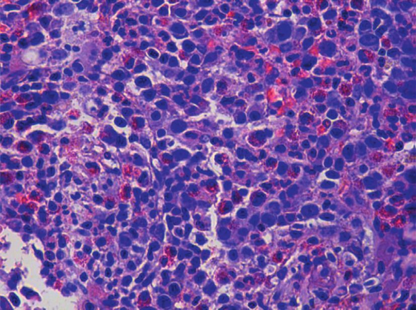
2 Case Reports in Hematology
Table 1: Laboratory values.
Laboratory analysis Patient’s values on initial hospitalization Patient’s values on second hospitalization Normal range
Hemoglobin 5.1 g/dl 7.4 g/dl 13–17
MCV 97 fl 90.4 fl 82–99
MCHC 31.5 g/dl 32.9 g/dl 31–37
WBC count 6 k/μL 5.1 k/μL 4–10
Platelet count 91 k/μL 90 k/μL 150–400
Absolute eosinophil count 2 k/μL 2 k/μL
Neutrophil 30% 30.8% 40–80
Lymphocyte 24% 23.5% 15–45
Eosinophil 33.5% 33.3% 0–6
Basophil 0% 0% 0–2
Glucose 140 mg/dl 117 mg/dl 70–99
BUN 16 mg/dl 14 mg/dl 7–21
Creatinine 0.87 mg/dl 0.83 mg/dl 0.67–1.17
AST 16 U/L 96 U/L 10–37
ALT 22 U/L 55 U/L 10–65
Alkaline phosphatase 475 U/L 130 U/L 50–136
Total bilirubin 0.8 mg/dl 21.3 mg/dl (direct 16.7 mg/dl) 0–1
Stool occult blood Negative
Haptoglobulin 214 mg/dl 30–200
Reticulocyte % 0.6 0.5–2.5
LDH 147 U/L 84–246
PT 12.8 s 27 s 9–12
INR 1.2 0.9–1.1
aPTT 33.1 s 77 s 23–34
Iron 202 μg/dl 65–175
TIBC 203 μg/dl 250–450
Iron saturation 100% 10–50
Ferritin 949.9 ng/ml 26–388
Vitamin B12 1680 pg/ml 193–986
Folate 19.6 ng/ml 8.7–55.4
TSH 1.48 uIU/ml 0.358–3.74
Fibrinogen 410
SPEP—protein 6.8 g/dl 6.4–8.2
Albumin 2.8 g/dl 1.3 g/dl 3.5–5.0
Alpha-1 globulin 0.5 g/dl 0.2–0.4
Alpha-2 globulin 1.0 g/dl 0.5–1.0
Beta globulin 0.7 g/dl 0.5–1.1
Gamma globulin 1.8 g/dl 0.6–1.5
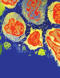
with increased number of neutrophils and eosinophils.
Erythropoiesis was markedly decreased with only very rare
proerythroblasts present (Figure 1). Megakaryocytosis with
dysmegakaryopoiesis was also appreciated. Several peri-
vascular fibrotic areas containing mast cell aggregates were
also identified (Figures 2 and 3). The mast cells were positive
for mast cell tryptase and aberrant expression of CD2 and
CD25 (Figures 4 and 5). c-KIT and D 816 V mutations were
detected. The above findings were suggestive of ASM. FISH,
cytogenetic, and flow cytometric analyses were unrevealing.
Parvovirus immunostain was negative.
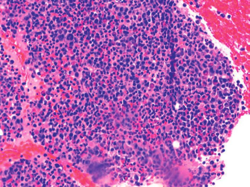
Figure 1: Area of hypercellular bone marrow with red cell aplasia.
2.3. Treatment. After the diagnosis of ASM and PRCA, the
patient left our hospital against medical advice. He became
completely transfusion-dependent and was receiving weekly social support to return to hospital for treatment. Peripheral
red cell transfusions from a nearby community hospital. He smear revealed normochromic normocytic anemia with
returned to our hospital after 5 weeks with worsening occasional ovalocyte, rare target cell, and dacrocyte. No
anemia, thrombocytopenia, liver function tests, and coa- nucleated red blood cell was identified. Granulocytes appeared
gulopathy. The patient reported lack of transportation and mature without abnormal granulation or segmentation.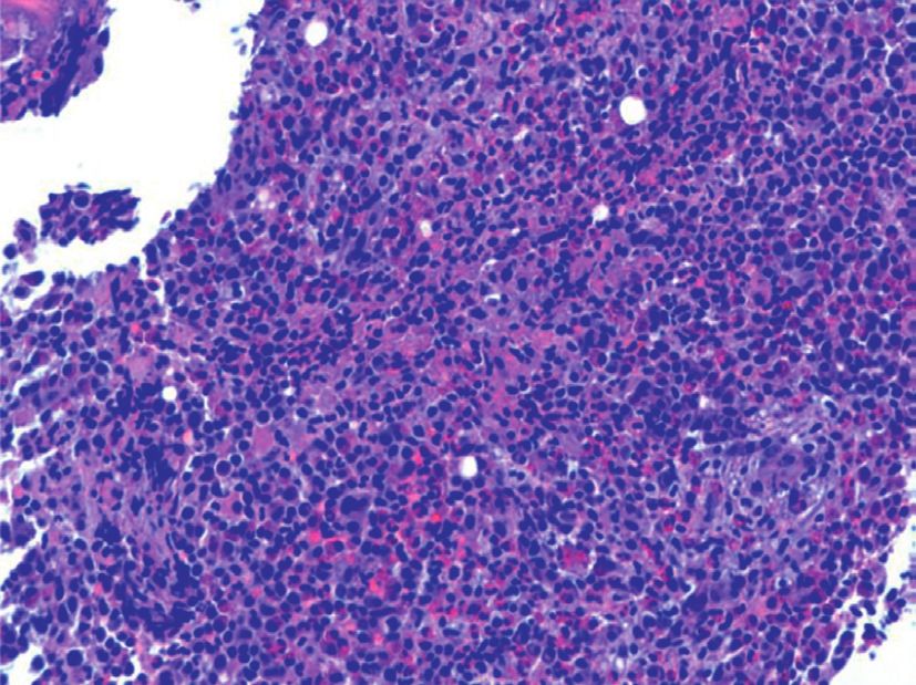
Case Reports in Hematology                                                                                                       3

                                                                    Eosinophils were increased (30% with an absolute number
                                                                    of 1400/mcL) without abnormal features. Monocytes were
                                                                    increased to 12.9% without absolute monocytosis. Imaging
                                                                    studies (ultrasound and CT scan of abdomen) revealed
                                                                    hepatomegaly (liver 19.7 cm in length) and splenomegaly
                                                                    (spleen 19.1 cm in length) with multiple retroperitoneal
                                                                    lymph nodes.
                                                                         With the patient’s prior history of depression and ongoing
                                                                    thrombocytopenia, interferon alfa and 2-chlorodeoxyadenosine
                                                                    were not recommended for treatment of ASM. New avenues
                                                                    of treatments were discussed. Since the mast cells were CD30+,
                                                                    brentuximab vedotin was administered at a dose of 1.8 mg/kg.
Figure 2: Bone marrow with hypercellularity and increased mast      After therapy, patient developed worsening neutropenia,
cells.
                                                                    despite filgrastim and worsening anemia, and thrombocy-
                                                                    topenia. The patient also developed Gram-negative bacteremia
                                                                    secondary to a urinary tract infection and became hypotensive
                                                                    and hypoxemic with lactic acidosis. The patient died 2 weeks
                                                                    later in the intensive care unit. Our patient lived for a short
                                                                    time after diagnosis of ASM; hence, there was not enough
                                                                    time for many sequential therapies. He did receive high-
                                                                    dose steroids before and concurrently with brentuximab
                                                                    therapy.

                                                                    2.4. Outcome. The patient deceased within 2 months of
                                                                    initial diagnosis.

Figure 3: Clot section which shows hypercellularity and increased   3. Discussion
mast cells.
                                                                    Aggressive systemic mastocytosis is an uncommon disorder
                                                                    characterized by neoplastic mast cell accumulation in var-
                                                                    ious organs. The skeletal system, bone marrow, gastroin-
                                                                    testinal tract, and spleen are commonly involved. Mast cell
                                                                    infiltration of bone marrow leads to cytopenias, the so-called
                                                                    “C” findings [5, 6]. Systemic mastocytosis patients have poor
                                                                    prognosis because of multiorgan involvement and dys-
                                                                    function [7].
                                                                        Our patient presented with systemic symptoms and
                                                                    severe anemia, workup of which led to diagnosis of un-
                                                                    derlying systemic mastocytosis and pure red cell aplasia. The
                                                                    diagnosis of ASM was made by a constellation of clinical,
                                                                    cytogenetic, and molecular analyses [8]. Pure red cell aplasia
                                                                    was confirmed by normochromic anemia with very low
Figure 4: Clot stained with CD2 which is positive in mast cells.    reticulocyte percentage in presence of normal white cell and
                                                                    platelet counts, along with the finding of cellular marrow
                                                                    that revealed normal myelopoiesis, lymphopoiesis, and
                                                                    megakaryocytopoiesis, but very rare, if any erythroid pre-
                                                                    cursors [9]. The association of mast cell disorder with pure
                                                                    red cell aplasia is rare and has been described only once in
                                                                    the literature [4].
                                                                        Our patient had first exposure to Agent Orange 45 years
                                                                    earlier and then for the next three years. Agent Orange is
                                                                    a mixture of two chemicals that are phenoxy herbicides: 2,4
                                                                    dichlorophenoxyacetic acid and 2,4,5 trichlorophenoxy-
                                                                    acetic acid (2,4,5 T). The 2,4,5 T in Agent Orange was
                                                                    contaminated with small amount of dioxins. The main di-
                                                                    oxin involved was 2,3,7,8-tetrachlorobenzo-p-dioxin or
                                                                    TCBD, which is one of the most toxic of dioxins and is
Figure 5: Clot stained with CD25 which is positive in mast cells.   classified as a human carcinogen by the U.S. Environmental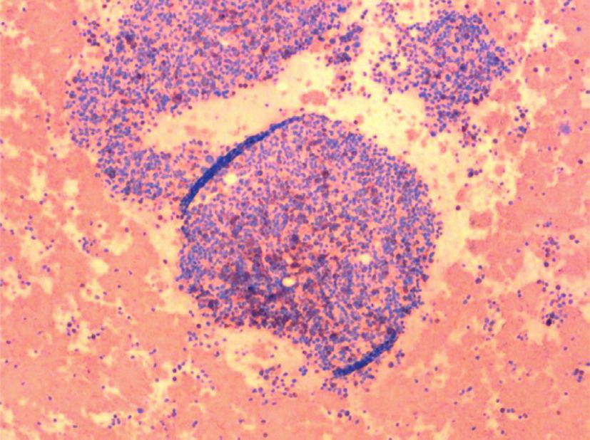
4                                                                                               Case Reports in Hematology

Protection Agency. In the Vietnam War, between 1962 and             The favoured diagnosis of bone marrow was ASM. The
1971, the US military sprayed these herbicides. The Centre      interpretation was supported by the presence of “C” findings
for Disease Control and Prevention notes that, in particular,   including cytopenia indicating bone marrow dysfunction, in
there are increased number of cases of acute/chronic leu-       association with elevated liver enzymes suggesting liver
kemias, Hodgkin’s and non-Hodgkin’s lymphomas, head             damage and splenomegaly probably associated with
and neck cancers, prostate cancer, lung/colon cancers, and      hypersplenism. The patient also had diarrhea on pre-
soft tissue sarcomas occurring in the exposed population.       sentation. As an infiltrative process, a proliferation of mast
Other reports have included multiple myeloma, AL amy-           cells in the intestinal submucosa causes malabsorption.
loidosis, and other benign hematologic changes like anemia,     Release of histamine, both locally and systemically, other
leukopenia, and thrombocytopenia. Role of Agent Orange          peptides, proteases, and generation of excessive quantities of
exposure in cytopenia or systemic mastocytosis in our pa-       mediators, such as prostaglandin D2, leukotriene C4, and
tient remains of concern but cannot be stated with certainty.   platelet-activating factor are likely to alter gastrointestinal
The complete blood count 2 years before diagnosis of ASM        function and motility.
was normal in our patient.                                          The patient presented with rapidly accumulating, ex-
    The etiology of anemia in our patient was probably          tremely high iron saturation, and raised ferritin level, with
multifactorial: anemia of chronic disease/malignancy, bone      negative mutation in HFE gene, which included C282 Y and
marrow mastocytosis, splenomegaly, and pure red cell            H63D. Of note, iron saturation and ferritin levels were
aplasia. Myelodysplastic syndrome (MDS) and autoimmune          normal 4 months before diagnosis of ASM. Other causes of
hemolytic anemia contributing to the patient’s anemia could     iron overload included ineffective erythropoiesis seen in
not completely be excluded. Serum protein electrophoresis       MDS/sideroblastic anemia, thalassemia, and congenital or
was negative for monoclonal gammopathy, and quantitative        acquired hemolytic anemia associated with multiple trans-
measurements of IgG, IgA, and IgM were normal. Direct           fusions. These conditions were unlikely in our patient be-
antiglobulin test was not performed initially. Despite fre-     cause of the short time course for iron accumulation. A
quent red cell transfusions, no crossmatching difficulties        number of acute and chronic liver diseases causing liver
were reported by the blood bank. But, 3 weeks prior to the      inflammation can release stored iron into circulation raising
patient’s demise, the blood bank reported difficulty in           serum ferritin [12]. One such inflammatory condition is
crossmatching for compatible red cells. A direct antiglobulin   hemophagocytosis syndrome (HPS). It is an extremely lethal
test performed then detected an IgG-negative complement-        condition in which excessive activation of immunity leads to
mediated positive test. The serum erythropoietin level was      tissue destruction. This condition was unlikely in our patient
elevated at 1234 IU/L. High-dose methylprednisone 80–           as the ferritin levels are usually over 5000 ng/ml in hemo-
100 mg IV was initiated over the next week. In view of the      phagocytosis syndrome, whereas it was below 1000 ng/ml in
severe reticulocytopenia and rising liver enzymes, coagul-      our patient. The other feature of HPS patients is the acuity of
opathy as a result of hepatic involvement of ASM, response      illness with multiorgan involvement. Though our patient
to steroids could not be determined. The patient remained       had multiorgan involvement from ASM, the cardinal fea-
transfusion-dependent and developed progressively severe        tures of HPS such as fever or rheumatologic symptoms were
pancytopenia.                                                   lacking, making the diagnosis unlikely in our patient. Ma-
    Similarly, the etiology of thrombocytopenia was also        lignancy can also be associated with elevated ferritin as
multifactorial: infiltrating mast cells in the bone marrow and   suggested by a clinical trial published in 2015 [13]. Our
splenic sequestration. Increased megakaryopoiesis as well as    patient had both an underlying malignancy and liver injury,
dysmegakaryopoiesis in bone marrow raise the possibility of     the latter most likely contributing to the increased ferritin
immune thrombocytopenia and MDS, respectively. MDS              levels.
mutational analysis and FISH was performed to evaluate for          Management of any form of mastocytosis involves 3
critical regions in myelodysplastic syndrome which included     different strategies: (a) general measures to prevent ana-
deletion of 5q31 and 7q31, enumeration of chromosome 8,         phylaxis, (b) antihistamine (cetirizine, hydroxyzine, and
and deletion of long arm of chromosome 20. The studies          doxepin) and antileukotriene therapy (montelukast and
were negative for all four regions, and JAK 2 study was also    zileuton) to treat symptoms associated with mast cell me-
negative. Hence, in absence of abnormal cytogenetics, FISH,     diator release, and (c) cytoreductive therapy for advanced
and flow cytometric studies, the diagnosis of MDS could not      disease [10, 14, 15]. Midostaurin is a KIT inhibitor, first-line
be confirmed. Flow cytometry did not demonstrate any             agent used in advanced disease, regardless of KIT mutation
increase in the blasts or immature cells. Immunostain for       status [16]. Our patient did not receive the drug as it was
CD34 was positive only in rare cells in the bone marrow.        approved by FDA only recently (April 2017). Tyrosine kinase
FISH analysis for t(9 : 22) BCR/ABL 1 translocation,            inhibitors (TKI) such as imatinib have been used in ASM
PDGFRA (4q12), FGFR 1(t 8 : 11), and PDGRB (5q33)               patients who do not have D816V mutation [17]. Our patient
rearrangement was negative. Mutational analysis for MDS         did express the D816V mutation and hence did not qualify
detected mutation of ASXL1 and EZH2 genes. These genes          for TKI therapy. At that time, the available cytoreductive
are not specific for MDS and have been reported in many          therapies were interferon alfa and 2-CDA (chlorodeox-
myeloid disorders as well as in ASM with unfavourable           yadenosine). These drugs tend to have significant side effects
prognosis [10, 11]. Mutations in IDH1, IDH2, KRAS, NRAS,        with response lasting for short duration. This has led to an
and TET 2 were negative.                                        increasing need for novel agents with longer response and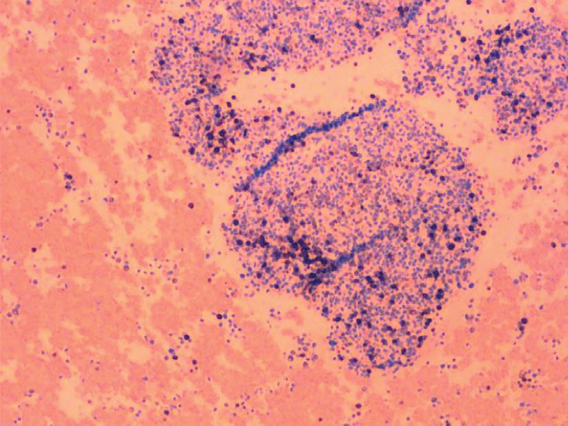
Case Reports in Hematology                                                                                                                  5

fewer side effects. Another potential therapeutic target CD30            [10] M. Jawhar, J. Schwaab, S. Schnittger et al., “Additional mu-
(Ki-1) antigen was identified in patients with advanced                       tations in SRSF2, ASXL1 and/or RUNX1 identify a high-risk
ASM, and brentuximab vedotin has been used as an alter-                      group of patients with KIT D816V+ advanced systemic
native therapy. Our patient received the same [18, 19].                      mastocytosis,” Leukemia, vol. 30, no. 1, p. 136, 2016.
Though brentuximab vedotin has a better safety profile, our              [11] F. Traina, V. Visconte, A. M. Jankowska et al., “Single nu-
                                                                             cleotide polymorphism array lesions, TET2, DNMT3A,
patient was unable to tolerate even a single dose.
                                                                             ASXL1 and CBL mutations are present in systemic masto-
     Cladribine is indicated in patients with rapidly pro-                   cytosis,” PloS One, vol. 7, no. 8, Article ID e43090, 2012.
gressive mastocytosis for rapid debulking and those who                 [12] D. Karam, S. Swiatkowski, P. Purohit, and B. Agrawal, “High-
failed to respond to midostaurin or TKI. Hydroxyurea is also                 dose steroids as a therapeutic option in the management of
used in ASM patients, especially those with leukocytosis                     spur cell haemolytic anaemia,” BMJ Case Reports, vol. 2018,
and/or splenomegaly-associated myeloproliferative neo-                       2018.
plasms [20, 21]. None of the above therapies could be ini-              [13] A. M. Schram, F. Campigotto, A. Mullally et al., “Marked
tiated in our patient due to worsening general condition                     hyperferritinemia does not predict for HLH in the adult
after brentuximab. Hematopoietic stem cell transplant is the                 population,” Blood, vol. 125, no. 10, pp. 1548–1552, 2015.
only curative option, though it is typically performed in               [14] J. Turk, J. A. Oates, and L. J. Roberts, “Intervention with
                                                                             epinephrine in hypotension associated with mastocytosis,”
younger adults [22]. PRCA, with or without ASM, is gen-
                                                                             Journal of Allergy and Clinical Immunology, vol. 71, no. 2,
erally managed with regular transfusions for anemia, fol-                    pp. 189–192, 1983.
lowed by immunosuppressive or cytotoxic therapy [23].                   [15] A. S. Worobec, “Treatment of systemic mast cell disorders,”
                                                                             Hematology/Oncology Clinics of North America, vol. 14, no. 3,
Data Availability                                                            pp. 659–687, 2000.
                                                                        [16] J. Gotlib, H. C. Kluin-Nelemans, T. I. George et al., “Efficacy
The published data used to support the findings of this study                 and safety of midostaurin in advanced systemic mastocytosis,”
(case report) are included within the article.                               New England Journal of Medicine, vol. 374, no. 26,
                                                                             pp. 2530–2541, 2016.
                                                                        [17] A. Vega-Ruiz, J. E. Cortes, M. Sever et al., “Phase II study of
Conflicts of Interest                                                        imatinib mesylate as therapy for patients with systemic
The authors declare that they have no conflicts of interest.                  mastocytosis,” Leukemia Research, vol. 33, no. 11, pp. 1481–
                                                                             1484, 2009.
                                                                        [18] A. Mehta, V. V. Reddy, and U. Borate, “Anti CD-30 antibody-
References                                                                   drug conjugate brentuximab vedotin (ADCETRIS®) may be
                                                                             a promising treatment option for systemic mastocytosis
 [1] D. D. Metcalfe, “Classification and diagnosis of mastocytosis:           (SM),” Blood, vol. 120, p. 2857, 2012.
     current status,” Journal of Investigative Dermatology, vol. 96,    [19] U. Borate, A. Mehta, V. Reddy, M. Tsai, N. Josephson, and
     no. 3, pp. S2–S4, 1991.                                                 I. Schnadig, “Treatment of CD30-positive systemic masto-
 [2] D. A. Arber, A. Orazi, R. Hasserjian et al., “Classification of          cytosis with brentuximab vedotin,” Leukemia Research,
     mastocytosis: a consensus proposal,” Leukemia Research,                 vol. 44, pp. 25–31, 2016.
     vol. 25, no. 7, pp. 603–625, 2001.                                 [20] K. H. Lim, A. Pardanani, J. H. Butterfield, C. Y. Li, and
 [3] J. W. Vardiman, “The 2016 revision to the World Health                  A. Tefferi, “Cytoreductive therapy in 108 adults with systemic
     Organization (WHO) classification of myeloid neoplasms and               mastocytosis: outcome analysis and response prediction
     acute leukemia,” Blood, vol. 127, no. 20, pp. 2391–2405, 2016.          during treatment with interferon-alpha, hydroxyurea, ima-
 [4] L. B. Afrin, “Mast cell activation disorder masquerading as             tinib mesylate or 2-chlorodeoxyadenosine,” American Journal
     pure red cell aplasia,” International Journal of Hematology,            of Hematology, vol. 84, no. 12, pp. 790–794, 2009.
     vol. 91, no. 5, pp. 907-908, 2010.                                 [21] A. Tefferi, “Treatment of systemic mast cell disease: beyond
 [5] P. Valent, C. Akin, W. R. Sperr et al., “Aggressive systemic            interferon,” Leukemia Research, vol. 28, no. 3, pp. 223-224,
     mastocytosis and related mast cell disorders: current treat-            2004.
     ment options and proposed response criteria,” Leukemia             [22] C. Ustun, A. Reiter, B. L. Scott et al., “Hematopoietic stem-cell
     Research, vol. 27, no. 7, pp. 635–641, 2003.                            transplantation for advanced systemic mastocytosis,” Journal
 [6] A. W. Hauswirth, I. Simonitsch-Klupp, M. Uffmann et al.,                 of Clinical Oncology, vol. 32, no. 29, pp. 3264–3274, 2014.
     “Response to therapy with interferon alpha-2b and pred-            [23] K. Sawada, M. Hirokawa, and N. Fujishima, “Diagnosis and
     nisolone in aggressive systemic mastocytosis: report of five             management of acquired pure red cell aplasia,” Hematology/
     cases and review of the literature,” Leukemia Research, vol. 28,        Oncology Clinics of North America, vol. 23, no. 2, pp. 249–259,
     no. 3, pp. 249–257, 2004.                                               2009.
 [7] L. Afrin, “Presentation, diagnosis, and management of mast
     cell activation syndrome,” in Mast Cells: Phenotypic Features,
     Biological Functions, and Role in Immunity, D. Murray, Ed.,
     Nova Science Publishers, Happauge, NY, USA, 2013.
 [8] L. B. Schwartz and A. M. Irani, “Serum tryptase and the
     laboratory diagnosis of systemic mastocytosis,” Hematology/
     Oncology Clinics of North America, vol. 14, no. 3, pp. 641–657,
     2000.
 [9] K. Sawada, N. Fujishima, and M. Hirokawa, “Acquired pure
     red cell aplasia: updated review of treatment,” British Journal
     of Haematology, vol. 142, no. 4, pp. 505–514, 2008.
MEDIATORS         of

                                                                                   INFLAMMATION

The Scientific                                 Gastroenterology                                                              Journal of
World Journal
Hindawi Publishing Corporation
                                               Research and Practice
                                               Hindawi
                                                                                   Hindawi
                                                                                                                             Diabetes Research
                                                                                                                             Hindawi
                                                                                                                                                              Disease Markers
                                                                                                                                                              Hindawi
                                                                                   www.hindawi.com        Volume 2018
http://www.hindawi.com
www.hindawi.com                  Volume 2018
                                        2013   www.hindawi.com   Volume 2018                                                 www.hindawi.com    Volume 2018   www.hindawi.com            Volume 2018

Journal of                                                                                                                                                    International Journal of
Immunology Research
Hindawi
                                                                                                                                                              Endocrinology
                                                                                                                                                              Hindawi
www.hindawi.com                  Volume 2018                                                                                                                  www.hindawi.com            Volume 2018

                                                                 Submit your manuscripts at
                                                                    www.hindawi.com

                                                                                                                                                              BioMed
PPAR Research
Hindawi
                                                                                                                                                              Research International
                                                                                                                                                              Hindawi
www.hindawi.com                  Volume 2018                                                                                                                  www.hindawi.com            Volume 2018

                                                                                                                             Journal of
                                                                                                                             Obesity

                                                                               Evidence-Based
Journal of                                     Stem Cells                      Complementary and                                                              Journal of
Ophthalmology
Hindawi
                                               International
                                               Hindawi
                                                                               Alternative Medicine
                                                                               Hindawi                                       Hindawi
                                                                                                                                                              Oncology
                                                                                                                                                              Hindawi
www.hindawi.com                  Volume 2018   www.hindawi.com   Volume 2018   www.hindawi.com                 Volume 2018   www.hindawi.com    Volume 2018   www.hindawi.com            Volume 2013

                                                                               Parkinson’s
                                                                               Disease

Computational and
Mathematical Methods
in Medicine
                                               Behavioural
                                               Neurology
                                                                                                                             AIDS
                                                                                                                             Research and Treatment
                                                                                                                                                              Oxidative Medicine and
                                                                                                                                                              Cellular Longevity
Hindawi                                        Hindawi                         Hindawi                                       Hindawi                          Hindawi
www.hindawi.com                  Volume 2018   www.hindawi.com   Volume 2018   www.hindawi.com                 Volume 2018   www.hindawi.com    Volume 2018   www.hindawi.com            Volume 2018

You can also read