Identification of the Immune-Related Genes in Tumor Microenvironment That Associated With the Recurrence of Head and Neck Squamous Cell Carcinoma
←
→
Page content transcription
If your browser does not render page correctly, please read the page content below
ORIGINAL RESEARCH
                                                                                                                                             published: 20 August 2021
                                                                                                                                        doi: 10.3389/fcell.2021.723721

                                            Identification of the Immune-Related
                                            Genes in Tumor Microenvironment
                                            That Associated With the Recurrence
                                            of Head and Neck Squamous Cell
                                            Carcinoma
                            Edited by:
                                            Liu Chengcheng 1 , Qi Wenwen 2,3 , Gong Ningyue 2 , Zhu Fangyuan 2 , Xu Runtong 2 ,
                                Na Luo,     Teng Zhenxiao 2 , Xu Fenglei 2 , Qin Yiming 4 , Zhao Miaoqing 5* † , Li Xiaoming 2* † and
                Nankai University, China    Xia Ming 2* †
                         Reviewed by:       1
                                             Department of Central Laboratory, Shandong Provincial Hospital Affiliated to Shandong First Medical University, Jinan,
                               Xingyi Li,   China, 2 Department of Otolaryngology, Shandong Provincial Hospital Affiliated to Shandong First Medical University, Jinan,
    Affiliated Eye Hospital of Wenzhou      China, 3 Cheeloo College of Medicine, Shandong Provincial Hospital, Shandong University, Jinan, China, 4 College
                 Medical College, China     of Chemistry, Chemical Engineering and Materials Science, Shandong Normal University, Jinan, China, 5 Department
                                  Lei Li,   of Pathology, Shandong Provincial Hospital Affiliated to Shandong First Medical University, Jinan, China
       Xi’an Jiaotong University, China
                   *Correspondence:
                        Zhao Miaoqing
                                            Head and neck squamous cell carcinomas (HNSCC) are still one of the most common
                  zhaomqsd@163.com          malignant tumors in China, with a high metastasis rate and poor prognosis. The tumor
                           Li Xiaoming
                                            immune microenvironment can affect the occurrence, development and prognosis of
                   lxmfmmu@sina.com
                              Xia Ming      tumors, but the underlying mechanism is still unclear. In this study, we tried to describe
                xiamingsdu@sohu.com         the correlation between the recurrence of HNSCC and the tumor microenvironment
     † These   authors have contributed     (TME). The expression data [estimate the level of tumor stromal and immune infiltration,
                    equally to this work
                                            expression data (ESTIMATE)] algorithm was used to identify and estimate highly
                    Specialty section:      correlated stromal cells, immune cells, and prognostic scores in 116 samples of
         This article was submitted to      head and neck cancer patients from The Cancer Genome Atlas (TCGA) dataset.
    Molecular and Cellular Pathology,
                a section of the journal    The functional enrichment analysis and protein-protein interaction (PPI) networks of
  Frontiers in Cell and Developmental       differential expressed genes (DEGs) were constructed. Subsequently, the abundance
                                Biology
                                            of various infiltrating immune cells was estimated with the tumor immune estimation
            Received: 11 June 2021
                                            resource (TIMER) and the infiltration pattern of immune cells were explored in HNSCC.
          Accepted: 03 August 2021
          Published: 20 August 2021         A total of 407 immune-related genes were identified to involve in the TME. We found that
                              Citation:     CCR5, CD3E, CD4, and HLA -DRB1 were the most obvious DEGs and the dendritic
         Chengcheng L, Wenwen Q,            cells (DCs) showed the highest abundance in the TME of HNSCC. In addition, the
 Ningyue G, Fangyuan Z, Runtong X,
     Zhenxiao T, Fenglei X, Yiming Q,       unsupervised cluster analysis determined 10 clusters of immune infiltration patterns, and
  Miaoqing Z, Xiaoming L and Ming X         indicated that immune infiltrated CD4 + T and B cells may be related to the prognosis of
           (2021) Identification of the
                                            HNSCC. In conclusion, our research determined the list of immune genes and immune
    Immune-Related Genes in Tumor
  Microenvironment That Associated          infiltrating cells related to the prognosis of HNSCC, and provided a perspective for
        With the Recurrence of Head         HNSCC evolution, anti-tumor drugs selection, and drug resistance research.
and Neck Squamous Cell Carcinoma.
      Front. Cell Dev. Biol. 9:723721.      Keywords: head and neck squamous cell carcinoma, recurrence, immune score, stromal score, immune
     doi: 10.3389/fcell.2021.723721         infiltrating

Frontiers in Cell and Developmental Biology | www.frontiersin.org                  1                                            August 2021 | Volume 9 | Article 723721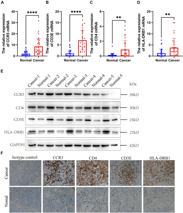
Chengcheng et al.                                                                                         Immune-Related Genes in HNSCC Tumor

INTRODUCTION                                                              the survival period, but also the immunotherapy response of
                                                                          different patient groups.
Head and neck squamous cell carcinomas (HNSCC), mainly                       In this study, we constructed an immune-related prognostic
including oral cavity, oropharyngeal, laryngopharyngeal,                  feature with patient information from The Cancer Genome
laryngeal cancers, and hypopharyngeal, are the sixth most                 Atlas (TCGA) data set, and further verified its prognostic value.
common malignant tumors with a 5-year overall survival                    In addition, we also determined the relationship between the
rate of less than 50% (Kanazawa et al., 2019; McDermott and               genetic characteristics, TME, and the prognosis of patients with
Bowles, 2019). These dismal malignant tumors affect more than             recurrent HNSCC. We further obtained four new prognostic
6,00,000 patients worldwide annually, and nearly half of the              molecular biomarkers, which was closely related to the immune
newly diagnosed cases are found at an advanced stage (Brenner             microenvironment of HNSCC. Finally, we verified the expression
et al., 2014). Though significant development in multimodal               of these four genes in tumors in a cohort study. The purpose
therapeutic strategies, including surgery, radiotherapy,                  of this study is to provide a new immune-related molecular
chemotherapy, immunotherapy, and combined treatment,                      biomarker of HNSCC, which can more effectively predict the
the 5-year survival rate of HNSCC has improved slightly in                prognosis of HNSCC patients and is closely related to the
the past decade due to the resistant nature of tumor cells to             immune microenvironment.
chemoradiotherapy (Paver et al., 2020). Recently, emerging
genomic evidence indicates that significant immunosuppression
and heterogeneous characteristics of HNSCC play vital roles               MATERIALS AND METHODS
in disease treatment and recurrence (Bhat et al., 2021).
However, the detailed roles and underlying mechanisms of                  Reagants
tumor heterogeneous on the relapse of HNSCC have not been                 The rabbit polyclonal to CCR5 (Cat. No. ab7346), rabbit
fully understood.                                                         monoclonal to CD3 epsilon (Cat. No. ab237721), and CD4
   Disturbance of the tumor microenvironment (TME) is one                 (Cat. No. ab183685) were purchased from abcam (Cambridge,
of the key causes of local immune dysfunction. It establishes a           United Kingdom). The mouse monoclonal to GAPDH (Cat.
stable ecological environment and space for cancer cells, a variety       No. ab8245) and HLA-DRB1 (Cat. No. ab215835) were also
of endothelial cells, immune cells, and extracellular components          purchased from abcam (Cambridge, United Kingdom).
(Hanahan and Coussens, 2012; Quail and Joyce, 2013). More
and more evidences show that TME is crucial in the occurrence             Gene Expression Data Extraction
and even recurrence of HNSCC. It has an adverse effect on the             The Cancer Genome Atlas dataset is downloaded from UCSC
prognosis by promoting the evolution of aggressive HNSCC and              Xena.1 RNA sequencing (RNA-seq) data is downloaded from
resistance to chemo- or radio-treatment (Sharma et al., 2017).            the TCGA data portal. Then convert the FPKM value of the
An in-depth understanding of the influencing factors of TME               fragment to the value of TPM, including tumor grade and
and the highly complex tumor-stromal interactions can promote             survival information. Our study included 116 tumor samples
the discovery of new therapeutic interventions for HNSCC.                 (Larynx HNSC) with complete prognostic information. We
Therefore, identifying the immune genes and active types of               defined patients with new tumors new tumor events occurring
immune cells related to TME can help to clarify the general               after surgery to be recurrent HNSCC. Supplementary Table 1
mechanism of HNSCC immunosuppression.                                     summarizes the characteristics of the patients in the training and
   Recent studies show that the regulation of the immune system           validation cohort.
played an important role in the prognosis of cancer patients,
and tumor molecular profiling could help to predict clinical
outcomes and determine the targets for treating tumors (Michot
                                                                          Calculation of Stromal and Immune
et al., 2016; Miyauchi et al., 2019; Bruni et al., 2020). It also         Score
revealed the prognostic characteristics of some genes in detail,          The expression data [estimate the level of tumor stromal and
which played an important role in the survival prediction of              immune infiltration, expression data (ESTIMATE)] analysis by
HNSCC (Zhou et al., 2019). Recently, researchers have identified          the “estimate” R software package was used to evaluate the
some important immune checkpoint components that promote                  stromal and immune scores in stromal cells and immune cells in
the tumor cells to evade from the immune surveillance, such               recurrent HNSCC tissues (Van Landuyt et al., 2011). According
as cytotoxic T lymphocyte-associated protein 4 (CTLA-4) and               to this algorithm, the tumor purity is calculated.
programmed cell death protein 1 (PD-1)/programmed cells
Death ligand 1 (PD-L1) (Brunner-Weinzierl and Rudd, 2018;                 Differential Expression Analysis
Lee et al., 2019). Subsequently, novel strategy for cancer therapy        According to the ESTIMATE analysis, we divided all patients
of the checkpoint immunosuppression was applied for multiple              into a high/low immune score group and a high/low stromal
malignant tumors, including HNSCC. However, checkpoint                    score group. Then, by using the “limma” package to analyze
blocking immunotherapy is not effective for all patients, and the         the differential expression between different immune groups, the
observed effective rate is only in the range of 16–25% (Medina            selection criteria are log2|fold change| greater than 1 and adj.p
and Adams, 2016). Therefore, there is an urgent need for an
immune-related prognostic feature that can predict not only               1
                                                                              https://xenabrowser.net/

Frontiers in Cell and Developmental Biology | www.frontiersin.org     2                                    August 2021 | Volume 9 | Article 723721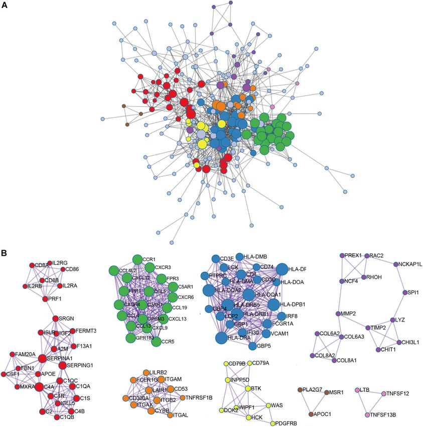
Chengcheng et al.                                                                                          Immune-Related Genes in HNSCC Tumor

less than 0.05. The corresponding heat maps and cluster maps              Declaration of Helsinki. The tumor and control tissues are
were generated using the “pheatmap” package.                              collected in RNase-free eppendorf tubes, and stored in −80◦ C.
                                                                          Then total RNA was isolated according to the manufacturer’s
Survival Analysis                                                         protocol (RNeasy Mini Kit, Qiagen). The extracted RNA was then
Kaplan–Meier analysis was performed to screen HNSCC with                  reverse transcribed using Superscript R reverse transcriptase.
recurrent characteristics. The survival curves were also applied to       Quantitative real time-PCR (qRT-PCR) was performed using
illustrate the relationship between the expression levels of these        cDNA as a template with Platinum SYBR Green (Invitrogen). To
genes and the recurrence of HNSCC.                                        obtain the best sensitivity and specificity of the primers, the cycle
                                                                          length of different PCR groups was adjusted to 40 cycles, and the
Gene Ontology and Kyoto Encyclopedia                                      annealing temperature was adjusted between 56 and 64◦ C. To
                                                                          analyze the qRT-PCR results, the 2-11Ct method was used, and
of Genes and Genomes Analysis
                                                                          β-actin served as a housekeeping gene. All PCRs were performed
Functional enrichment analysis was performed by the
                                                                          in triplicate. Specific primer sequences are recorded below:
“clusterProfiler” package to determine the functions and
                                                                          CCR5-forward        primer     (AGGAATCATCTTTACCAGAT),
pathways of the screened genes (Yu et al., 2012). Functional
                                                                          reverse primer (ATGACAAGCAGCGGCA); CD3E-forward
enhancements to gene ontology (GO) terms include cell
                                                                          primer (TCCCAACCCAGACTATGAGC), reverse primer
component (CC), biological processes (BPs), molecular function
                                                                          (CAAGACTAGCCCAGGAAACAG); CD4-forward primer
categories (MF), and the Kyoto Encyclopedia of Genes and
                                                                          (AGTCCAAGGGGTAAAAACAT), reverse primer (AAGGAG
Genomes (KEGG) approach in the Kyoto Protocol. The cut-off
                                                                          AACTCCACCTGTTC); HLA-DRB1-forward primer (AGAG
value was false discovery rate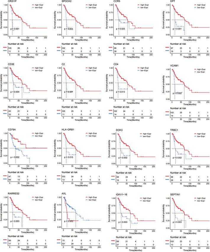
Chengcheng et al.                                                                                                         Immune-Related Genes in HNSCC Tumor

was applied for heat map generation and visualization. For                           Identification of Immune-Related Genes
multiple groups, one-way analysis of variance (ANOVA) was                            in Recurrent HNSCC
selected as the parameter method for comparing means, and
                                                                                     All patients were divided into a high or low immune score
the Kruskal–Wallis test analysis was performed by the non-
                                                                                     group and a high or low stroma score group according to the
parametric method. The Wilcoxon test was used to compare non-
                                                                                     results of ESTIMATE analysis. Subsequently, gene differential
normally distributed variables. The unpaired Student t-test was
                                                                                     expression analysis was performed to identify the DEGs between
used to compare normally distributed variables. The Shapiro–
                                                                                     the high or low groups of immune score and stroma score.
Wilk normality test was used to test the normality of the variables.
                                                                                     The heat map shows the gene expression pattern of DEGs
All tests are two-tailed tests, and a P-value of less than 0.05 was
                                                                                     between the high and low immune score or stroma score groups
considered statistically significance.
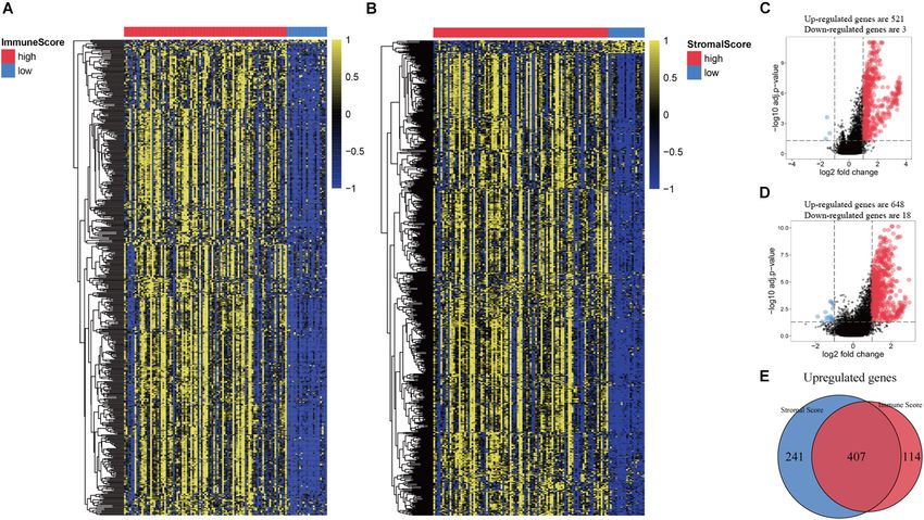
                                                                                     (Figures 2A,B). Then, gene expression levels in the high and
                                                                                     low immune score groups were compared. There was 521 up-
                                                                                     regulated genes and 3 down-regulated genes in the high immune
RESULTS
                                                                                     score group (Figure 2C). Similarly, 648 genes were up-regulated
                                                                                     and 18 genes were down-regulated in the high stroma score
Association of Immune and Stromal                                                    group (Figure 2D). As shown in the Venn diagram (Figure 2E),
Scores With Pathological Characteristics                                             we selected the up-regulated overlapping genes as candidate
of Recurrent HNSCC                                                                   immune-related genes (407 up-regulated genes), because there is
A total of 116 eligible HNSCC data, including clinicopathological                    no overlap of the down-regulated genes in the immune group
and prognostic informations, were extracted from the TCGA                            and the stroma group. These crossover genes will be used for
database. In general, 65.5% of the patients were over 60 years old,                  subsequent analysis.
43.1% were alive, and 75.9% were in stage III/IV at the time of
diagnosis (Supplementary Table 1). Using ESTIMATE analysis,                          Module-Related Central Gene Network
immune score and stroma score were performed according to                            In order to further identify the DEGs related to TME in
age, tumor grade, and survival status and divided all patients into                  HNSCC, we constructed a modular-centric gene network. We
high- and low-score groups (Supplementary Table 1). Kaplan–                          also calculated a PPI network with 212 nodes and 2,016 edges
Meier analysis was performed and the result indicated that                           to explore the interaction between the candidate immune-related
the immune and stroma scores were obviously correlated with                          genes of tumor prognosis (Figure 3A). It can be seen that the
the prognosis of recurrent HNSCC patients (Figures 1A,B).                            more a gene is associated with other genes in the network,
In addition, we also tried to explore the relationship between                       the more relevant the module is. In addition, MCODE analysis
immune and stromal scores and the age, tumor grade, and                              identified 8 modules with at least 3 nodes (Figure 3B). Among
treatment methods of patients with recurrent HNSCC. The                              the 8 modules, the red module had 29 nodes and 372 edges, which
analysis showed that immunity and stromal scores have a certain                      scored the highest among these modules, and the smallest brown
relationship with age and tumor grade, but there is no significant                   and pink module had only 3 nodes and 3 edges. To some extent,
correlation with treatment (Supplementary Figure 1).                                 there are fewer nodes with a large number and more nodes with

  FIGURE 1 | Immune and stromal scores are significantly related to the prognosis of patients with recurrent HNSCC. Based on the immune/stromal scores, recurrent
  HNSCC patients were divided into high/low score groups. (A) Kaplan–Meier curve of the high and low immune score groups. (B) Kaplan–Meier curve of high and
  low stromal score groups.

Frontiers in Cell and Developmental Biology | www.frontiersin.org                4                                          August 2021 | Volume 9 | Article 723721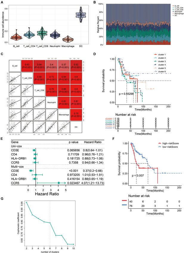
Chengcheng et al.                                                                                                         Immune-Related Genes in HNSCC Tumor

  FIGURE 2 | Comparison of gene expression profiles based on immune score and stromal score in recurrent HNSCC. (A) Heat map of genes that are significantly
  expressed differently based on immune scores. (B) Heat map of genes that are significantly differently expressed based on stromal scores. (C) Volcano map of
  differential expressed genes (DEGs) according to the immune score. (D) Volcano map of DEGs according to the stromal scores. (E) Venn diagram analysis of
  abnormally expressed genes according to the immune and stromal scores.

a small number, which is consistent with the characteristics of                      Survival Analysis of Immune-Related
biological networks.                                                                 Genes in Recurrent HNSCC
                                                                                     To further investigate the prognostic value of these immune-
Function Richness Analysis of                                                        related genes, Kaplan–Meier analysis was performed and survival
Immune-Related Genes in Recurrent                                                    curves were illustrated to further investigate the relationship
HNSCC                                                                                between the expression levels of these immune-related genes and
In order to explore the potential molecular mechanisms of 407                        the survival of recurrent HNSCC patients. The results indicated
immune-related genes, we performed a functional enrichment                           that a total of 206 genes were significantly associated with
analysis of these genes. As shown in Figure 3 and Supplementary                      the survival of the recurrent HNSCC among which, OR2I1P,
Table 2, the BP of GO analysis shows that immune-related genes                       SPOCK2, CCR5, DPT, CD3E, C2, CD4, VCAM1, CD79A, HLA-
are mainly enriched in the BP of leukocyte migration, immune                         DRB1, DOK3, TRBC1, RARRES2, AXL, and IGKV1-16 are the
response-regulating cell surface receptor signaling pathway                          most significant prognostic factors in recurrent HNSCC patients
and immune response-activating cell surface receptor signaling                       (Figure 5 and Supplementary Table 3). We therefore selected
pathway (Figure 4A). In the CC group genes are mainly enriched                       these prognostic immune-related genes for further analysis.
in the extracellular matrix, side of membrane, and collagen-
containing extracellular matrix (Figure 4B). MF enriches antigen                     The Immune Landscape of Recurrent
binding, extracellular matrix structural constituent, peptide                        HNSCC TME
binding (Figure 4C). In addition, KEGG analysis showed that                          In order to evaluate the TME in recurrent HNSCC, we
immune-related genes are related to staphylococcus aureus                            estimated the abundance of tumor infiltrating immune cells,
infection, hematopoietic cell lineage, cell adhesion molecules                       and explored the impact of these infiltrating immune cells on
(CAMs), rheumatoid arthritis, viral protein interaction with                         the clinical outcome in recurrent HNSCC patients. Among the
cytokine and cytokine receptor, phagosome, and cytokine-                             six types of immune cells, DCs have the highest abundance
cytokine receptor interaction (Figure 4D and Supplementary                           in the microenvironment of HNSCC (Figure 6A). Figure 6B
Table 2). The above results indicate that immune-related genes                       summarizes the proportion of immune cells in each cluster.
are related to the extracellular stroma, bacterial infections, and                   Subsequently, we analyzed the abundance matrix of different
cell interactions of the TME.                                                        tumor infiltrating immune cells and found that the abundance

Frontiers in Cell and Developmental Biology | www.frontiersin.org                5                                          August 2021 | Volume 9 | Article 723721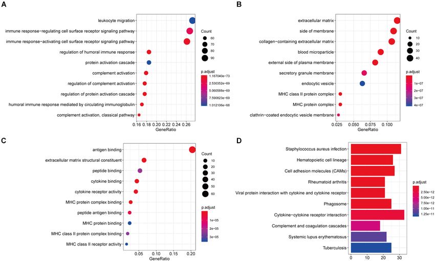
Chengcheng et al.                                                                                                                 Immune-Related Genes in HNSCC Tumor

  FIGURE 3 | Module-related central genes network in recurrent HNSCC. (A) STRING database and Cytoscape software were used to construct the protein-protein
  interaction (PPI) network module. In the PPI network, the color of the node reflects the logarithmic (FC) value, the size of the node indicates the number of interacting
  protein. (B) Through Molecular Complex Detection (MCODE) analysis, eight modules with at least three nodes are determined. The more a gene is associated with
  other genes in the network, the more relevant the module is.

of different infiltrating cells was weakly correlated (Figure 6C                         of the PPI network (Figure 3A) and the p-value less than 0.05
and Supplementary Table 4). Then, we further studied the                                 in the survival analysis to screen out four genes CCR5, CD3E,
relationship between different immune cell infiltration patterns                         CD4, and HLA-DRB1. After grouping these four genes into
and recurrence HNSCC survival. As shown in Figure 6D, cluster                            high/low risk scores, the survival risk assessment was performed
3, defined by high levels of CD4 + T cells and low levels of B cells                     again, and it was found that CCR5, CD3E, CD4, and HLA-
is associated with a better prognosis, while cluster 5 (defined by                       DRB1 genes significantly affected the survival risk of patients
low levels of CD4 + T cells and low levels of B cells) is associated                     (Figures 6E,F and Supplementary Table 5). Considering the
with better prognosis. Cell definition had the worst outcomes.                           unique distribution of individual infiltrating immune cells, all
The above results indicate that changes in the abundance of                              patients were subjected to unsupervised clustering of k-means
tumor infiltrating immune cells may be related to the prognosis                          algorithm according to the proportion of immune cells, and the
of recurrent HNSCC.                                                                      optimal number of clusters was ten (k-means = 10, Figure 6G and
                                                                                         Supplementary Table 6).
Expression Verification of Four Screened                                                    To further verify the results, the relative mRNA expressions of
Immune-Related Gene                                                                      CCR5, CD3E, CD4, and HLA-DRB1 in HNSCC tumor tissue and
In order to find the genes that are most critical to the TME                             adjacent non-tumor tissues were tested. The results showed that
in HNSCC, we combined the genes located in the middle node                               the expression of CCR5, CD3E, CD4, and HLA-DRB1 mRNAs

Frontiers in Cell and Developmental Biology | www.frontiersin.org                    6                                             August 2021 | Volume 9 | Article 723721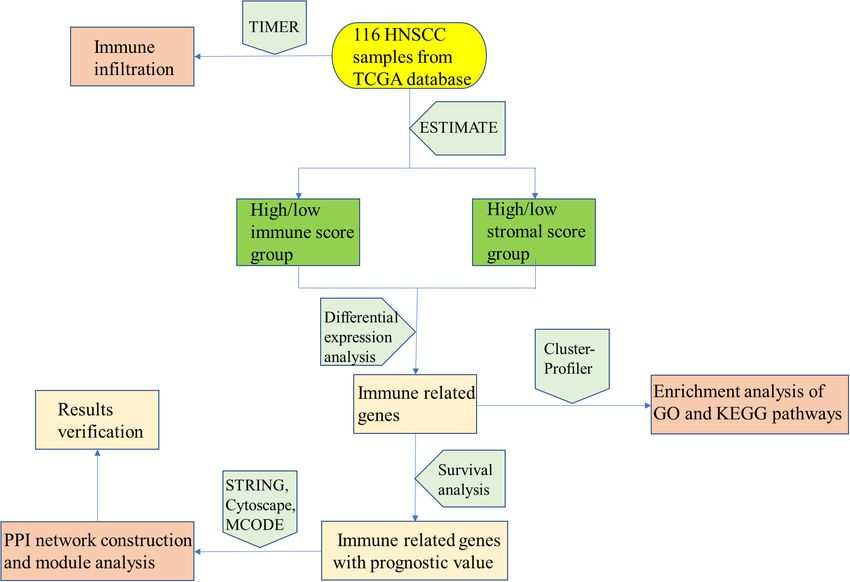
Chengcheng et al.                                                                                                     Immune-Related Genes in HNSCC Tumor

  FIGURE 4 | Function richness analysis of immune-related genes in recurrent HNSCC. (A) The biological process (BP) of genetic ontology (GO) of prognostic
  immune-related genes. (B) Cellular components of the gene ontology (GO) of prognostic immune-related genes. (C) The molecular function (MF) of the GO of
  prognostic immune-related genes. (D) Kyoto Encyclopedia of Genes and Genomes (KEGG) pathway enrichment analysis of prognostic immune-related genes. The
  color of the bar indicates –log10 (p-value). The intersection of the KEGG pathway and GO Term is enriched by the number of genes.

in tumor tissues was significantly higher than that of normal                     treatment, new treatment methods have emerged. Cetuximab is a
tissues adjacent to the tumor (Figures 7A–D). Our western blot                    monoclonal antibody (mAb) that targets epidermal growth factor
analysis showed that compared with normal tissues, HNSCC                          receptor (EGFR), which was introduced into platinum-based
tissues showed relatively higher expression levels of CCR5, CD3E,                 therapy for HNSCC (Cohen et al., 2019). Immunotherapy attracts
CD4, and HLA-DRB1 proteins (Figure 7E). In addition, IHC                          people’s attention due to it fights tumors by activating one’s own
analysis was performed to determine the protein expression levels                 immune cells (Poggio et al., 2019). Although immunotherapy
of CCR5, CD3E, CD4, and HLA-DRB1 in HNSCC. From the                               provides HNSCC patients with a cancer treatment option, and
immunostaining, we can observe that the expression of these four                  HNSCC have a wide range of immune-related genomic features,
proteins is increased in tumor tissues compared with neighboring                  the immune molecular mechanism of HNSCC is still little known.
normal tissues (Figure 7F). The above results all indicate that                   Therefore, we use TCGA databases to screen prognostic immune-
CCR5, CD3E, CD4, and HLA-DRB1 are highly expressed in                             related biomarkers through ESTIMATE and TIMER algorithms,
HNSCC tumor tissues.                                                              and provide new insights for the treatment of HNSCC.
                                                                                     Increasing studies have shown that the regulation of TME
                                                                                  is not only related to immunotherapy but also related to the
DISCUSSION                                                                        prognosis of tumor patients (Chen et al., 2019; Liu G. et al.,
                                                                                  2021). To ESTIMATE was used to model and test the estimation
Head and neck squamous cell carcinomas is one of the most                         of stromal cells and immune cells in HNSCC tissue to calculate
common malignant tumors in the world, with a mortality rate                       the purity of the tumor, stromal, and immune score through
of 40–50% (McDermott and Bowles, 2019). Considering the                           expression profiles, and give an overall view of the TME. We
difficulty of early diagnosis, most HNSCC patients are already                    found that the immunity and stromal scores were significantly
at an advanced stage at diagnosis. In these advanced HNSCC                        related to the prognosis of HNSCC patient. Then, we performed
patients, local metastasis and recurrence of cancer cells are                     differential expression analysis between the high/low immune
common and in need of improved treatment (Paver et al.,                           score groups and between the high/low stroma score groups,
2020). The prognosis is even worse for relapsed HNSCC patients                    and identified 407 immune-related genes (Figure 2). In our
with progressive disease after platinum therapy (Burtness et al.,                 results, almost all DEGs are up-regulated, we speculate that the
2019). In order to solve these difficulties faced by HNSCC                        possible reason is that tumor cells have faster energy metabolism

Frontiers in Cell and Developmental Biology | www.frontiersin.org             7                                        August 2021 | Volume 9 | Article 723721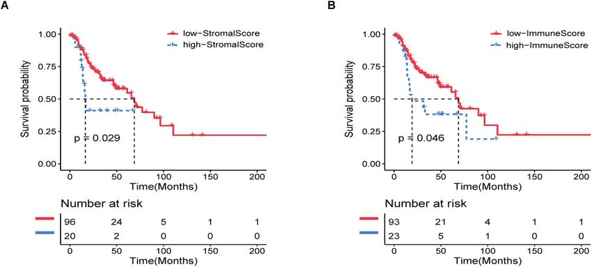
Chengcheng et al.                                                                                                          Immune-Related Genes in HNSCC Tumor

  FIGURE 5 | Kaplan–Meier survival curve shows the survival analysis of immune-related genes in recurrent HNSCC. The expression level of 16 immune-related genes
  to affect the overall survival are selected randomly, and compare the overall survival of the high and low gene expression groups. The red and blue dashed lines
  represent the upper and lower limits of the 95% confidence interval for gene expression, respectively.

and proliferation, which leads to more active gene transcription.                    matrix in the TME has a strong correlation with immune
Subsequently, GO and KEGG were used to analyze these                                 infiltrating cells. In addition, we constructed a PPI module, which
immune-related genes to reveal their functions. As expected,                         is used to reveal the relationship and gene function between
most of the immune-related genes are involved in immune                              DEGs (Figure 4). The nodes with rich connectivity in the module
processes (Figure 3). These results indicate that the extracellular                  are also related to immune/inflammatory response.

Frontiers in Cell and Developmental Biology | www.frontiersin.org                8                                          August 2021 | Volume 9 | Article 723721
Chengcheng et al.                                                                                                             Immune-Related Genes in HNSCC Tumor

  FIGURE 6 | The immune landscape of recurrent HNSCC tumor microenvironment (TME). (A) Abundance of immune infiltrating cells in recurrent HNSCC.
  (B) Stacked bar graph of samples arranged in the order of cluster assignment, and unsupervised clustering of all samples based on the proportion of immune cells.
  (C) Correlation stroma of the abundance of six immune cells. (D) Summary of survival analysis of patients in different groups. (E) Uni-Cox and Multi-Cox regression
  analysis of CCR5, CD3E, CD4, and HLA-DRB1 four genes. (F) Survival analysis of CCR5, CD3E, CD4, and HLA-DRB1 four genes in the high-risk group and the
  low-risk group. (G) Optimal number of clusters by unsupervised clustering analysis.

Frontiers in Cell and Developmental Biology | www.frontiersin.org                  9                                           August 2021 | Volume 9 | Article 723721
Chengcheng et al.                                                                                                  Immune-Related Genes in HNSCC Tumor

  FIGURE 7 | Expression verification of four screened immune-related gene. (A–D) The mRNA levels of CCR5, CD3E, CD4, and HLA-DRB1 in HNSCC samples.
  (E) The protein levels of CCR5, CD3E, CD4, and HLA-DRB1 in HNSCC samples. (F) Representative immunohistochemical images of CCR5, CD3E, CD4, and
  HLA-DRB1. Original magnification: ×200. Bar = 50 um. **P < 0.01, ****P < 0.0001.

    To better explore the impact of immune infiltrating cells                   also related to tumor prognosis in HNSCC patients (Djaldetti and
and extracellular matrix related genes on prognosis, we                         Bessler, 2014; Tamminga et al., 2020).
performed Kaplan–Meier analysis to explore potential regulatory                    In order to investigate effects of the immune-related genes
mechanisms. Survival analysis was performed according to the                    on HNSCC prognosis, we further screened these prognostic
407 differential immune-related genes and 206 genes were closely                immune-related genes and explored the potential regulatory
correlated to the survival rate of the recurrent HNSCC patients                 mechanisms. A total of four genes (CCR5, CD3E, CD4, and
(Figure 5). To further explore the relationship between these                   HLA-DRB1) were identified that related to the immune matrix
DEGs and immune cells, we performed the TIMER method to                         and immune cells. We found that HNSCC patients with high
estimate the proportion of different immune infiltrating cells.                 expression of these four immune-related genes (P > 0.05) had
Furthermore, unsupervised cluster analysis is also used to identify             a worse prognosis (P < 0.05). To further verify the accuracy, we
the immune infiltrating patterns in HNSCC. The results were                     conducted verification in HNSCC patient tissues, and found that
consistent with other studies that monocyte immune cells such as                the mRNA expression of CCR5, CD3E, CD4, and HLA-DRB1
B cells, CD4 + T cells, CD8 + T cells, macrophages, and natural                 were significantly upregulated in HNSCC tissues comparing
killer (NK) cells played a key role in tumor immunity and were                  with that of the controlled tissues. The protein levels of these

Frontiers in Cell and Developmental Biology | www.frontiersin.org          10                                       August 2021 | Volume 9 | Article 723721
Chengcheng et al. Immune-Related Genes in HNSCC Tumor
FIGURE 8 | Analysis process structure diagram.
genes were also identical to that (Figure 7). The immune above analysis enables us to better understand the TEM and
microenvironment can reflect the immune status of tumor immune characteristics of HNSCC, and helps us to predict the
patients to a certain extent, which provides a reference for us prognosis of HNSCC patients through immune-related genes.
to treat tumors more accurately. Many studies have shown that
CCR5, CD3E, CD4, and HLA-DRB1 play an important role in the
development of tumors. Anti-CCR5 therapy in cancer patients DATA AVAILABILITY STATEMENT
can effectively target tumor immune cells in colorectal cancer
The datasets presented in this study can be found in online
metastasis (Jiao et al., 2019). As an indicator of bladder cancer
repositories. The names of the repository/repositories
TME regulation, CD3E is closely related to immune infiltration
and accession number(s) can be found in the article/
(Liu Y. et al., 2021). The tumor antigen-specific CD4 + T
Supplementary Material.
cells in cancer immunity are a key strategy for tumor prognosis
and treatment (Protti et al., 2014). HLA-DRB1 expression can
predict the prognosis of patients with non-small cell lung cancer ETHICS STATEMENT
who receive PD-1/PD-L1 immune checkpoint blockade (Correale
et al., 2020). In our study, a total of 10 clusters were formed, and The studies involving human participants were reviewed
further survival analysis showed that changes in the abundance and approved by the Biomedical Research Ethic Committee
of CD4 + T cells and B cells may be the main prognostic of Shandong Provincial Hospital. The patients/participants
factors for HNSCC. In summary, these results indicate that the provided their written informed consent to participate in
heterogeneity of immune infiltration in HNSCC can be used as a this study.
prognostic indicator and therapeutic target for immunotherapy.
In conclusion, as shown in Figure 8, our analysis process
was mainly performed based on the ESTIMATE algorithm to AUTHOR CONTRIBUTIONS
calculate the immune as well as stromal scores and further
LC drafted the manuscript, performed the experiments, and
detect the correlations between the immune/stromal scores and
analyzed the data. QW, GN, ZF, XR, TZ, XF, and QY designed
clinical parameters. According to the immune/stromal scores,
the study and analyzed the data. XM, LX, and ZM revised the
we divided the HNSCC patient obtained in the TCGA database
manuscript. All authors contributed to the article and approved
into high or low score immune group and matrix group to
the submitted version.
determine the DEGs. Subsequently, the results of functional
enrichment analysis showed that these genes are mainly involved
in the immune/inflammatory response, and the PPI network FUNDING
further demonstrated this discovery. Finally, we calculated the
abundance of each type of immune cells in the TME to explore This present study was supported by funds from the National
the relationship between immune scores and immune cells. The Natural Science Foundation of China (Grant Nos. 81900922 to
Frontiers in Cell and Developmental Biology | www.frontiersin.org 11 August 2021 | Volume 9 | Article 723721Chengcheng et al. Immune-Related Genes in HNSCC Tumor
LC and 81770979 to XM) and the Natural Science Foundation of associations of immune and stromal scores with the treatment of
Shandong Province (Grant No. ZR2019BH019 to LC). recurrent HNSCC patients.
Supplementary Table 1 | The characteristics of the patients in the training and
validation cohort.
Supplementary Table 2 | Gene ontology (GO) and Kyoto Encyclopedia of Genes
SUPPLEMENTARY MATERIAL and Genomes (KEGG) analysis of 407 immune-related genes.
Supplementary Table 3 | Survival analysis of immune-related genes in recurrent
The Supplementary Material for this article can be found
head and neck squamous cell carcinomas (HNSCC).
online at: https://www.frontiersin.org/articles/10.3389/fcell.2021.
723721/full#supplementary-material Supplementary Table 4 | The abundance stroma of different tumor infiltrating
immune cells in recurrent HNSCC.
Supplementary Figure 1 | Correlation between immune and stromal scores and Supplementary Table 5 | Survival analysis of CCR5, CD3E, CD4, and
characteristics of patients with recurrent head and neck squamous cell HLA-DRB1 genes in the high-risk group and the low-risk group.
carcinomas (HNSCC). (A) Immune scores’ distribution for agesChengcheng et al. Immune-Related Genes in HNSCC Tumor Sharma, P., Hu-Lieskovan, S., Wargo, J. A., and Ribas, A. (2017). Primary, adaptive, spermatogenesis. Cell Mol. Life Sci. 76, 2681–2695. doi: 10.1007/s00018-019- and acquired resistance to cancer immunotherapy. Cell 168, 707–723. doi: 03101-9 10.1016/j.cell.2017.01.017 Tamminga, M., Hiltermann, T. J. N., Schuuring, E., Timens, W., Fehrmann, Conflict of Interest: The authors declare that the research was conducted in the R. S. N., and Groen, H. J. M. (2020). Immune microenvironment composition absence of any commercial or financial relationships that could be construed as a in non-small cell lung cancer and its association with survival. Clin. Transl. potential conflict of interest. Immunol. 9:e1142. Van Landuyt, K. L., Nawrot, T., Geebelen, B., De Munck, J., Snauwaert, J., Publisher’s Note: All claims expressed in this article are solely those of the authors Yoshihara, K., et al. (2011). How much do resin-based dental materials release? and do not necessarily represent those of their affiliated organizations, or those of A meta-analytical approach. Dent. Mater. 27, 723–747. doi: 10.1016/j.dental. the publisher, the editors and the reviewers. Any product that may be evaluated in 2011.05.001 this article, or claim that may be made by its manufacturer, is not guaranteed or Yang, S., Liu, T., Cheng, Y., Bai, Y., and Liang, G. (2019). Immune endorsed by the publisher. cell infiltration as a biomarker for the diagnosis and prognosis of digestive system cancer. Cancer Sci. 110, 3639–3649. doi: 10.1111/cas. Copyright © 2021 Chengcheng, Wenwen, Ningyue, Fangyuan, Runtong, Zhenxiao, 14216 Fenglei, Yiming, Miaoqing, Xiaoming and Ming. This is an open-access article Yu, G., Wang, L. G., Han, Y., and He, Q. Y. (2012). clusterProfiler: an R package distributed under the terms of the Creative Commons Attribution License (CC BY). for comparing biological themes among gene clusters. OMICS 16, 284–287. The use, distribution or reproduction in other forums is permitted, provided the doi: 10.1089/omi.2011.0118 original author(s) and the copyright owner(s) are credited and that the original Zhou, R., Wu, J., Liu, B., Jiang, Y., Chen, W., Li, J., et al. (2019). The publication in this journal is cited, in accordance with accepted academic practice. No roles and mechanisms of Leydig cells and myoid cells in regulating use, distribution or reproduction is permitted which does not comply with these terms. Frontiers in Cell and Developmental Biology | www.frontiersin.org 13 August 2021 | Volume 9 | Article 723721
You can also read


