Value and limitation of immunohistochemical expression of HMGA2 in mesenchymal tumors: about a series of 1052 cases - Nature
←
→
Page content transcription
If your browser does not render page correctly, please read the page content below

Modern Pathology (2010) 23, 1657–1666
                                                                  & 2010 USCAP, Inc. All rights reserved 0893-3952/10 $32.00   1657

Value and limitation of immunohistochemical
expression of HMGA2 in mesenchymal
tumors: about a series of 1052 cases
Nadège Dreux1, Marion Marty1, Frédéric Chibon1,2, Valérie Vélasco1, Isabelle Hostein1,2,
Dominique Ranchère-Vince3, Philippe Terrier4 and Jean-Michel Coindre1,2,5
1
  Department of Pathology, Institut Bergonié, 229 cours de l’Argonne, Bordeaux Cédex, France; 2INSERM
U916, Institut Bergonié, 229 cours de l’Argonne, Bordeaux Cédex, France; 3Department of Pathology, Centre
Léon Bérard, Lyon, France; 4Department of Pathology, Institut Gustave Roussy, Villejuif, France and
5
  Laboratory of Pathology, Université Victor Segalen Bordeaux 2, 146 rue Léo-Saignat, Bordeaux Cédex, France

    The high mobility group A (HMGA2) gene encodes a protein that alters chromatin structure and regulates the
    transcription of many genes; it is implicated in both benign and malignant neoplasias, but its rearrangements
    are a feature of development of several mesenchymal tumors. Given its implication in these tumors and
    particularly adipocytic tumors, and the availability of antibodies usable on paraffin-embedded tissues, we
    evaluated the immunohistochemical expression of this gene in a series of 1052 mesenchymal tumors. The
    objective was to define the value and limitations of HMGA2 immunohistochemical expression for histotyping,
    and compare with molecular data reported in the literature. We thus analyzed 880 cases on tissue microarray
    and 182 cases on whole sections (211 adipocytic tumors, 628 sarcomas, 213 benign mesenchymal tumors, and
    10 normal adipose tissues). A nuclear immunostaining was detected in 86% of conventional and intramuscular
    lipomas, in 86% of well-differentiated liposarcomas and in 67% of dedifferentiated liposarcomas, as opposed to
    16% of other benign adipose tumors and to 15% of non-well-differentiated liposarcoma/dedifferentiated
    liposarcoma sarcomas. Among benign mesenchymal tumors and lesions, it was detected in 90% of nodular
    fasciitis and in 88% of benign fibrous histiocytomas with respective specificities of 85 and 100%, and in 90% of
    aggressive angiomyxoma, contrary to other vulvovaginal tumor types, which expressed HMGA2 only rarely. The
    normal adipose tissue was always negative for HMGA2. Although not specific, immunohistochemical detection
    of the HMGA2 protein is helpful for the distinction of normal adipose tissue from well-differentiated lesions,
    particularly on biopsy or on re-excision. It is less sensitive than MDM2/CDK4 for dedifferentiated liposarcomas
    diagnosis, but it appears more specific to distinguish dedifferentiated liposarcomas from other poorly
    differentiated sarcomas. Finally, and may be more importantly, HMGA2 is useful for the diagnosis of benign
    fibrous histiocytoma, nodular fasciitis and vulvovaginal benign mesenchymal tumors.
    Modern Pathology (2010) 23, 1657–1666; doi:10.1038/modpathol.2010.174; published online 10 September 2010

    Keywords: HMGA2; immunohistochemistry; mesenchymal tumor; tissue microarray

The human high mobility group A (HMGA2) gene                      HMGA2 can modify chromatin structure enabling a
encodes a non-histone chromatin protein that                      fine tuning of transcription in a variety of cellular
belongs to a family of the HMG proteins including                 processes such as differentiation and oncogenesis.3–6
also HMGA1a, HMGA1b, HMGA1c, which result                         With a length of 160 kb, it is located at the
from alternative splicing of the HMGA1 mRNA.1,2                   chromosome band 12q14.3 and consists of five
                                                                  exons. Each of the first three exons codes for an
                                                                  AT hook domain, which is separated from the fourth
Correspondence: Professor J-M Coindre, MD, Department of          exon by a large intronic sequence (140 kB), whereas
Pathology, Institut Bergonié, 229 Cours de l’Argonne, Bordeaux   the fifth exon codes for the acidic C-terminal
Cédex 33076, France.
E-mail: coindre@bergonie.org
                                                                  domain. The 30 UTR of HMGA2 contains binding
Received 10 May 2010; revised 28 June 2010; accepted 11 July      sites for the let-7 family of microRNAs that inhibit
2010; published online 10 September 2010                          gene expression at the posttranscriptional level.7,8

www.modernpathology.org
HMGA2 in mesenchymal tumors
1658                                                      N Dreux et al

          HMGA2 is at high levels during embryonic devel-                 peripheral nerve sheath tumors, 10 angiosarcomas,
          opment,9,10 but it is not detectable at any significant         10 dermatofibrosarcoma protuberans, 9 osteosarco-
          level in adult tissues.11,12 Recent studies report              mas, 8 primitive neuroectodermal tumors, 6 epithe-
          increased expression of HMGA2 in several human                  lioid sarcomas, 6 clear cell sarcomas, 5 soft part
          malignancies, including lung and pancreatic                     alveolar sarcomas, 4 extra-skeletal myxoid chondro-
          tumors, oral squamous cell carcinomas and a subset              sarcomas, 4 Kaposi’s sarcomas, 3 epithelioid he-
          of breast cancers.13–18 This expression represents              mangioendotheliomas, 3 low-grade fibromyxoid
          a poor prognostic index as overexpression often                 sarcomas, 3 desmoplastic small round cell tumors,
          correlates with the presence of metastasis. This                2 malignant mesenchymomas, 1 malignant solitary
          supports the idea of the implication of HMGA2 in                fibrous tumor; (3) benign mesenchymal tumors
          pathogenesis, although its function is not yet well             or lesions with 176 cases including 81 desmoid
          understood in these neoplasms.4 However, this gene              tumors, 20 nodular fasciitis, 17 benign fibrous histio-
          is also a target of chromosomal rearrangements in               cytomas, 13 uterine leiomyomas, 10 intramuscular
          some benign solid tumors19,20 such as mesenchymal               myxomas, 5 angiomas, 5 angiolipomas, 5 schwan-
          tumors:21–24 in particular, HMGA2 is a prominent                nomas, 3 neurofibromas, 3 solitary fibrous tumors,
          feature of conventional lipomas as 75% of these                 3 myofibromas, 2 angiomyolipomas, 2 perineuriomas,
          harbor translocation t(3;12) involving it,3,25,26 but           1 angioleiomyoma, 1 granular cell tumor, 1 giant
          it is also overexpressed or amplified in atypical               cell tumor of soft tissue and 4 reactive fibrosis; and
          lipomatous tumor/well-differentiated27–29 and ded-              (4) benign mesenchymal vulvovaginal tumors with
          ifferentiated liposarcomas. Given the involvement of            30 cases including 11 fibroepithelial stromal polyps,
          HMGA2 in mesenchymal tumors and particularly                    10 aggressive angiomyxomas, 4 angiomyofibroblas-
          adipocytic tumors and the availability of antibodies            tomas, 2 cellular angiofibromas and 3 leiomyomas.
          usable on paraffin-embedded tissues, we evaluated               We also analyzed 10 samples of normal adipose
          immunohistochemical expression of this gene in a                tissue.
          large series of mesenchymal tumors in order to                     Eight hundred and eighty mesenchymal tumors
          define its value and limitations for histotyping.               were analyzed on tissue microarray, divided into 18
                                                                          paraffin blocks. Each case was represented by three
                                                                          spots of 1 mm. The other 182 cases were analyzed on
          Materials and methods                                           whole sections. In particular, this concerned the 10
                                                                          samples of normal adipose tissues, the benign
          We selected 1052 mesenchymal tumors from 1014                   adipocytic tumors (excluding spindle cell lipomas)
          patients. Cases were issued from the archives of the            and atypical lipomatous tumor/well-differentiated
          Department of Pathology of Institut Bergonié and               because of their properties with tissue unsuitable for
          from the database of the French Sarcoma Group                   tissue microarray. To this we added some samples
          (http://www.conticabase.org) with the participation             of benign tumors including some cases of fasciitis,
          of Paul Papin Center, Angers; University hospital,              or benign fibrous histiocytomas, solitary fibrous
          Caen; Henri Mondor Hospital, Créteil; Georges-                 tumors, mesenchymal vulvovaginal tumors and
          Francois Leclerc Center, Dijon; University Institute            leiomyomas to supplement the number of cases
          of Pathology, Lausanne; Oscard Lambret Center,                  treated by this method to improve the representa-
          Lille; Leon-Berard Center, Lyon; Val d’Aurelle                  tiveness of the results.
          Center, Montpellier; Alexis Vautrin Center, Nancy;
          René Huguenin Center, Saint-Cloud; Claudius
          Regaud Institute, Toulouse and Gustave Roussy                   Immunohistochemistry
          Institute, Villejuif. These cases have been collegially
          reviewed and classified according to the latest                 A total of 4 mm-thick paraffin sections were cut and
          World Health Organization classification for bone               mounted on glass slides (Superfrost þ Menzel
          and soft tissue tumors.                                         Glazer). Subsequently, the preparations were en-
             The tumors selected belong to four main cate-                tirely treated on automate VENTANA-Benchmark-
          gories: (1) adipocytic tumors with 218 cases in-                XTs. The primary antibody was a polyclonal
          cluding 73 benign tumors (31 conventional lipomas,              anti-rabbit antibody from BIOCHECK laboratories
          5 intramuscular lipomas, 34 spindle cell and                    (Foster City, Ref. 59170AP) and was used at a 1:500
          pleomorphic lipomas, 2 hibernomas and 1 myoli-                  dilution. Sections were incubated at room tempera-
          poma) and 145 liposarcomas (43 atypical lipoma-                 ture in standard CCI buffer (Ventana) and were
          tous tumor/well-differentiated, 69 dedifferentiated             revealed with ‘Ultra ViewTr’ Universal DAB kit, after
          liposarcomas, 19 pleomorphic liposarcomas and                   that they stained with Hematoxylin ReaDi Ventana
          14 myxoid/round cell liposarcomas); (2) non-adipocytic          solution (Ref. 760-2021).
          sarcomas with 628 cases including 217 leiomyosar-                  A tumor was considered as HMGA2 positive
          comas, 179 unclassified sarcomas, 84 myxofibrosar-              when at least one-cell nuclei stained per high-power
          comas, 35 rhabdomyosarcomas (18 pleomorphic,                    field. The number of positive cells was counted in
          11 embryonal and 6 alveolar), 14 synovial sarcomas,             medium-power fields for each histology section and
          13 gastro-intestinal stromal tumors, 12 malignant               quantified as a percentage. For each case derived

Modern Pathology (2010) 23, 1657–1666
HMGA2 in mesenchymal tumors
                                                                     N Dreux et al                                                          1659

from tissue microarray, we used the average value of                 whereas other tumors were rarely positive (six cases
the three spots.                                                     of spindle cell and pleomorphic lipomas, no case of
                                                                     hibernoma or myolipoma, ie 16% of cases). Among
                                                                     liposarcomas (Table 1), HMGA2 was expressed
Statistical Analysis                                                 almost exclusively by atypical lipomatous tumor/
                                                                     well-differentiated and dedifferentiated liposarco-
Specificity and sensitivity for HMGA2 immuno-
                                                                     mas (86 and 67% of cases, respectively), with only two
stainings were evaluated in the well-differentiated
                                                                     positive cases among pleomorphic liposarcomas and
liposarcoma subgroup and compared with that
                                                                     no cases for myxoid/round cell liposarcomas (6%).
of the normal adipose tissue, and evaluated in
                                                                        As opposed to 67% positivity in dedifferentiated
the well-differentiated/dedifferentiated liposarcoma
                                                                     liposarcomas, only 16% of potential dedifferen-
and compared with that of the so-called ‘others
                                                                     tiated liposarcomas simulators were positive, espe-
sarcoma’ type subgroup. Sensitivity for HMGA2 was
                                                                     cially in myxofibrosarcomas (26%), and embryonal
calculated as the ratio of positively stained atypical
                                                                     rhabdomyosarcomas (36%). Only 5 out of 78 (8%) of
lipomatous tumor/well-differentiated/dedifferentiated
                                                                     other sarcomas were positive (Table 2).
liposarcomas to the number of atypical lipo-
matous tumor/well-differentiated/dedifferentiated
liposarcomas examined. Specificity for HMGA2                         Table 2 Immunohistochemistry results for the high mobility
was calculated as the ratio of unstained non-atypical                group A (HMGA2) gene in dedifferentiated liposarcomas, poten-
                                                                     tial simulators and other sarcomas
lipomatous tumor/well-differentiated/dedifferentiated
liposarcomas tumors to the total number of non-                      Type of sarcoma                Number HMGA2 %              Non-
atypical lipomatous tumor/well-differentiated/ded-                                                  of cases positivity         inter-
ifferentiated liposarcomas. Among the benign tumors                                                                            pretable
or the vulvovaginal area, sensitivities and specifi-
cities were evaluated on the same principle with the                 Dedifferentiated                  69          46     67      0
                                                                     liposarcomas
aggressive angiomyxomas as the referent positive
tumor. According to cytogenetic data, we hypothe-                    Simulators                       583          93     16     12
sized that conventional lipomas, atypical lipoma-                      Unclassified sarcoma           179          32     18      1
tous tumor/well-differentiated and dedifferentiated                    Leiomyosarcoma                 217          28     13      4
                                                                       Myxofibrosarcoma                84          22     26      1
liposarcomas would be positive as opposed to                           Pleomorphic                     19           2     11      1
normal adipose tissue and to others sarcomas.                          liposarcoma
                                                                       Pleomorphic                     18           3     17      0
                                                                       rhabdomyosarcoma
Results                                                                Myxoid/round cell               14           0     0       0
                                                                       liposarcoma
HMGA2 staining according to tumor histotype is                         Gastro-intestinal               13           1     8       0
summarized in Tables 1–3. Among benign adipocy-                        stromal tumor
                                                                       Malignant peripheral            12           1     14      5
tic tumors (Table 1), HMGA2 was expressed in                           nerve sheath tumor
86% of conventional and intramuscular lipomas,                         Embryonal                       11           4     36      0
                                                                       rhabdomyosarcoma
                                                                       Alveolar rhabdomyosarcoma        6           0     0       0
                                                                       Malignant solitary               1           0     0       0
Table 1 Immunohistochemistry results for the high mobility             fibrous tumor
group A (HMGA2) gene in adipocytic tumors                              Osteosarcoma                     9           0     0       0
Type of tumor                       Number HMGA2 %         Non-      Others                            78           5      8     17
                                    of cases positivity    inter-      Synovial sarcoma                14           1     14      7
                                                          pretable     Desmoplastic small round         3           2     67      0
                                                                       cell tumor
Benign adipocytic tumors               73      37   51       0         Epithelioid sarcoma              6           2     33      0
  Conventional lipoma                  31      26   84       0         Clear cell sarcoma               6           0      0      0
  Intramuscular lipoma                  5       5   100      0         Soft part alveolar sarcoma       5           0      0      2
  Spindle cell and pleomorphic                                         Angiosarcoma                    10           0      0      0
    Lipoma                             34       6   18       0         Epithelioid                      3           0      0      0
    Hibernoma                           2       0    0       0         hemangioendothelioma
    Myolipoma                           1       0    0       0         Kaposi’s sarcoma                 4           0     0       0
                                                                       Primitive neuroectodermal        8           0     0       2
Liposarcomas                          145      85   59       0         tumor
  Atypical lipomatous tumor—           43      37   86       0         Malignant mesenchymoma           2           0     0       0
  well-differentiated liposarcoma                                      Dermatofibrosarcoma             10           0     0       4
  Dedifferentiated liposarcoma         69      46   67       0         protuberans
  Pleomorphic liposarcoma              19       2   11       1         Myxoid chondrosarcoma            4           0     0       0
  Myxoid/round cell liposarcoma        14       0    0       0         Low-grade fibromyxoid            3           0     0       2
                                                                       sarcoma
Total                                 218     122   56       1
Normal adipose tissue                  10       0    0       0       Total                            730         144     21     29

                                                                                                            Modern Pathology (2010) 23, 1657–1666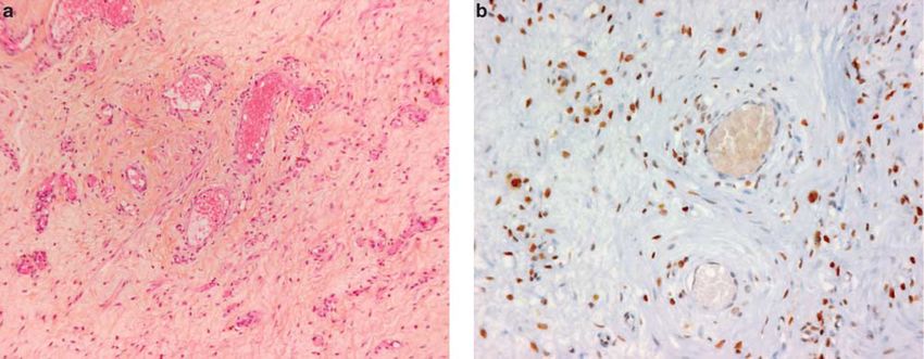
HMGA2 in mesenchymal tumors
1660                                                            N Dreux et al

          Table 3 Immunohistochemistry results for the high mobility            cases showed o50% of positive cells. The propor-
          group A (HMGA2) gene in benign mesenchymal tumors/lesions             tion of positive cells in leiomyomas varies widely
          Type of tumor                     Number HMGA2 %          Non-
                                                                                between 10 and 100% of cells. Among vulvovaginal
                                            of cases positivity     inter-      tumors, 90% of aggressive angiomyxomas expressed
                                                                   pretable     HMGA2 with an intense nuclear positivity invol-
                                                                                ving at least 75% of cells in all cases (Figure 6).
          Spindle cell tumors                148       52     35       0        Nearly 30% of fibroepithelial stromal polyps ex-
            Benign fibrous histiocytoma       17       15     88       0        pressed this antibody, but the immunostaining was
            Nodular fasciitis                 20       18     90       0        less intense in fewer cells. Table 4 summarizes the
            Desmoid tumor                     81       13     16       0
            Leiomyoma                         13        4     31       0        statistical analysis regarding sensitivity and specifi-
            Schwannoma                         5        1     20       0        city of HMGA2.
            Solitary fibrous tumor             3        0      0       0
            Myofibroma                         3        1     33       0
            Neurofibroma                       3        0      0       0
            Perineurioma                       2        0      0       0        Discussion
            Angioleiomyoma                     1        0      0       0
                                                                                This study of immunohistochemical expression of
          Vulvovaginal tumors                 30       13     43       0        HMGA2 in a large series of mesenchymal tumors
            Aggressive angiomyxoma            10        9     90       0        showed a strong positivity in most conventional and
            Fibroepithelial stromal polyp     11        3     27       0
            Cellular angiofibroma              2        0      0       0        intramuscular lipomas and in atypical lipomatous
            Angiomyofibroblastoma              4        0      0       0        tumor/well-differentiated. These results are in
            Leiomyoma                          3        1     33       0        agreement with our cytogenetics knowledge as such
                                                                                tumors show rearrangements in the 12q13–15 region
          Others                              28        4     14       0
            Myxoma                            10        4     40       0
                                                                                in which the HMGA2 gene is the target. Transloca-
            Angioma                            5        0      0       0        tion (3;12) is the most common cytogenetic aberra-
            Angiolipoma                        5        0      0       0        tion in conventional lipomas, and leads to a loss of
            Angiomyolipoma                     2        0      0       0        C-terminal region and the formation of a fusion gene
            Granular cell tumor                1        0      0       0        HMGA2-LPP. HMGA2 is certainly the important
            Giant cell tumor                   1        0      0       0
            Reactive fibrosis                  4        0      0       0        gene of this rearrangement in lipomas3,4 as there are
                                                                                no fewer than 41 other chromosomal loci as partners
          Total                              206       69     33       0        of HMGA2.3,30,31 However, conventional lipomas
                                                                                with normal karyotypes31 or with aberrations in
                                                                                6p21 or on chromosome 13q have been reported.
             Among non-adipocytic benign mesenchymal                            These data could explain that in our series, roughly
          tumors, HMGA2 was expressed in 88% of benign                          14% of conventional lipomas do not express
          fibrous histiocytomas, in 90% of nodular fasciitis                    HMGA2. Moreover, although the amplification of
          and in 30% of leiomyomas. Among vulvovaginal                          MDM2 and CDK4 genes is considered to be the major
          mesenchymal tumors, we obtained a positive                            event in atypical lipomatous tumor/well-differen-
          staining in 90% of aggressive angiomyxoma, con-                       tiated/dedifferentiated liposarcomas, overexpres-
          trary to other tumor types, which expressed HMGA2                     sion of HMGA2 in these tumors (86 and 67%,
          rarely (Table 3). In our study, none of the 10 cases of               respectively) has been suggested a possible function
          normal adipose tissue was positive for HMGA2.                         of HMGA2 in atypical lipomatous tumor/well-
             HMGA2 shows a nuclear positivity with a variable                   differentiated.32
          number of positive cells: in 128 cases (48% of                           HMGA2 does not allow us to distinguish a lipoma
          positive cases), at least 50% of tumor cells were                     from an atypical lipomatous tumor/well-differen-
          immunostained for HMGA2, especially in lipomas,                       tiated. Although it is not more sensitive than
          atypical     lipomatous      tumor/well-differentiated                MDM2/CDK4 for diagnosing dedifferentiated lipo-
          liposarcoma and dedifferentiated liposarcomas                         sarcomas, with a positivity of 67% of cases col-
          (Figures 1–3).                                                        lected, HMGA2 can be useful for the diagnosis of
             Regarding atypical lipomatous tumor/well-differ-                   dedifferentiated liposarcomas, especially on biopsy,
          entiated, we analyzed expression according to                         or for distinguishing areas of atypical lipomatous
          histologic subtype: 86% of atypical lipomatous                        tumor/well-differentiated from normal adipose tis-
          tumor/well-differentiated lipoma-like subtypes ex-                    sue. However, we should keep in mind that the gold
          pressed HMGA2 with only 4% of cases showing                           standard for the diagnosis of difficult cases of
          450% of positive cells. Eighty-three percent of                       atypical     lipomatous    tumor/well-differentiated
          atypical lipomatous tumor/well-differentiated scler-                  and dedifferentiated liposarcomas is currently the
          osing subtypes expressed HMGA2 with half of them                      demonstration of MDM2/CDK4 amplification by
          showing 450% of labeled cells. In other HMGA2-                        FISH.33
          positive sarcomas, between 5 and 30% of the cells                        In our study, the most frequent potential simula-
          were stained. Finally, among nodular fasciitis and                    tors of dedifferentiated liposarcomas, which are
          benign fibrous histiocytomas, which frequently                        HMGA2 positive are myxofibrosarcomas, poorly
          expressed HMGA2 (Figures 4 and 5), only three                         differentiated sarcomas and leiomyosarcomas with

Modern Pathology (2010) 23, 1657–1666
HMGA2 in mesenchymal tumors
N Dreux et al 1661
Figure 1 Nuclear positivity of HMGA2 in a conventional lipoma (a) and in an intramuscular lipoma (b).
Figure 2 Atypical lipomatous tumor/well-differentiated liposarcoma. Presence of a few atypical cells in a fibrous septa (H&E) (a).
Positivity of HMGA2 in numerous cells in the fibrous septa (b) and in a few cells in the lipoma-like areas (c).
Modern Pathology (2010) 23, 1657–1666HMGA2 in mesenchymal tumors
1662 N Dreux et al
Figure 3 Dedifferentiated liposarcoma. Histological aspect of a poorly differentiated spindle cell sarcoma (H&E) (a). Nuclear positivity of
MDM2 in a few tumor cells (b) and positivity of numerous cells for HMGA2 (c).
Figure 4 Nodular fasciitis. Histological aspect of nodular fasciitis composed of bland myofibroblasts (H&E) (a). HMGA2 is strongly and
diffusely positive (b).
Modern Pathology (2010) 23, 1657–1666HMGA2 in mesenchymal tumors
N Dreux et al 1663
Figure 5 Benign fibrous histiocytoma. Conventional form (H&E) (a) with a positivity of numerous cells for HMGA2 (b).
Figure 6 Aggressive angiomyxoma. Histological aspect (H&E) (a). This lesion shows a nuclear immunopositivity for HMGA2, whereas
the nuclei of endothelial cells from blood vessels are negative (b).
a positivity of 26, 18 and 13%, respectively. There- benign mesenchymal lesions. It is positive in almost
fore, in an individual case, HMGA2 expression is of 90% of benign fibrous histiocytoma and negative in
rather limited value in the differential diagnosis of dermatofibrosarcoma protuberans. This result is in
dedifferentiated liposarcoma. agreement with a recent study, which reported that
Our results are similar to those reported by only 1 case out of 14 dermatofibrosarcoma protuber-
Bartuma,27 showing that the expression levels of ans focally overexpressed HMGA2 and that 96% of
HMGA2 evaluated by RT–PCR were correlated with benign fibrous histiocytoma expressed HMGA2
morphological and cytogenetic subgroups of adipo- intensely and diffusely; the difference was statisti-
cytic tumors. These data suggest a practical interest cally significant.34 In our study, 90% of nodular
of HMGA2 immunohistochemistry for the diagnosis fasciitis, also overexpressed HMGA2. In contrast,
in three situations: for distinguishing normal adi- only 16% of desmoid tumors were positive. There-
pocytic tissue from a mature adipocytic tumor when fore, this marker can be useful when dealing with
dealing with a small quantity of tissue; for evaluat- nodular fasciitis. HMGA2 gene has a function in the
ing the margin status in atypical lipomatous tumor/ development and differentiation of soft tissues35,36
well-differentiated and for separating dedifferen- and its dysregulated expression has been reported in
tiated liposarcomas from simulators. benign mesenchymal lesions.37,38 Given the physio-
This study also showed that HMGA2 immunohis- logical function of HMGA2, its overexpression can
tochemistry can be useful for the diagnosis of some be the cause of cellular proliferation. On the other
Modern Pathology (2010) 23, 1657–1666HMGA2 in mesenchymal tumors
1664 N Dreux et al
Table 4 Statistical analysis results: sensitivity and specificity of other tumors (18 angiomyofibroblastomas, 6 cellular
the high mobility group A (HMGA2) gene angiofibromas, 5 fibroepithelial stromal polyps and
5 leiomyomas).49 The resulting immunohistochem-
Type of tumor Sensitivity Specificity
ical application presents a clear interest in practice.
Atypical lipomatous tumor-well- 0.86 0.54
Although the correlation between rearrangements of
differentiated liposarcoma vs the HMGA2 protein and its expression is not clearly
benign adipocytic tumors established in these lesions, the literature reports an
Well-differentiated adipocytic tumors 0.60 1 overexpression in about 50% of cases of aggressive
vs normal adipose tissue angiomyxoma.50 Our results corroborated these data
Dedifferentiated liposarcoma vs 0.67 0.95
other sarcomas with an intense and diffuse positivity in 9 out of 10
Dedifferentiated liposarcoma vs 0.67 0.84 cases of aggressive angiomyxoma as opposed to
simulators other differential diagnoses such as cellular angiofi-
Angiomyxoma vs fibroepithelial 0.90 0.80 broma and angiomyofibroblastoma, which are al-
stromal polyp, angiomyofibroblastoma,
myofibroblastoma, leiomyoma
ways negative for this marker. However, HMGA2
and angiofibroma positivity is not specific as a third of fibroepithelial
Nodular fasciitis vs leiomyosarcoma, 0.90 0.85 stromal polyps and leiomyomas cases of this
myxofibrosarcoma, dermatofibrosarcoma location express this protein; but in these cases,
protuberans, synovial sarcoma, there are other morphologic criteria for diagnosis,
desmoid tumor
Benign fibrous histiocytoma vs 0.88 1 except when dealing with an extensively myxoid
dermatofibrosarcoma protuberans leiomyoma, which is very similar to aggressive
angiomyxoma and, therefore, represents one of the
most difficult differential diagnosis for this tumor.
hand, HMGA2 expression can be induced by growth This marker is also potentially interesting for
factors such as platelet-derived growth factor39 and, assessing surgical margins of atypical lipomatous
therefore, could be the consequence rather than the tumor/well-differentiated as this is challenging at
cause of cellular proliferation, particularly in reac- the histological level.
tive lesions such as nodular fasciitis. As nodular In conclusion, although not specific of a line of
fasciitis and cellular fibrous histiocytoma are easily differentiation, immunohistochemical detection of
confused, this overexpression of HMGA2 in both HMGA2 is a potential valuable aid in the diagnosis
lesions may make the differential diagnosis more of mesenchymal tumors. In particular, it is useful for
difficult. distinguishing well-differentiated adipocytic tumors
Among vulvovaginal benign mesenchymal tu- from normal tissue, for margin assessment in
mors, aggressive angiomyxoma also overexpressed atypical lipomatous tumor/well-differentiated, for
HMGA2 in 90% of cases. However, fibroepithelial dedifferentiated liposarcomas and, potentially more
stromal polyps and leiomyomas were positive in 27 importantly, for the diagnosis of benign fibrous
and 33% of our cases. histiocytoma, nodular fasciitis and vulvovaginal
This HMGA2 overexpression may certainly be benign mesenchymal tumors.
explained by the complex cytogenetic rearrange-
ments of HMGA2 in benign lesions. The most
frequent cytogenetic abnormalities in leiomyomas Acknowledgements
(especially in uterus) are on the one hand a
reciprocal translocation t(12;14) (q15; q23–24) with We are grateful to the following pathologists for
an extragenic breakpoint at the 50 part of HMGA2 contributing cases and paraffin blocks: A Carbo-
and on the other hand, a deletion of the long arm nelle, Créteil; MC Chateau, Montpellier; B Chetaille,
of chromosome 7.23,40 But FISH showed complex Marseille; F Collin, Dijon; L Guillou, Lausanne; A
rearrangements on chromosomes 3, 6, 10 and 12.41,42 Leroux, Nancy; B Marques, Toulouse; JJ Michels,
Cytogenetic abnormalities of HMGA2 are reported in Caen; F Mishellany, Clermont-Ferrand; YM Robin,
40–50% of uterine leiomyomas. These data are Lille; M Trassard, Saint-Cloud; I Valo, Angers and to
significantly correlated with immunohistochemical Pippa McKelvie-Sebileau for help with the English
expression of HMGA2 in our series and in several manuscript.
other studies.42–44 Among vulvovaginal lesions,
aggressive angiomyxomas also have rearrangements
of 12q13–15 region with translocations involving
Disclosure/conflict of interest
chromosomes 1, 7, 8 and 21.45–48 But HMGA2 was The authors declare no conflict of interest.
identified as the only regulated gene, and RT–PCR
techniques have reported breakpoints in HMGA2
at different levels: preferentially in the 50 part of
gene and also described in intron 3. More widely, a
References
study of the characterization of rearrangements of 1 Giancotti V, Bandiera A, Buratti E, et al. Comparison of
HMGA2 in 90 genital tumors showed their presence multiple forms of the high mobility group I proteins in
in 33% of aggressive angiomyxomas, but not in rodent and human cells. Identification of the human
Modern Pathology (2010) 23, 1657–1666HMGA2 in mesenchymal tumors
N Dreux et al 1665
high mobility group I-C protein. Eur J Biochem 21 Dahlen A, Mertens F, Rydholm A, et al. Fusion,
1991;198:211–216. disruption, and expression of HMGA2 in bone
2 Johnson KR, Cook SA, Davisson MT. Chromosomal and soft tissue chondromas. Mod Pathol 2003;16:
localization of the murine gene and two related 1132–1140.
sequences encoding high-mobility-group I and Y 22 Ingraham SE, Lynch RA, Surti U, et al. Identification
proteins. Genomics 1992;12:503–509. and characterization of novel human transcripts
3 Fedele M, Battista S, Manfioletti G, et al. Role of the embedded within HMGA2 in t(12;14)(q15;q24.1) uterine
high mobility group A proteins in human lipomas. leiomyoma. Mutat Res 2006;602:43–53.
Carcinogenesis 2001;22:1583–1591. 23 Mine N, Kurose K, Nagai H, et al. Gene fusion
4 Fusco A, Fedele M. Roles of HMGA proteins in cancer. involving HMGIC is a frequent aberration in uterine
Nat Rev Cancer 2007;7:899–910. leiomyomas. J Hum Genet 2001;46:408–412.
5 Narita M, Krizhanovsky V, Nunez S, et al. A novel role 24 Ashar HR, Tkachenko A, Shah P, et al. HMGA2 is
for high-mobility group a proteins in cellular senes- expressed in an allele-specific manner in human
cence and heterochromatin formation. Cell 2006;126: lipomas. Cancer Genet Cytogenet 2003;143:160–168.
503–514. 25 Nilsson M, Mertens F, Hoglund M, et al. Truncation
6 Reeves R. Molecular biology of HMGA proteins: hubs and fusion of HMGA2 in lipomas with rearrangements
of nuclear function. Gene 2001;277:63–81. of 5q32–4q33 and 12q14–4q15. Cytogenet Genome
7 Lee YS, Dutta A. The tumor suppressor microRNA Res 2006;112:60–66.
let-7 represses the HMGA2 oncogene. Genes Dev 26 Petit MM, Schoenmakers EF, Huysmans C, et al. LHFP,
2007;21:1025–1030. a novel translocation partner gene of HMGIC in a
8 Mayr C, Hemann MT, Bartel DP. Disrupting the pairing lipoma, is a member of a new family of LHFP-like
between let-7 and Hmga2 enhances oncogenic trans- genes. Genomics 1999;57:438–441.
formation. Science 2007;315:1576–1579. 27 Bartuma H, Panagopoulos I, Collin A, et al. Expression
9 Chiappetta G, Avantaggiato V, Visconti R, et al. High levels of HMGA2 in adipocytic tumors correlate with
level expression of the HMGI (Y) gene during embryo- morphologic and cytogenetic subgroups. Mol Cancer
nic development. Oncogene 1996;13:2439–2446. 2009;8:36.
10 Hirning-Folz U, Wilda M, Rippe V, et al. The expres- 28 Dei Tos AP, Doglioni C, Piccinin S, et al. Coordinated
sion pattern of the Hmgic gene during development. expression and amplification of the MDM2, CDK4, and
Genes Chromosomes Cancer 1998;23:350–357. HMGI-C genes in atypical lipomatous tumours. J
11 Li O, Li J, Droge P. DNA architectural factor and proto- Pathol 2000;190:531–536.
oncogene HMGA2 regulates key developmental genes 29 Tallini G, Dal Cin P, Rhoden KJ, et al. Expression of
in pluripotent human embryonic stem cells. FEBS Lett HMGI-C and HMGI(Y) in ordinary lipoma and atypical
2007;581:3533–3537. lipomatous tumors: immunohistochemical reactivity
12 Rogalla P, Drechsler K, Frey G, et al. HMGI-C correlates with karyotypic alterations. Am J Pathol
expression patterns in human tissues. Implications 1997;151:37–43.
for the genesis of frequent mesenchymal tumors. Am J 30 Arlotta P, Tai AK, Manfioletti G, et al. Transgenic mice
Pathol 1996;149:775–779. expressing a truncated form of the high mobility group
13 Abe N, Watanabe T, Masaki T, et al. Pancreatic duct I-C protein develop adiposity and an abnormally
cell carcinomas express high levels of high mobility high prevalence of lipomas. J Biol Chem 2000;275:
group I(Y) proteins. Cancer Res 2000;60:3117–3122. 14394–14400.
14 Fabjani G, Tong D, Wolf A, et al. HMGA2 is associated 31 Bartuma H, Hallor KH, Panagopoulos I, et al. Assess-
with invasiveness but not a suitable marker for the ment of the clinical and molecular impact of different
detection of circulating tumor cells in breast cancer. cytogenetic subgroups in a series of 272 lipomas with
Oncol Rep 2005;14:737–741. abnormal karyotype. Genes Chromosomes Cancer
15 Hristov AC, Cope L, Reyes MD, et al. HMGA2 protein 2007;46:594–606.
expression correlates with lymph node metastasis and 32 Italiano A, Bianchini L, Keslair F, et al. HMGA2 is the
increased tumor grade in pancreatic ductal adenocar- partner of MDM2 in well-differentiated and dediffer-
cinoma. Mod Pathol 2009;22:43–49. entiated liposarcomas whereas CDK4 belongs to a
16 Meyer B, Loeschke S, Schultze A, et al. HMGA2 distinct inconsistent amplicon. Int J Cancer 2008;122:
overexpression in non-small cell lung cancer. Mol 2233–2241.
Carcinog 2007;46:503–511. 33 Sirvent N, Coindre JM, Maire G, et al. Detection
17 Motoyama K, Inoue H, Nakamura Y, et al. Clinical of MDM2-CDK4 amplification by fluorescence
significance of high mobility group A2 in human in situ hybridization in 200 paraffin-embedded tumor
gastric cancer and its relationship to let-7 microRNA samples utility in diagnosing adipocytic lesions
family. Clin Cancer Res 2008;14:2334–2340. and comparison with immunohistochemistry
18 Miyazawa J, Mitoro A, Kawashiri S, et al. Expression of and real-time PCR. Am J Surg Pathol 2007;31:
mesenchyme-specific gene HMGA2 in squamous cell 1476–1489.
carcinomas of the oral cavity. Cancer Res 2004;64: 34 Li N, McNiff J, Hui P, et al. Differential expression of
2024–2029. HMGA1 and HMGA2 in dermatofibroma and derma-
19 Hess JL. Chromosomal translocations in benign tofibrosarcoma protuberans: potential diagnostic ap-
tumors: the HMGI proteins. Am J Clin Pathol 1998;109: plications, and comparison with histologic findings,
251–261. CD34, and factor XIIIa immunoreactivity. Am J
20 Schoenmakers EF, Geurts JM, Kools PF, et al. A 6-Mb Dermatopathol 2004;26:267–272.
yeast artificial chromosome contig and long-range 35 Zhou X, Benson KF, Ashar HR, et al. Mutation
physical map encompassing the region on chromo- responsible for the mouse pygmy phenotype in the
some 12q15 frequently rearranged in a variety of developmentally regulated factor HMGI-C. Nature
benign solid tumors. Genomics 1995;29:665–678. 1995;376:771–774.
Modern Pathology (2010) 23, 1657–1666HMGA2 in mesenchymal tumors
1666 N Dreux et al
36 Battista S, Fidanza V, Fedele M, et al. The expression quantitative analysis and tissue culture studies. Genes
of truncated HMGI-C gene induces gigantism Chromosomes Cancer 2003;38:68–79.
associated with lipomatosis. Cancer Res 1999;59: 44 Peng Y, Laser J, Shi G, et al. Antiproliferative effects by
4793–4797. Let-7 repression of high-mobility group A2 in uterine
37 Schoenmakers EF, Wanschura S, Mols R, et al. leiomyoma. Mol Cancer Res 2008;6:663–673.
Recurrent rearrangements in the high mobility group 45 Horsman D, Berean K. Aggressive angiomyxoma of the
protein gene, HMGI-C, in benign mesenchymal pelvis: cytogenetic findings in a single case. Cancer
tumors. Nat Genet 1995;10:436–444. Genet and Cytogenet. [Abstract] 1991;56:130.
38 Tallini G, Dal Cin P. HMGI(Y) and HMGI-C dysregula- 46 Betz J, Meloni A, Moore G. Cytogenetics findings in a
tion: a common occurrence in human tumors. Adv case of angiomyxoma of the vaginal wall. Cancer Genet
Anat Pathol 1999;6:237–246. Cytogenet 1995 Abstract 84:157.
39 Ayoubi TA, Jansen E, Meulemans SM, et al. Regulation 47 Kazmierczak B, Dal Cin P, Wanschura S. Involvement
of HMGIC expression : an architectural transcription of the MAR genes in two rares benign mesenchymal
factor involved in growth control and development. tumors: a hamartoma of the breast and a aggressive
Oncogene 1999;18:5076–5087. angiomyxoma. Cancer Genet Cytogenet. [Abstract]
40 Kazmierczak B, Pohnke Y, Bullerdiek J. Fusion 1995;84:148.
transcripts between the HMGIC gene and RTVL-H- 48 Nucci MR, Weremowicz S, Neskey DM, et al. Chromo-
related sequences in mesenchymal tumors without somal translocation t(8;12) Induces aberrant HMGIC
cytogenetic aberrations. Genomics 1996;38:223–226. expression in aggressive angiomyxoma of the vulva.
41 Nilbert M, Heim S, Mandahl N, et al. Characteristic Genes Chromosomes Cancer 2001;32:172–176.
chromosome abnormalities, including rearrangements 49 Medeiros F, Erickson-Johnson MR, Keeney GL, et al.
of 6p, del(7q), +12, and t(12;14), in 44 uterine Frequency and characterization of HMGA2 and
leiomyomas. Hum Genet 1990;85:605–611. HMGA1 rearrangements in mesenchymal tumors of
42 Rein MS, Friedman AJ, Barbieri RL, et al. Cytogenetic the lower genital tract. Genes Chromosomes Cancer
abnormalities in uterine leiomyomata. Obstet Gynecol 2007;46:981–990.
1991;77:923–926. 50 Medeiros F, Oliveira A, Llyod R. HMGA2 expression
43 Gross KL, Neskey DM, Manchanda N, et al. HMGA2 was a biomarker for aggressive angiomyxoma. Mod
expression in uterine leiomyomata and myometrium: Pathol. [Abstract]. 2008;21:214A.
Modern Pathology (2010) 23, 1657–1666You can also read