Cytocompatibility of Bone Substitute Materials and Membranes
←
→
Page content transcription
If your browser does not render page correctly, please read the page content below
in vivo 35: 2035-2040 (2021)
doi:10.21873/invivo.12472

Cytocompatibility of Bone Substitute Materials and Membranes
    SOGAND SCHAFER1,2*, HAYDER AL-QADDO1*, MARTIN GOSAU1, RALF SMEETS1,2, PHILIP HARTJEN1,
       REINHARD E. FRIEDRICH1, OLA A. NADA1, TOBIAS VOLLKOMMER1 and ASHKAN RASHAD3

                                            1Department
                                                 of Oral and Maxillofacial Surgery,
                        University Medical Center Hamburg-Eppendorf, Hamburg, Germany;
              2Department of Oral and Maxillofacial Surgery, Division of Regenerative Orofacial Medicine,

                            University Hospital Hamburg- Eppendorf, Hamburg, Germany;
                            3Department of Oral, Maxillofacial and Facial Plastic Surgery,

                                RWTH Aachen University Hospital, Aachen, Germany

Abstract. Background/Aim: With the demographic change                 As a result of the demographic change among western countries,
and associated chronic bone loss, the need for                        the percentage of the population older than 65 years will increase
cytocompatible bone replacement materials arise in modern             by 21% in OECD-countries (Organisation for Economic Co-
medicine. The aim of this in vitro study was to investigate           operation and Development, Paris, France) and even by 29% by
the cytocompatibility of eleven different bone substitute             2030 in Germany. The new challenges arising from the shift
materials and membranes. Materials and Methods: Seven                 towards demographic ageing include an increasing need for
bone substitute materials and four membranes were assessed            osseous regeneration and replacement (1, 2).
in vitro. The specimens were tested based on their                       Osseous regeneration approaches and grafting procedures
interaction with MC3T3 pre-osteoblasts, through the                   were already conducted in early 1900s by Vittorio Putti and
utilization of viability, proliferation, and cytotoxicity assays.     contemporaries, who established a significant foundation for
Cell vitality was evaluated using live-dead staining. Results:        further research in this field (3, 4). Today, the specialties of
Although we found minor differences in cytocompatibility              oral and maxillofacial surgery and periodontology have a
among the assessed materials, all tested materials can be             great demand for autogenous bone grafts, allografts, and in
considered as cytocompatible with a viability of more than            particular bone substitute materials (BSM) (5). In contrast to
70% of the negative control, which indicates the non-toxic            extensive bony defect situations after trauma or ablative
range as defined in current, international standards (DIN EN          tumor surgery, defects that are limited to the dentoalveolar
ISO 10993-5:2009, German Institute for Standardization,               field have great relevance as implantological cases.
Berlin, Germany). Direct live-dead staining assays                    Examples include: intra-bony/contained defects, vertical
confirmed satisfactory cytocompatibility of all tested                alveolar crest defects, maxillary sinus defects, periodontal
membranes. Conclusion: All examined bone substitute                   and peri-implant defects, alveolar socket/ridge preservation.
materials and membranes were found to be cytocompatible.                 Furthermore, the importance of BSMs is emphasized with
In order to assess whether the observed minor differences             due regard to the burgeoning global market for dental bone
can impact regenerative processes, further in vivo studies            substitute materials and membranes, which prospectively
need to be conducted.                                                 will have doubled the value from US $419 million in 2015
                                                                      to US $922.6 million by 2024 (2, 6).
                                                                         Looking into the question of which grafting material is
                                                                      suitable for a particular indication, it is crucial to decide not
This article is freely accessible online.                             only based on the patient’s individual case but also with the
                                                                      different material properties in mind. Many clinical studies
*These Authors contributed equally to this work.                      have examined autologous bones as the preferred gold
                                                                      standard material followed by allogenous grafts (5, 7, 8).
Correspondence to: Sogand Schäfer, Department of Oral and             However, there are reports of donor site morbidity associated
Maxillofacial Surgery, University Medical Center Hamburg-Eppendorf,
                                                                      with autografts and concerns regarding the transmission of
Martinistraße 52, Hamburg, Germany. Tel: +49 (0)40741053254, Fax:
+49 (0)40741055467, e-mail: sog.schaefer@uke.de
                                                                      diseases from allografts. In this context, bone substitute
                                                                      materials serve as a promising alternative. BSMs are usually
Key Words: Bone substitute materials, guided bone regeneration,       classified in three main groups: natural, synthetic, and
guided tissue regeneration, cytocompatibility, biocompatibility.      composite materials (Table I) (5, 7, 8).

                                                                                                                                  2035
in vivo 35: 2035-2040 (2021)
Table I. Tested bone substitute materials and membranes with respective properties due to manufacturer’s information.
Main Origin Specimens Properties Resorbability
Category
Natural Bovine BIOVIn1 Osteoconductive Resorbable
(autologous, allogenic, Bio-Oss2 Osteoconductive Resorbable
xenogenic, phytogenic) Cerabone3 Osteoconductive Resorbable
Membrane-BIOVIn1 Osteoconductive Resorbable
Membrane-Bio-Gide2 Osteoconductive Resorbable
Porcine Membrane-CollProtect3 Osteoconductive Resorbable
Membrane-Jason3 Osteoconductive Resorbable
Synthetic β-Tricalcium Phosphate+ Cerasorb4 Osteoconductive Resorbable
(ceramics, cements, polymers) Hydroxylapatite OTOss1 Osteoconductive Resorbable
Maxresorb3 Osteoconductive Resorbable
Composite Hydroxylapatite+ Nanobone5 Osteoconductive Resorbable
(organic and inorganic Silicon dioxide (SiO2) Osteoconductive
mixture of synthetic materials
plus growth factors)
1OT medical, Bremen, Germany; 2Geistlich Biomaterials GmbH, Baden-Baden, Germany; 3Botiss biomaterials GmbH, Zossen, Germany; 4Curasan
Inc, Wake Forest, NC, USA; 5ARTOSS GmbH, Rostock, Germany.
Table II. Biological properties of bone grafts.
Term Definition
Osteogenic Material or tissue which has the ability to build new bone with its containing cells
Osteoinductive Material or tissue which due to its structure or contained growth factors is able to initiate and promote bone formation
Osteoconductive Material or tissue which promotes the formation of new bone on its own surface
due to its specific surface structure or chemical composition
In order to guarantee a sustainable bone regeneration, (GBR/GTR) were evaluated in this study. The selection
BSMs should meet specified standards. Standards such as included materials of bovine Bio-Oss (Geistlich Biomaterials
those described by Kolk et al. and other studies include GmbH, Baden-Baden, Germany), Cerabone (Botiss
biomaterials GmbH, Zossen, Germany), Membrane-BIOVIn
biocompatibility, osteoinduction or osteoconduction (Table
(OT medical GmbH, Bremen, Germany), Membrane-Bio-Gide
II), stability under stress, porosity, uncomplicated handling, (Geistlich Biomaterials GmbH), BIOVIn (OT medical GmbH,
resorbability/degradability, cost efficiency, plasticity, safety Bremen, Germany), porcine Membrane-CollProtect (Botiss),
& sterility, long-term stable integration, and successful Membrane-Jason (Botiss), synthetic Cerasorb (Curasan Inc.,
implantation (2, 5, 7-9). For long-term success and clinical Wake Forest, NC, USA), OTOss (OT medical), Maxresorb
approval, biocompatibility is one of the key factors. The term (Botiss), and Composite origin Nanobone (ARTOSS GmbH,
describes the ability of the material to develop beneficial Rostock, Germany). A detailed overview of the test samples is
given in Table I.
features and, at the same time, to behave in a non-toxic, non-
carcinogenic, and non-teratogenic manner (2, 5, 7-9). Reference materials (toxic and non-toxic controls). Ten μM
The path to the clinical or pre-clinical study of every tributyltin chloride (TBTC, Sigma-Aldrich, St. Louis, MO, USA)
material and medical device usually begins with in vitro was used as a toxic control substance. For the indirect assays,
cytocompatibility testing (10). Hence, the aim of the following medium that was incubated in the absence of specimens was used
in vitro study was to investigate the cytocompatibility of as a non-toxic control. For the live-dead staining assay, tissue
different bone substitute materials and membranes, each from culture coverslips (TCC) (Sarstedt, Nümbrecht, Germany, Cat.
No. 83.1840.002) were used as a non-toxic control material. As
a subgroup according to the scheme presented in Table I.
a toxic control for the live-dead staining assay, cells were seeded
on TC coverslips, allowed to attach and subsequently TBTC was
Materials and Methods added to the attached cells at a final concentration of 10 μM.
Samples of TCC plastic sheets with the same identical surface
Bone substitute materials and membranes. Seven BSM and areas as the material specimens were cut and sterilized as
five membranes applicable for guided bone/tissue regeneration described for the specimens.
2036Schafer et al: Cytocompatibility of Bone Substitute Materials and Membranes
Figure 1. Cytocompatibility of bone substitute materials. A) Viability B) Cytotoxicity. The mean absorbance of controls without cells was subtracted
from the mean absorbances. Columns represent mean values of quadruplicate measurements; error bars represent the standard deviation. The dotted
line shows 70% of the negative control. Assays were performed following a 24 h incubation of MC3T3 cells with extracts of the materials. *, **
and ***: Significantly different from the negative control at p≤0.05, p≤0.01 and p≤0.001, respectively.
Cell culture. MC3T3 pre-osteoblasts were obtained from the Cytotoxicity assay. Cytotoxicity was determined using a “LDH-
European Collection of Cell Culture, ECACC (Salisbury, UK). Cytotoxicity Assay Kit II” (BioVision, Milpitas, CA, USA)
Cells were cultured in MEM-alpha supplemented with 10% fetal according to the manufacturer’s instructions. Briefly, 10 μl of the
bovine serum and penicillin/streptomycin (100 U/ml each) (all cell supernatants were incubated with 100 μl LDH reaction mix for
from Life Technologies, Carlsbad, USA), in the following referred 30 min at room temperature. After addition of stopping solution,
to as cell culture medium, at 37˚C, 5% CO2 and 95% humidity (cell absorbances were measured using a scanning multi-well
culture conditions). Cells were passaged when they reached about spectrophotometer (ELISA reader) with filters for 450 nm and 650
80% confluency. nm (reference wavelength).
Extraction. The bone substitute materials were extracted by saturation Live-dead staining assay. In order to perform live-dead cell staining
with cell culture medium and subsequent addition of medium to a on the surfaces of the membrane specimens, 60 μl per ml medium
ratio of 0.1 g/ml. Membrane-specimens were extracted with 3 cm2/ml propidium iodide (PI) stock solution (50 μg/ml in PBS) and 500 μl
of cell culture medium. All materials were extracted for 72 h at 37˚C, per ml medium fresh fluorescein diacetate (FDA) working solution
5% CO2 and 95% humidity (cell culture conditions). (20 μg/ml in PBS from 5 mg/ml FDA in acetone stock solution)
were added to each well (12 well plate). After a brief incubation for
Indirect assay procedure. Assays and in vitro settings were applied 3 min at room temperature, specimens were rinsed in prewarmed
as described in our previous work (11), except that MC3T3 cells PBS and immediately examined with an upright fluorescence
were used. In brief, 96-well plates (Sarstedt, Nümbrecht, Germany) microscope (Nikon ECLIPSE Ti-S/L100, Nikon GmbH, Düsseldorf,
were seeded with 1×10⁴ cells/well in 100 μl cell culture medium Germany) equipped with a filter for parallel detection of red and
and incubated under cell culture conditions for 24 h. Thereafter, cell green fluorescence.
culture medium was discarded and 100 μl of extract was added to
each well. Cells were further incubated for 24 h and then subjected Data evaluation. The mean absorbance of the controls without cells
to the XTT-assay, while the supernatants were subjected to the was subtracted from the mean absorbances. Statistical analysis was
LDH-assay. performed using the software Graphpad Prism 5 (GraphPad
Software, Inc., La Jolla, CA, USA). For differences between each
Viability and proliferation assay. Cells incubated with the extracts test specimens and the negative control, unpaired t-tests were
were subjected to an XTT-assay. The “Cell Proliferation Kit II” performed. All tests were two-tailed and the statistically significant
(XTT) (Roche Diagnostics, Mannheim, Germany) was used level was set at 0.05.
according to the manufacturer’s instructions. Briefly, the electron-
coupling reagent was mixed with XTT labeling reagent (1:50 Results
dilution) and 50 μl of the mixture was added to the cells. After 4 h
of incubation under cell culture conditions, substrate conversion was
quantified by measuring the absorbance of 100 μl aliquots in a new
In vitro characterization of bone substitute materials. All
96-well plate using a scanning multi-well spectrophotometer tested bone substitute materials showed satisfactory
(Microplate Reader, Bio-Rad Laboratories, Inc., CA, USA) with cytocompatibility with viability >70% of the negative
filters for 450 nm and 650 nm (reference wavelength). control in the indirect assay, which indicates the non-toxic
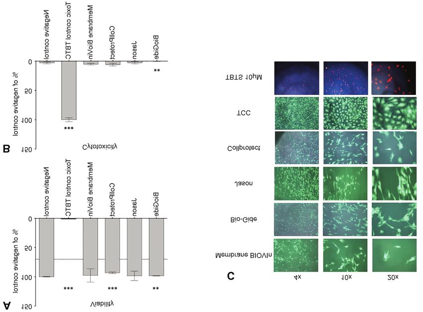
2037in vivo 35: 2035-2040 (2021) Figure 2. Cytocompatibility of membranes. A) Viability B) Cytotoxicity. The mean absorbance of controls without cells was subtracted from the mean absorbances. Columns represent mean values of quadruplicate measurements; error bars represent the standard deviation. The dotted line shows 70% of the negative control. Assays were performed following a 24 h incubation of MC3T3 cells with extracts of the materials. *, ** and ***: Significantly different from the negative control at p≤0.05, p≤0.01 and p≤0.001, respectively. C) Live-dead staining assay. Double staining with fluoresceindiacetate (FDA) and propidium iodide (PI) was performed on MC3T3 cells that were directly seeded and cultured for 24 h on the membrane specimens. The green dye FDA exclusively stains viable cells, dead cells with compromised plasma membrane integrity are stained by the red dye PI. Left, middle and right panels show images generated using a 4×, 10×, and a 20× objective, respectively. range as defined in DIN EN ISO 10993-5:2009 (Figure different from the negative control (p≤0.001), they still fall 1A). In concordance, no cytotoxicity was detected for any within the non-toxic range. of the tested bone substitute materials. The values in the LDH-assay were in a similar range as the negative control In vitro characterization of membranes. All tested membranes (cells incubated with cell culture medium) (Figure 1B). showed satisfactory cytocompatibility with viability >70% of Although cytotoxicity was low for all tested bone the negative control in the indirect assay (Figure 2A). In substitute materials (range=3-10% of the toxic control), concordance, no cytotoxicity was detected for any of the tested BIOVIn, OTOss and Nanobone were significantly different membranes in the indirect LDH assay. The values in the LDH- from the negative control with p≤0.05, p≤0.001 and assay were in a similar range as the negative control (cells p≤0.05, respectively. With viability around 70% of the incubated with cell culture medium) (Figure 2B). Although negative control, cells incubated in extracts of two of the viability was very similar to the negative control for all tested assessed bone substitute materials (BIOVin, OTOss) materials (range=93-98%), Collprotect showed significantly showed lower viability than cells incubated in extracts of lower viability (p≤0.001) and Bio-Gide showed significantly all other tested materials (around 100% of the negative lower viability (p≤0.01) as well as a significantly lower control). Even though these values are significantly cytotoxicity (p≤0.01) when compared to the negative control. 2038
Schafer et al: Cytocompatibility of Bone Substitute Materials and Membranes

  Large numbers of green fluorescein diacetate (FDA)                5 weeks and a complete degradation of the material after
positive vital cells and only sporadic red propidium iodide         eight months (17). In clinical practice, the use of BSMs and
(PI) positive dead cells were visible on all tested membranes       membranes can provide adequate bone regeneration
and on the non-toxic control material TCC (Figure 2C),              depending on the prevailing bone and volume defect (19).
confirming good cytocompatibility. The cells on all                 Despite the individual decision of which material can offer
membranes exhibited elongated, cell type characteristic             the best treatment to different patients’ indication, protection
morphologies, suggesting firm adherence to the materials.           against inflammatory reactions should be of first priority.
                                                                       Our study is limited in scope to an in vitro assessment of
Discussion                                                          bone substitute materials and membranes. To assess whether
                                                                    the observed minor differences can impact regenerative
Clinical success of biomaterials is strongly associated with        processes, further in vivo studies need to be conducted.
a sufficient biocompatibility (12). In this study, we assessed
7 different bone substitute materials and 4 membranes using         Conclusion
present, international standards (DIN EN ISO 10993-5:2009)
and an established pre-osteoblast cell line (MC3T3). EN ISO         The increasing demand for bone substitute materials in oral
10993-5:2009 defines viability ≥70% relative to the negative        and maxillofacial surgery is the reason for expanding research
control as nontoxic. Although we found minor differences in         into developing biocompatible bone substitute materials and
cytocompatibility among the assessed materials, all tested          membranes that can reduce donor site morbidity and infection
materials can be considered as cytocompatible.                      risk. The prerequisite for the clinical use of the materials are
   The examined BSMs are of different origin, mechanical and        standardized tests to assess the materials’ required
structural property. A unifying factor is their common objective    harmlessness in clinical application and biocompatibility in
to regenerate human bone in structure and function. Human           application. This preclinical investigation provides evidence
bone is comprised of 25% organic substances (collagen type          of cytocompatibility of the materials examined.
I), 65% inorganic matrix (hydroxyapatite) and 10% water, all
together contributing to its unique qualities (7, 8).               Conflicts of Interest
   As indicated above, autologous bone grafts have a stand-
alone position in bone regeneration approaches. Nevertheless,       OT Medical GmbH provided the test specimens for the experimental
                                                                    investigation. The design, documentation, and analyses of this study
promising natural alternatives are allogenic and xenogenic
                                                                    were completed entirely independent of OT Medical. The Authors
grafts. Former grafts can be derived from either living donations   declare that they have no conflicts of interest to report.
or corpses. The tested specimens from BIOVin, Bio-Oss and
Cerabone belong to the latter, xenogenic category. They are         Authors’ Contributions
produced from bovine bones and therefore, mainly consist of
natural and mechanically stable inorganic hydroxyapatite (HA).      Schäfer S: data analysis; interpretation; drafted the manuscript. Al-
The feared risk of a “slow virus” transmission, e.g., Bovine        Qaddo H: data collection; critical revision; approval of the article;
spongiform encephalopathy (BSE) is almost excluded due to           Gosau M: critical revision; approval of the article. Smeets R:
their manufacturing process. Notably, some xenogenic materials      concept; critical revision; approval of the article. Hartjen P: concept;
                                                                    design; data collection; data analysis; critical revision of the article;
can contain additives that may enhance their properties, e.g.,
                                                                    Friedrich R: critical revision; approval of the article. Nada OA: data
BIOVin includes based on manufacturer’s information a               collection; data analysis; critical revision; approval of the article.
bioactive microcoat, composed of polymers and cell nutrients.       Vollkommer T: critical revision; approval of the article. Rashad A:
   Moreover, the synthetic specimens used in our study such         critical revision; approval of the article.
as Cerasorb, Otoss, and Maxresorb have a foundation of β-
Tricalcium-phosphate and ceramics. Natural and synthetic            Acknowledgements
BSMs can therefore serve as leading scaffolds for osteoblasts
and thus, act osteoconductively (7, 8). Still, they have minor      The Authors gratefully acknowledge Jane Rehberg for her excellent
osteogenic and osteoinductive properties (13-15) (Table II).        technical support.
                                                                       The Authors thank OT Medical GmbH (Bremen, Germany) for
   In contrast, composite materials were created to overcome
                                                                    providing the materials and funding for the experimental investigation.
disadvantages of common synthetic materials, by ideally
promoting osteoinductive properties. They are comprised of
a carrier material such as an osteoconductive scaffold,
                                                                    References
osteogenic cells and/or bone morphogenic proteins (BMPs)            1 Historical population data and projections (1950-2050).
(14, 16-18). Nanobone is a composite material, containing             Organisation for economic cooperation and development 2015.
76% HA and 24% silicon dioxide (SiO2). In vivo experiments            Available at: https://stats.oecd.org/Index.aspx?DataSetCode=
for Nanobone observed the formation of trabecular bone after          POP_PROJ [Last accessed on April 10, 2021]

                                                                                                                                       2039
in vivo 35: 2035-2040 (2021) 2 Trajkovski B, Jaunich M, Müller WD, Beuer F, Zafiropoulos GG 12 Murray PE, García Godoy C and García Godoy F: How is the and Houshmand A: Hydrophilicity, viscoelastic, and biocompatibilty of dental biomaterials evaluated? Med Oral physicochemical properties variations in Dental Bone Grafting Patol Oral Cir Bucal 12(3): E258-E266, 2007. PMID: 17468726. Substitutes. Materials (Basel) 11(2): 215, 2018. PMID: 13 Takagi S, Chow LC, Markovic M, Friedman CD and Costantino 29385747. DOI: 10.3390/ma11020215 PD: Morphological and phase characterizations of retrieved 3 Griffin K, Davis K, Mckinley T, Anglen J, Chu T, Boerckel J calcium phosphate cement implants. J Biomed Mater Res 58(1): and Kacena M: Evolution of Bone Grafting: bone grafts and 36-41, 2001. PMID: 11152995. DOI: 10.1002/1097- tissue engineering strategies for vascularized bone regeneration. 4636(2001)58:13.0.co;2-# Clinical Reviews in Bone and Mineral Metabolism 13(4): 232- 14 Cipitria A, Reichert JC, Epari DR, Saifzadeh S, Berner A, Schell 244, 2020. DOI: 10.1007/s12018-015-9194-9 H, Mehta M, Schuetz MA, Duda GN and Hutmacher DW: 4 Donati D, Zolezzi C, Tomba P and Viganò A: Bone grafting: Polycaprolactone scaffold and reduced rhBMP-7 dose for the historical and conceptual review, starting with an old manuscript regeneration of critical-sized defects in sheep tibiae. by Vittorio Putti. Acta Orthop 78(1): 19-25, 2007. PMID: Biomaterials 34(38): 9960-9968, 2013. PMID: 24075478. DOI: 17453388. DOI: 10.1080/17453670610013376 10.1016/j.biomaterials.2013.09.011 5 Kolk A, Handschel J, Drescher W, Rothamel D, Kloss F, 15 Schliephake H, Zghoul N, Jäger V, van Griensven M, Zeichen J, Blessmann M, Heiland M, Wolff KD and Smeets R: Current Gelinsky M and Szubtarsky N: Bone formation in trabecular trends and future perspectives of bone substitute materials - from bone cell seeded scaffolds used for reconstruction of the rat space holders to innovative biomaterials. J Craniomaxillofac mandible. Int J Oral Maxillofac Surg 38(2): 166-172, 2009. Surg 40(8): 706-718, 2012. PMID: 22297272. DOI: 10.1016/ PMID: 19121923. DOI: 10.1016/j.ijom.2008.11.018 j.jcms.2012.01.002 16 Mai R, Reinstorf A, Pilling E, Hlawitschka M, Jung R, Gelinsky 6 TMR: Dental membrane and bone graft substitutes market. M, Schneider M, Loukota R, Pompe W, Eckelt U and Stadlinger Available at: https://www.transparencymarketresearch.com/ B: Histologic study of incorporation and resorption of a bone dental-membrane-bone-graft-substitutes-market.html [Last cement-collagen composite: an in vivo study in the minipig. Oral accessed on April 21, 2021] Surg Oral Med Oral Pathol Oral Radiol Endod 105(3): e9-14, 7 Smeets R, Hanken H, Jung O, Rothamel D, Handschel J, Al-dam A, 2008. PMID: 18280955. DOI: 10.1016/j.tripleo.2007.09.016 Blessmann M, Heiland M and Kolk A: Knochenersatzmaterialien. 17 Abshagen K, Schrodi I, Gerber T and Vollmar B: In vivo Der Freie Zahnarzt 58(10): 78-88, 2020. DOI: 10.1007/s12614-014- analysis of biocompatibility and vascularization of the synthetic 1989-4 bone grafting substitute NanoBone. J Biomed Mater Res A 8 Smeets R, Hanken H, Beck-broichsitter B, Gröbe A, Precht C, 91(2): 557-566, 2009. PMID: 18985779. DOI: 10.1002/ Heiland M and Jung O: Knochenersatzmaterialien. Der Freie jbm.a.32237 Zahnarzt 60(5): 76-87, 2020. DOI: 10.1007/s12614-015-5461-x 18 Ren J, Blackwood KA, Doustgani A, Poh PP, Steck R, Stevens 9 Sheikh Z, Javaid MA, Hamdan N and Hashmi R: Bone MM and Woodruff MA: Melt-electrospun polycaprolactone regeneration using bone morphogenetic proteins and various strontium-substituted bioactive glass scaffolds for bone biomaterial carriers. Materials (Basel) 8(4): 1778-1816, 2015. regeneration. J Biomed Mater Res A 102(9): 3140-3153, 2014. PMID: 28788032. DOI: 10.3390/ma8041778 PMID: 24133006. DOI: 10.1002/jbm.a.34985 10 Bhatia SK and Yetter AB: Correlation of visual in vitro 19 Kaneko A, Marukawa E and Harada H: Hydroxyapatite cytotoxicity ratings of biomaterials with quantitative in vitro cell nanoparticles as injectable bone substitute material in a vertical viability measurements. Cell Biol Toxicol 24(4): 315-319, 2008. bone augmentation model. In Vivo 34(3): 1053-1061, 2020. PMID: 17932777. DOI: 10.1007/s10565-007-9040-z PMID: 32354892. DOI: 10.21873/invivo.11875 11 Jung O, Smeets R, Porchetta D, Kopp A, Ptock C, Müller U, Heiland M, Schwade M, Behr B, Kröger N, Kluwe L, Hanken H and Hartjen P: Optimized in vitro procedure for assessing the cytocompatibility of magnesium-based biomaterials. Acta Received March 11, 2021 Biomater 23: 354-363, 2015. PMID: 26073090. DOI: 10.1016/ Revised April 20, 2021 j.actbio.2015.06.005 Accepted April 21, 2021 2040
You can also read