Open Fractures Ivan S. Tarkin, MD Professor, Chief of Orthopaedic Trauma University of Pittsburgh Medical Center (UPMC) - Orthopaedic Trauma ...
←
→
Page content transcription
If your browser does not render page correctly, please read the page content below
Open Fractures
               Ivan S. Tarkin, MD
    Professor, Chief of Orthopaedic Trauma
University of Pittsburgh Medical Center (UPMC)

                                                 Core Curriculum V5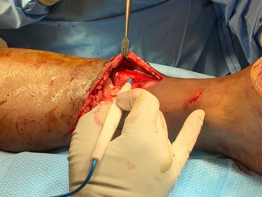
Objectives
    Open Fracture “Take-Home Points”
• Modern medicosurgical management has improved outcomes for
  patients with open fractures
• Open fractures, however, are infamous for complications
    • I.e., Infection, nonunion, malunion, chronic pain, physical and mental dysfunction
    • Increased risk with higher grade and compromised host
• Timely antibiotic administration, debridement, and fracture stabilization are of paramount
  importance
• Orthoplastic reconstruction is required for severe open fracture with soft tissue and/or bone loss
• Standard and innovative treatment strategies are required for limb salvage
• Amputation should be considered as part of the reconstructive armamentarium

                                                                                      Core Curriculum V5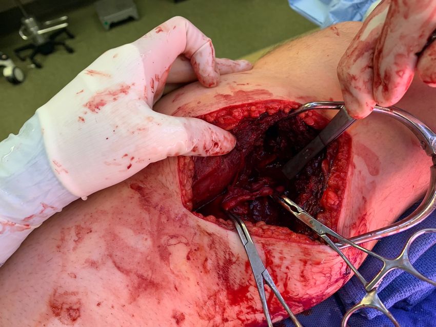
History, BUT… We have made progress
• Open fractures were associated with high (~40%) mortality rate due to sepsis
  (Billroth 1866). In his series of 93 open tibia fractures: 36 deaths, 28 amputations.
• Today, timely antibiotics, thorough fracture debridement, and orthoplastic
  reconstruction can yield successful limb salvage for severe open fractures
• Recent large database study showed that amputation rate for open tibia fracture
  between 2-3% (Mundy Orthopedics 2021). If mortality occurs after open tibia
  fracture today, often due to other injuries or medical complication.
                                                                Two young men with 3B
                                                                open tibia fractures s/p
                                                                orthoplastic reconstruction
                                                                sharing their experiences of
                                                                 limb salvage

                                                                                  Core Curriculum V5
Mechanism - Low Energy
                                                                             Geriatric open distal third
• Frailty                                                                    tibia fracture requiring soft
                                                                             tissue coverage procedure
• Poor skin quality                                                          for limb salvage

• Osteoporosis

                      • Geriatric open ankle fracture/dislocation (lateral
                        malleolar insufficiency fracture & deltoid tear)
                        with typical transverse medial wound
                      • Valgus moment on thin skin causing tearing and
                        herniation of distal tibia
                                                                                       Core Curriculum V5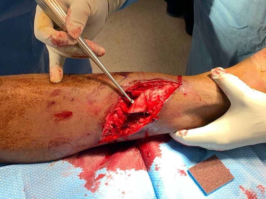
Open femur

  Mechanism - High Energy     fracture after
                              high-speed traffic
                              accident with
• Young or old patient        critical-sized bone
                              defect and soft
• I.e., MVA, MCA, ped vs      tissue loss after
  motor vehicle               open fracture care

• Large zone of injury
• Devitalized bone and soft
  tissue

                                 Core Curriculum V5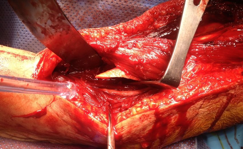
• Grossly
Open Fracture from Lawn Mower                                                     contaminated
                                                                                • Severe bone and
                                                                                  soft tissue injury

  Open midfoot fracture/dislocation from lawn mower injury managed with serial
  I&D, limited internal fixation, trans-articular wire fixation, and hybrid ex fix   Core Curriculum V5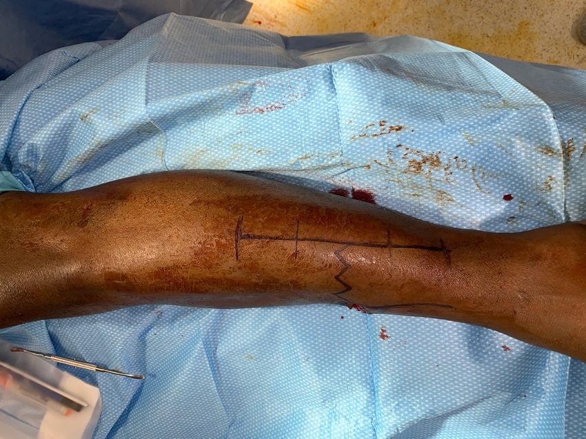
Open Fracture from Gunshot Wound (GSW)
• Variable degree of bone and
  soft tissue injury based on
  energy/velocity of projectile

-Low-energy gunshot treated with
local wound care/fracture
stabilization
-High-energy gunshot treated with
formal I&D with removal of all
devitalized soft tissue and bone

         Ayoub & Tarkin. Best care paradigm to optimize functionality after extra-articular distal humeral
         fractures in the young patient. J Clin Orthop Trauma 2018 (with permission from Elsevier)           Core Curriculum V5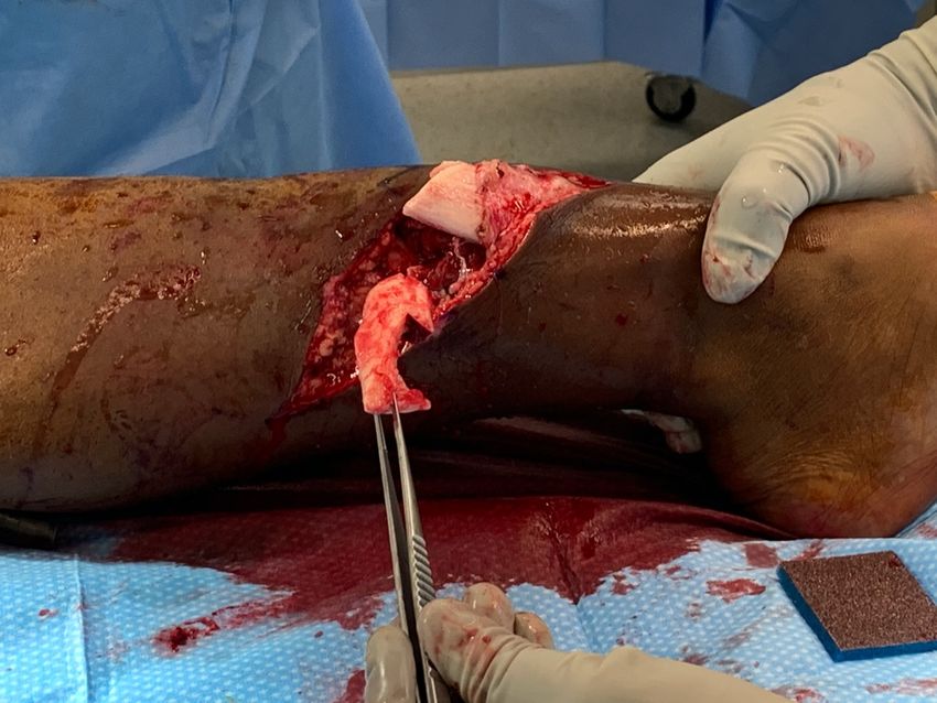
High-Energy GSW
               35yo s/p high-energy GSW treated with I&D (x3), nail, antibiotic beads
               Delayed autogenous bone grafting/supplemental plate fixation

50-caliber
muzzleloader rifle at
short range with
sabot (plastic) from
projectile in thigh

                                                                                        Core Curriculum V5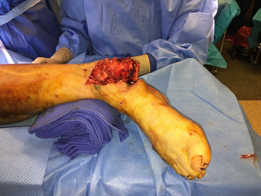
Impending Open Fracture
Urgent reduction/fracture stabilization
necessary to avoid open fracture

           Skin buttonholed in                                    Open humeral shaft fracture after
           proximal humeral fracture                              attempted closed management using
                                                                  Sarmiento brace in alcoholic

      Skin tenting from clavicle fracture can   Wawrose, Tarkin et al. Temporizing external fixation vs splinting
      cause necrosis and open fracture          following ankle fracture dislocation. Foot Ankle Int 2020.
                                                                                             Core Curriculum V5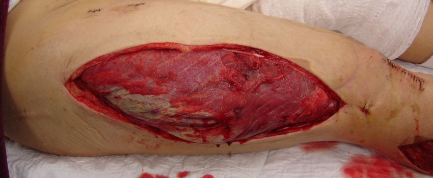
Open Fracture Mechanisms
Inside Out vs Outside In

Fracture displacement causes penetration of the
soft tissue envelope and open wound
vs.
Environmental object directly penetrates skin and
soft tissue sleeve causing fracture/wound

                                         Inside out -
                                         Multiply co-morbid patient with low-
                                         energy open tibia fracture

                                         Predisposition to injury based on poor
                                         bone and skin quality

                             Outside in -
                             Dirt bike handlebar                                  Core Curriculum V5
Epidemiology
• Open fracture mechanisms
(Weber et al. Eur J Trauma Emerg Surg
2019 & FLOW Investigators NEJM 2015)
• MVC ~30%
• Motorcycle ~20%
• Fall ~20%
• MVC vs pedestrian ~15%
Open fracture more common when subcutaneous bones
involved (i.e., tibia, ankle)
Core Curriculum V5Classification of Open Fractures Gustilo-Anderson (1976 & 1984) “The Classic” • Type I – wound
Disadvantages of Gustilo-Anderson Open
    Fracture Classification
• Meant to be applied after initial debridement!
• Interobserver reliability ~60%
• Only directly applicable to tibia fractures
• Does not consider degree of bone loss after open fracture   Gustilo-Anderson Type???
  care
• Does not consider host factors related to healing/immune
  response

                                                                              Core Curriculum V5
Gustilo-Anderson Type I & II Open Fractures
The difference in Type I vs II is not only wound size
Type II injuries will have greater degree of soft tissue and bone injury
Type II open tibia fractures
Rate of complications such as infection and nonunion for Type I-
II injuries, however, are significantly less than Type III Injuries
Core Curriculum V5Gustilo-Anderson Type III Open Fractures
High-energy injuries with significant damage to the soft tissue sleeve and bone
Heightened risk of complication (infection, nonunion, chronic pain/dysfunction)
IIIA open tibia fractures will have
sufficient soft tissue coverage
IIIB open tibia fractures will require
IIIB soft tissue coverage, typically
rotational or free flap
Significant debridement of crushed dysvascular
tissue, particularly muscle, is typically the rule!
Core Curriculum V5Type IIIC - Open Fracture with Vascular Injury
Critically ill polytrauma patient s/p MCA with 3C
open contaminated tibia fracture with significant
bone loss, profound degree of nonviable muscle,
and pulseless foot secondary to vascular injury
Amputation performed as definitive
reconstructive option using multi-disciplinary
decision-making process
Core Curriculum V5Classification of Open Fractures (OTA)
OTA classification
(OTA Study Group- JOT 2010)
• Based on 5 categories:
1) Skin injury
2) Muscle injury
3) Arterial injury
4) Contamination
5) Bone loss
May be superior to Gustilo-Anderson in predicting outcomes (Hao et al. JOT 2016)
Core Curriculum V5Open Fracture-Associated Conditions
Nerve Injury
Peripheral nerves are vulnerable to injury, with greater fracture displacement
typically seen with open fractures secondary to direct injury and/or stretch
Type I open segmental humeral fracture associated radial nerve neuropraxia
Radial nerve injury infamously at risk with distal 1/3 fractures (Holstein-Lewis)
Core Curriculum V5Open Fracture-Associated Conditions
Vascular Injury
Reverse saphenous vein graft for arterial
injury after open elbow fracture/dislocation
Core Curriculum V5Open Fracture-Associated Conditions
Tendon and Ligament Injuries
• Beyond critical neurovascular
structures, optimal
functionality predicated on
uneventful healing of tendon
and ligament injuries
• Either acute or delayed
reconstruction indicated to
promote best outcomes
Core Curriculum V5Open Fracture-Associated Conditions
Compartment Syndrome Necrotic muscle from
delayed compartment
Open fractures are at increased risk release in open tibia fracture
for CS necessitating fasciotomy
Low threshold to perform fasciotomy in case of
—especially high-energy cases high-energy open fracture to avoid
compressive ischemia of traumatized muscle
Floating knee (open fractures
of the femur and tibia)
managed with I&D of open
wounds, retrograde femoral
nail, antegrade tibial nail, and
fasciotomies of the thigh and
leg for associated compartment
syndrome
Core Curriculum V5Antibiotics – Timing & Type • Crucial to start antibiotics early • Obremskey et al. JOT 2014 – 86% of orthopaedic trauma surgeons responded that
Antibiotics - Duration
24 hours adequate for Type I and II open fractures (Hoff et al. J Trauma 2011)

                                                         Longer for Type III (72 hours?),
                                                         though length is controversial
                                                         (Chang et al. JBJS Rev 2015)

                                                      Contaminated Type III fractures of distal
                                                      femur, proximal tibia, and tibial pilon
                                                      given Zosyn within 1 hour of presentation
                                                      and continued for 72 hours

   Judgment!... Duration of therapy dictated on host and injury factors (ie severity of wound, patient co-morbidities)
                                                                                                  Core Curriculum V5
Antibiotics - Local
May be role for local antibiotics as well (Morgenstern et al. Bone Joint Res
2018)
Can be with or without carrier (e.g. beads/spacer)
Open femur Bone healing after
fracture from delayed bone
industrial grinder grafting/plating
accident (grossly over retained nail
contaminated
with bone defect)
Staged by on-call
surgeon with
cement beads in
bone defect as
antibiotic carrier
Core Curriculum V5Initial Care in Emergency Department
• Antibiotics! Tetanus, betadine/saline gauze, temporizing
immobilization (splint, skeletal traction, external fixation)
Field splint by EMS
with traumatic
wound covered
with betadine-
impregnated gauze
Type I???
Determine after debridement! “Halt the Cycle of Injury!”
Mehta et al 2010 JAAOS
Core Curriculum V5Timing of Operative Debridement
Earlier the better! from the “Journal of Common Sense”
As long as competent resources are available and patient physiologically stable
The Science
• “6-hour rule” - based on guinea pig
study in 1898
• No difference in infection rates
between early and late debridement
(Schenker et al. JBJS 2012)
• However, no argument that highly
contaminated wounds should go to OR
ASAP
Core Curriculum V5Principles of Open Fracture Wound Care • Extend the traumatic wound to appreciate zone of injury/facilitate comprehensive debridement • Preserve vascularity to the traumatic wound • AVOID sharp angles when extending wound (
Debride Open Fracture Through Surgical Wound

• Extension of traumatic wound commonplace for
  open fracture care
• However, if wound over exposed bone and
  cannot be closed primarily or delayed, flap
  coverage necessary

Alternative- close traumatic wound(s) over
exposed bone and make surgical wound to
debride open fracture, which typically will require
a split-thickness skin graft (STSG)

                                                      Core Curriculum V5
Debridement of Open Fracture
Remove any dysvascular,
nonviable tissue that will
ultimately serve as nidus for
infection (skin, subcutaneous
tissue, MUSCLE, and bone)
Healthy muscle will contract with
electrocautery stimulation
-less reliable after muscle has produced
granulation tissue in response to NPWT
Core Curriculum V5Osseous Debridement
Remove bone without meaningful soft tissue attachment
Debride IM canal (hidden environmental contamination)
“Tug Test”
(Southam et al.
JOT 2017)
Novel Technique- Lindvall E
JOT. 2015 Dec;29(12):558-62.
Critical-sized bone defect after debridement
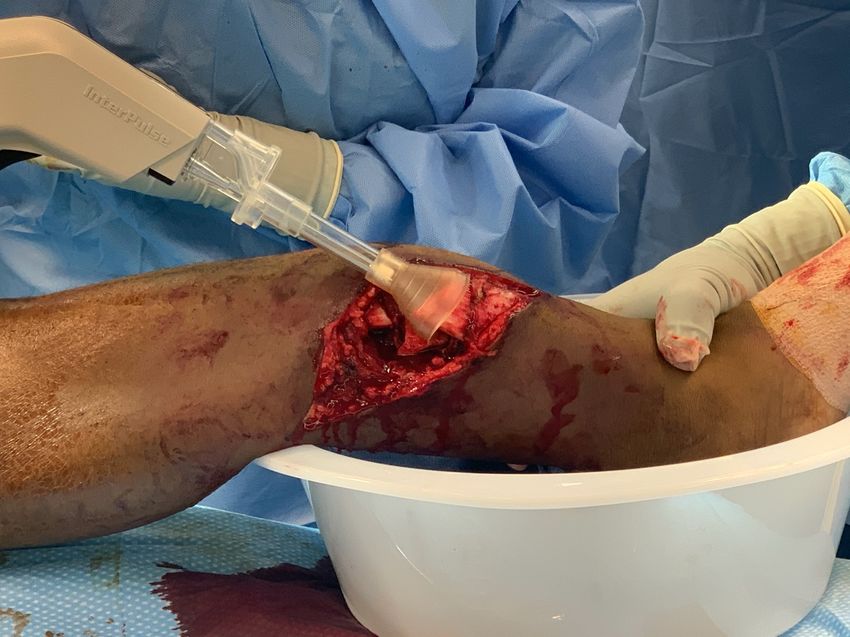
Core Curriculum V5Debridement and Irrigation
FLOW Investigators NEJM 2015 -RCT
• Low-pressure irrigation an
acceptable alternative to high
flow.
• Reoperation rate lower in saline
group (vs soap)
Pendulum has swung toward low-pressure irrigation
due to concerns about…
• Bone damage (Bhandari et al. JOT 1999) Traditional Teaching
• Damage to soft tissue envelope (Boyd et al. CORR 3L for Type I
2004, Chiramonti et al. JBJS 2017)
• Propagation of bacteria into soft tissue with high- 6L for Type II
pressure irrigation (Hassinger et al. CORR 2005) 9L for Type III
Core Curriculum V5Fracture Reduction
Take advantage of traumatic wound to
obtain fracture reduction
Core Curriculum V5Traumatic Wound Care
Primary closure preferred if skin edges are free of
significant tension after suture repair (Scharfenberger et al. JOT 2017)
Core Curriculum V5Zone of Injury
Case 2
• Open femur fracture/traumatic
knee arthrotomy in young woman
after MCA
• Traumatic wounds extended to
appreciate extent of bone, joint,
and soft tissue injury
• Organized debridement to remove
all devitalized soft tissue and bone
• Bone ends delivered to debride IM
canal
Core Curriculum V5Principles of Open Fracture Care
Nonviable tissue (including bone) serve as a nidus for infection
and should be excised
Type IIIA open distal
Level of aggressiveness debatable
femur fracture after MCA
BUT… “When in Doubt Take It Out”
• Bone without
meaningful vascular Dugan, Tarkin, et al. Open supracondylar femur
attachment removed fractures with bone loss in the polytraumatized
patient–Timing is everything! Injury 2013.
Ricci et al. A comparison of more and less
aggressive bone debridement protocols for the
treatment of open supracondylar femur
fractures. JOT 2013.
Core Curriculum V5Remove Environmental Contamination
• Extent of contamination may
not be obvious
• The entire zone of injury needs
to be explored to remove
environmental debris in
addition to nonviable tissue
• IM canals need to be exposed
and debried as well
Core Curriculum V5Skeletal Stabilization of Open Fracture - External Fixation
• Polytrauma - damage-control orthopaedics (DCO)
• Complex peri-articular fractures
• Critically ill, under-resuscitated
polytrauma patient with
multiple open fractures
• Rapid I&D/ex fix as temporizing
skeletal stabilization
Pape, Tarkin et al. Timing of fracture fixation in
multitrauma patients: the role of early total care and
damage control surgery. JAAOS 2009.
Core Curriculum V5External fixation
Definitive skeletal stabilization for severe open fracture
Indications:
• Gross environmental
contamination
• Contraindication to internal
fixation present
• Anticipated need for
lengthening procedure
Core Curriculum V5Internal Fixation for Open Fracture
• If a clean wound bed achievable after open fracture care, definitive
fixation is INDICATED at index procedure
• Intramedullary nailing is the “workhorse” for most open diaphyseal
and extra-articular meta-diaphyseal cases of the lower extremity
• Plate osteosynthesis is typically employed for open peri-articular cases
Exacting
reduction of
intra-articular
fractures is of
paramount
importance for
best outcomes
Core Curriculum V5Intramedullary Nailing for Open Fracture
Nailing limits surgical exposure necessary for fracture stabilization - “biologic fixation”
Avoids tension on soft tissue sleeve (vs plating), thereby facilitating traumatic wound closure
42yo with Type II open tibia fracture after MCA
Traumatic wound closed primarily after I&D with reamed nail
Core Curriculum V5Intramedullary Nailing for Open Fracture
Excellent bridging device for open diaphyseal fractures with bone loss
Core Curriculum V5Intramedullary Nailing for Open Fracture
Positioning plate as adjunct in open tibia
Reduction with open manipulation through wound
69yo s/p fall down steps with Type II open distal 1/3 tibia fracture
Core Curriculum V5Segmental Open Tibia Fracture
Nailing adjuncts
• Small fragment plate used
as preliminary reduction
tool prior to nailing
• Plate prevents segmental
piece from “spinning”
during reaming process
• Increases construct
rigidity
Core Curriculum V5Nailing for Open Femur Fracture
• Retrograde nail is an
effective and efficient
technique for open femur
fracture in the polytrauma
patient
• Zone of injury is explored
through extension of
traumatic wound
• Nonviable skin,
subcutaneous tissue,
fascia, MUSCLE, and bone
excised
18yo polytrauma patient with Type I open femur fracture after MVC • Bone ends harvested and
s/p retrograde IMN with uneventful healing IM canals debrided
Core Curriculum V5Open Peri-Articular Fracture - Goals of Care
• Anatomic joint reconstruction (with intra-articular involvement)
• Alignment of reconstructed articular block to shaft
• Restoration of length, alignment, rotation of articular block to shaft
Core Curriculum V5Managing Bone and Soft Tissue Defects
after Open Fracture
• Early primary closure should be
attempted if soft tissues allow
• Soft tissue coverage should be obtained
within 7-10 days (Melvin et al. JAAOS
2010) if critical-sized soft tissue defect
• Bone grafting strategies should typically
be performed delayed once soft tissue
envelope has healed/revascularized
without signs of infection
Core Curriculum V5Open Fracture Wound Management
• Early primary closure of the
traumatic open fracture
wound preferred
• Healing the traumatic
wound integral to avoiding
infectious complications
• Caveat - Skin edges
vascularized and closure
without excessive tension
Core Curriculum V5Negative Pressure Wound Therapy (NPWT)
Temporizing option for critical-sized soft tissue defect associated with open
fracture
• 3B open tibia fracture in
• Protects/seals open fracture polytrauma patient
wound from environmental • Wound vac/ex fix used as
contamination temporizing measure as part of
damage-control orthopaedics
(nosocomial infection)
• Negative pressure applied to
traumatic wound promotes
neovascularization/wound
granulation and removes
deleterious edema fluid
• Prepares tissue bed for soft
tissue coverage (e.g., flap) Tarkin. The versatility of negative pressure wound therapy with
reticulated open cell foam for soft tissue management after severe
musculoskeletal trauma. JOT 2008.
Core Curriculum V5Soft Tissue Coverage after Open Fracture
• Options depend on location of injury
and presence of exposed bone (Boss
and Buehler JAAOS 1994)
• For tibia fracture with exposed bone
and/or joint
• Proximal third - gastrocnemius
rotational
• Middle third - soleus rotational
• Distal third - free flap
Timing of coverage after open tibia < 7 days
with interval vac coverage recommended
D'Alleyrand JOT 2014, Rotational flap/STSG used to cover knee and
Hou J Trauma 2011 fractures of the proximal/middle tibia
Pincus JOT 2019
Core Curriculum V5Management of Bone Defect Associated with Open Fracture
Autogenous bone grafting (delayed!)
Autogenous bone grafting strategies are reliable after…
• Soft tissue envelope has recovered (re-vascularized)
• Rigid mechanical environment present
Reamer Irrigator
Aspirator (RIA)
method
as alternative
Iliac crest bone graft—
gold standard Quintero, Tarkin, Pape. Technical
• Osteoconductive tricks when using the reamer
irrigator aspirator technique for
• Osteoinductive autologous bone graft harvesting.
JOT 2010.
• Osteogenic
Core Curriculum V5Posterolateral Bone Grafting
• Classic strategy for bone grafting open tibia fracture with nonunion
and/or critical-sized bone defect
• Nonunion debrided of fibrous scar
Nonunion after open fracture initially
• Medial portion of fibula decorticated managed with nail
• Graft deposited on
interosseous membrane from
fibula to tibia
• “Creation of synostosis”
Core Curriculum V5Management of Bone Defect after Open Fracture
Masquelet technique
• Critical-sized bone defect often realized after open fracture care
• Masquelet technique uses antibiotic spacer (Masquelet et al. Orthop Clin North Am
2010)
• Sterilization of previously contaminated environment
• Creates of vascularized space/envelope ideal for delayed bone grafting
• Open ulna fracture with critical-sized
bone defect after open fracture care
managed initially with plate
osteosynthesis/antibiotic spacer
• Tricortical grafting then performed in
delayed fashion
Core Curriculum V5Open Distal 1/3 Tibia with Bone Defect
Induced membrane technique
26yo s/p multiple GSW • Critical-sized defect after • Removal of beads
debridement • Autograft applied in
• IM nail as bridging device vascularized pocket
• Antibiotic beads applied to • Plate over retained nail
defect Core Curriculum V5Open Supracondylar Femur Fracture
Masquelet technique
Figures from – Dugan, Tarkin et
al. Open supracondylar femur
fractures with bone loss in the
polytraumatized patient–Timing
is everything! Injury 2013
(with permission from Elsevier)
Core Curriculum V5Management of Bone Defect after Open Fracture
Acute shortening (+/- lengthening)
• Technique used to simplify management of open fracture with both bone and
soft tissue defects
• Typically reserved for severe cases in lower-demand hosts
• Option for delayed lengthening procedure
• Open pilon fracture with • Bone transport options include
metaphyseal bone loss those such as the Ilizarov
and transverse medial technique (Cattaneo et al. CORR
wound 1992) or newer techniques using
magnetic intramedullary limb
• Traumatic wound could lengthening system (Barinaga et al.
be closed without JAAOS 2018)
tension after shortening
Core Curriculum V5Salvage for Severe Open Fracture
(limb threatening)
• Requires individualized treatment plan based on injury, host factors, patient
goals and expectations
• Standard, unconventional, and innovative treatment options
• Think “Out of the Box” when standard treatments not reliable (Cole 2006)
• Intramedullary nailing
• Open reduction internal fixation
Clinical judgment!!! • External fixation
• Selected fusion
• Arthroplasty
• Combinations of above or OTHER
Amputation (i.e., BKA, AKA) should also be considered as part of
the treatment armamentarium (as opposed to “treatment failure”) Core Curriculum V5Arthroplasty for Open Fracture
Lower extremity
Used for primary reconstruction or for nonunion, malunion, arthrosis
after open peri-articular fracture - esp peri-prosthetic in geriatric host
TEAMWORK!!!
78yo with open femur fracture after
horseback riding accident failed ORIF
with infected nonunion - staged DFR
Hoellwarth, Tarkin et al. Equivalent mortality and complication rates following periprosthetic distal femur
fractures managed with either lateral locked plating or a distal femoral replacement. Injury 2018.
Chen, Tarkin, et al. Primary versus secondary distal femoral arthroplasty for treatment of total knee
arthroplasty periprosthetic femur fractures. J Arthroplasty 2013.
Pushilin & Tarkin. Management of distal femoral nonunions. Operative Techniques in Orthopaedics 2018.
Moloney, Tarkin et al. Geriatric distal femur fracture: are we underestimating the rate of local and systemic
complications? Injury 2016.
Core Curriculum V5Innovative Treatment Solutions for Open Fracture
Metal talus?
25yo s/p close-range GSW to talus,
which was not amenable to ORIF.
Patient did not want to pursue TTC
fusion.
Core Curriculum V5Select open peri-articular fractures
Formal Arthrodesis with profound cartilage injury or loss
for open peri-articular fracture
Severe open talus fracture
with bone/joint loss after MCC
managed with TTC fusion and
delayed bone grafting
Tricortical graft from pelvis
to span tibia to calcaneus
Tarkin & Fourman. Retrograde hindfoot nailing for
acute trauma. Curr Rev Musculoskelet Med 2018.
Core Curriculum V5Unconventional Open Fracture Stabilization
Temporary trans-articular fixation
Open distal radius fracture (transverse volar wound) with diminutive articular block treated with open fracture
care and dorsal spanning plate to internally splint the fracture and soft tissue wound)
Core Curriculum V5Decision for Limb Salvage
The decision for amputation vs
salvage is typically not as clear
as in this extreme case
Core Curriculum V5Amputation
Definitive reconstructive option after severe open fracture (ieBKA)
LEAP Study
• 601 patients
• Multicenter
• No significant difference
in functional outcomes for
limb salvage vs amputation
MacKenzie EJ, Bosse MJ. Factors influencing
outcome following limb-threatening lower BKA with posterior
limb trauma: lessons learned from the
Lower Extremity Assessment Project (LEAP).
myofascial cutaneous flap
JAAOS 2006
S/p open distal femur fracture and mangled leg
managed with open fracture care after boating
propeller - distal femur ORIF and BKA.
Patient returned to water skiing with prosthesis Core Curriculum V5Complications after Open Fracture
• Unfortunately common
• Infection
• Nonunion
• Malunion
• Arthrosis
• Chronic pain
• Physical dysfunction
• Psychosocial distress
Core Curriculum V5Open Fracture Complications
Infection
Etiology of infection after open
fracture secondary to…
environmental contamination,
compromised soft tissue envelope
(i.e., vascularity), delayed or frank
failure of wound healing, and/or host
factors (polytrauma, co-morbid
conditions, nutritional deficiency)
Core Curriculum V5Open Fracture Complications
Nonunion Sands, Siska, Tarkin. Reconstructive strategies for skeletal complications in the polytrauma
patient. In The Poly-Traumatized Patient with Fractures 2011 (pp. 333-344). Springer, Berlin,
Heidelberg.
Ineffective healing
response after
bridge plating
“Anticipated” nonunion -
Critical-sized bone defect
initially managed with
antibiotic beads
Core Curriculum V5Open Fracture Complications
Deformity/Malunion
• Patient referred after treatment for open tibia fracture
with ambulatory dysfunction secondary to varus
malunion
• Post-traumatic reconstruction (osteotomy) required to
restore mechanical axis
Infected nonunion/malunion (varus collapse) treated with
serial debridements, interval external fixation, osteotomy,
and tricortical wedge graft/Internal fixation
Tarkin et al. Soft tissue and biomechanical challenges encountered with the
management of distal tibia nonunions. Orthop Clin 2010.
Core Curriculum V5Open Fracture Outcomes Bowen CORR 2005
N=195 open fractures
As compared to Type A (healthy) Hosts
Type B had 2.86X infection risk
Infection, nonunion, and secondary amputation Type C had 5.72X infection risk
predicted by host factors and severity of injury
Westgeest JOT 2016
N=791 open fractures
• Infection and higher Gustillo Grade
Functional outcomes improving, but associated with risk of fx nonunion
significant impacts on health-related
quality of life Giannoudis at al. Injury 2009- Grade 3B/C open tibia fractures
associated with ongoing pain and difficulties with activities
Outcomes of severe open fractures most commonly predicated
on “economic, social, and personal resources”
MacKenzie & Bosse. [LEAP] JAAOS 2006
Core Curriculum V5Summary
• Open fractures are a surgical urgency requiring thorough
debridement/irrigation and skeketal stabilization
• External and internal fixation, amputation, as well as innovative strategies
are necessary for skeletal reconstruction of open fractures
• Orthoplastic reconstruction is often necessary for open fracture with soft
tissue loss
• Complications are commonplace with open fracture including infection and
nonunion as well as physical and psychosocial dysfunction
• Host, injury pattern, and treatment plan will influence outcome after open
fracture
Core Curriculum V5Acknowledgement
Special thanks to Dr. Peter Mittwede
for assistance with literature review
and presentation edits
Core Curriculum V5Thank you for your attention…
s/p IIIB open tibia fracture
enjoying his grandchildren
Core Curriculum V5You can also read


