MiR 146b correlates with increased disease activity and psoriatic tissue inflammation and promotes keratinocyte proliferation in psoriasis
←
→
Page content transcription
If your browser does not render page correctly, please read the page content below
EXPERIMENTAL AND THERAPEUTIC MEDICINE 21: 296, 2021

               miR‑146b correlates with increased disease activity
                and psoriatic tissue inflammation and promotes
                    keratinocyte proliferation in psoriasis
                      LING ZHANG1, SHENGLAN ZHANG2, JIANBIN WANG1 and XIAOJING LI1

                      1
                       Department of Dermatology, Affiliated Hospital of Hebei University of Engineering;
                       2
                       Medical Department, Handan Central Hospital, Handan, Hebei 056002, P.R. China

                                        Received May 24, 2019; Accepted October 1, 2020

                                                    DOI: 10.3892/etm.2021.9727

Abstract. The present study aimed to investigate the expres‑          (95% CI: 0.720‑0.843). In addition, miR‑146b expression in
sion of microRNA (miR)‑146b in psoriatic tissue and its               psoriatic tissue was correlated with an increased PASI score in
correlation with psoriasis activity and inflammation. The             patients with psoriasis. miR‑146b expression in psoriatic tissue
effect of miR‑146b overexpression on keratinocyte prolif‑             was positively correlated with TNF‑ α, interleukin (IL)‑6
eration and apoptosis was also explored. The expression of            and IL‑17 mRNA levels. In vitro, miR‑146b overexpression
miR‑146b in the psoriasis‑affected tissue and non‑affected            enhanced HaCaT cell proliferation and suppressed apoptosis
tissue of 110 patients was determined via reverse transcrip‑          as well as TRAIL‑induced apoptosis when compared with that
tion‑quantitative (RT‑q)PCR. The psoriasis‑affected body              in control‑transfected HaCaT cells. In conclusion, miR‑146b
surface area and psoriasis area severity index (PASI) score           was positively correlated with disease activity and psoriatic
were recorded for evaluating disease activity. The expression         tissue inflammation. Keratinocyte proliferation was also
of various inflammatory cytokines in psoriasis‑affected tissue        promoted in psoriasis.
was also detected via RT‑qPCR. miR‑146b overexpression and
control plasmids were constructed and transfected into HaCaT          Introduction
cells in vitro. Subsequently, cell proliferation, apoptosis and
tumor necrosis factor (TNF)‑related apoptosis‑inducing                Psoriasis is a common autoimmune disease that is char‑
ligand (TRAIL)‑induced cell apoptosis were determined. The            acterized by thick erythematous skin plaques, affecting
results revealed that the expression of miR‑146b was increased        0.91‑8.5% of the global population (1,2). The high preva‑
in psoriasis‑affected tissue compared with that in unaffected         lence and long disease course of psoriasis cause substantial
tissue. The results obtained from a receiver operating charac‑        economic and psychological burdens to individuals and their
teristic curve analysis demonstrated that miR‑146b levels were        families (3,4). In addition, psoriasis is a type of chronic inflam‑
able to discriminate between psoriasis‑affected tissue and            matory condition and is classified as a T‑helper (Th)1 disease,
unaffected tissue, with an area under the curve value of 0.781        as the cytokines involved in the Th1 pathway [including tumor
                                                                      necrosis factor α (TNF‑α), interleukin (IL)‑6, IL‑17 and IL‑22
                                                                      expression] are increased in the lesional skin and peripheral
                                                                      blood of patients (5). Although several therapeutic approaches
Correspondence      to: Professor Xiaojing Li, Department             (including topical therapy, phototherapy, systemic non‑biologic
of Dermatology, Affiliated Hospital of Hebei University of            treatments and biologic treatment) are able to decrease the
Engineering, 81 Congtai Road, Handan, Hebei 056002, P.R. China        disease activity in the majority of circumstances, there is still
E‑mail: huan91439141@163.com                                          no cure for psoriasis (6,7). Therefore, it is paramount to inves‑
                                                                      tigate the underlying mechanisms of the pathology of psoriasis
Abbreviations: miRNAs, microRNAs; RA, rheumatoid arthritis;           to further explore potential treatment options.
ATD, autoimmune thyroid diseases; BMI, body mass index; BSA,
                                                                          MicroRNAs (miRNAs/miRs) are a group of small
body surface area; PASI, psoriasis area and severity index; TNF‑α,
                                                                      non‑coding RNAs comprised of 22 nucleotides (8).
tumor necrosis factor α; IL, interleukin; TRAIL, TNF‑related
apoptosis‑inducing ligand; ROC, receiver operating characteristic;    Accumulating studies have revealed that miRNAs are widely
AUC, area under curve; TRAF6, TNF receptor‑associated factor 6;       distributed in animals, plants and certain viruses, and they
IRAK‑1, IL‑1 receptor‑associated kinase 1; imDCs, immature            have been determined to be key regulators of numerous
dendritic cells; mDCs, mature DCs                                     important biological processes, including cell prolifera‑
                                                                      tion, differentiation and inflammation (9‑11). As one of the
Key   words: psoriasis,      microRNA‑146b,     disease   activity,   commonly investigated miRNAs, miR‑146b has been impli‑
inflammation, keratinocyte                                            cated in the malignant proliferation of various cancer cell
                                                                      types (including ovarian and papillary thyroid cancer cells)
                                                                      and several immune activities, including innate immune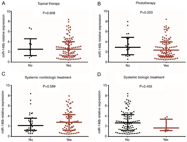
2                                               ZHANG et al: miR-146b IN PSORIASIS

sensing and pro‑inflammatory cytokine release (12‑16).              was performed using the following program: 95˚C for 60 sec,
Several studies have revealed that miR‑146b expression is           followed by 40 cycles of 95˚C for 15 sec and 61˚C for 30 sec.
increased in several autoimmune diseases [including rheuma‑
toid arthritis (RA), multiple sclerosis and autoimmune thyroid      Cell culture and transfection. The spontaneously immortal‑
diseases (ATD)] (17‑19). Considering that miR‑146b promotes         ized human keratinocyte cell line HaCaT was purchased
cell proliferation and has a role in the pathology of several       from Kunming Cell Bank and cultured in Dulbecco's modi‑
autoimmune diseases, and that psoriasis is a typical autoim‑        fied Eagle's medium (Gibco; Thermo Fisher Scientific, Inc.)
mune disease associated with malignant proliferation of             supplemented with 10% inactivated fetal bovine serum
keratinocytes, the present study hypothesized that miR‑146b         (Gibco; Thermo Fisher Scientific, Inc.), penicillin (100 U/ml)
may participate in the development and progression of psori‑        and streptomycin (100 mg/ml; both from Sigma‑Aldrich;
asis. Of note, the role of miR‑146b in the pathology of psoriasis   Merck KGaA). miR‑146b overexpression and control over‑
has so far remained elusive. Therefore, the present study aimed     expression plasmids (with non‑coding DNA fragment) were
to explore the expression of miR‑146b in psoriatic tissue and       constructed by Shanghai QeeJen Bio‑Tech Co., Ltd. and then
to determine its association with psoriasis activity and inflam‑    transfected into HaCaT cells, which were correspondingly
mation. In addition, the present study explored the effect of       designated as the miR‑146b+ group and the Control+ group.
miR‑146b overexpression on the regulation of keratinocyte           The relative expression of miR‑146b was detected at 24 h
proliferation and apoptosis.                                        post‑transfection via RT‑qPCR as described above. All cell
                                                                    experiments were performed in triplicate.
Materials and methods
                                                                    Measurement of cell proliferation and apoptosis. After
Patients. A total of 110 patients with psoriasis were enrolled      transfection, cell proliferation was detected at 0, 24, 48
at the Affiliated Hospital of Hebei University of Engineering       and 72 h using a Cell Counting Kit‑8 (Dojindo Molecular
(Handan, China) between January 2017 and June 2018.                 Technologies, Inc.). The rate of cell apoptosis was also
The inclusion criteria were as follows: i) Diagnosed with           detected at 24 h following transfection with an FITC
psoriasis vulgaris; ii) age >18 years; iii) consent to donate       Annexin V Apoptosis Detection kit II (BD Biosciences).
psoriasis‑affected skin tissue samples and normal skin tissue       The flow cytometer (BD Biosciences) was used for the
samples. The exclusion criteria were as follows: i) Complicated     Annexin V assay. All procedures were performed according
with inflammatory skin diseases other than psoriasis;               to the manufacturers' protocols.
ii) complicated with other systemic immune or inflammatory
diseases, including systemic lupus erythematosus or inflam‑         Measurement of TNF‑related apoptosis‑inducing ligand
matory bowel disease; iii) complicated with malignancies.           (TRAIL)‑induced apoptosis. At 48 h post‑transfection,
The present study was approved by the Ethics Review Board           200 ng/ml TRAIL (Sigma‑Aldrich; Merck KGaA) was added
of the Affiliated Hospital of Hebei University of Engineering       to HaCaT cells and the cells were incubated for a further 24 h.
(Handan, China). All patients provided written informed             The rate of cell apoptosis was then detected using an FITC
consent.                                                            Annexin V Apoptosis Detection kit II (BD Biosciences) to
                                                                    determine TRAIL‑induced apoptosis.
Disease assessment. After enrollment, the characteristics
of the patients, including their age, gender, body mass             Statistical analysis. SPSS Software 23.0 (IBM Corp.) was
index (BMI), disease duration and treatment options, were           used for statistical analysis and GraphPad Prism Software 7.01
documented. The psoriasis‑affected body surface area (BSA)          (GraphPad, Inc.) was used for visualization of quantitative
and psoriasis area severity index (PASI) scores were subse‑         results. Comparisons between two groups were performed
quently determined (20,21).                                         using Wilcoxon's signed rank‑sum tests. Comparisons
                                                                    between two individual groups were determined by
Sample collection and reverse transcription‑quantitative            Wilcoxon's rank‑sum tests and Student's t‑tests. Differences
(RT‑q)PCR. Psoriasis‑affected skin and normal skin tissue           in miR‑146b expression among groups were determined
samples were obtained via punch biopsy. The expression of           using the Kruskal‑Wallis H‑test. Correlations between two
miR‑146b, TNF‑ α, IL‑6, IL‑17 and IL‑22 in each sample              parameters were determined by Spearman's correlation test.
were measured via RT‑qPCR. Total RNA was extracted using            In addition, receiver operating characteristic (ROC) curves
TRIzol reagent (Invitrogen; Thermo Fisher Scientific, Inc.),        were drawn and the area under the curve (AUC) was calcu‑
after which the concentration, purity and integrity of samples      lated to determine the ability of the parameter to distinguish
was determined. Complementary DNA was then produced                 psoriasis‑affected skin tissue from normal skin tissue. P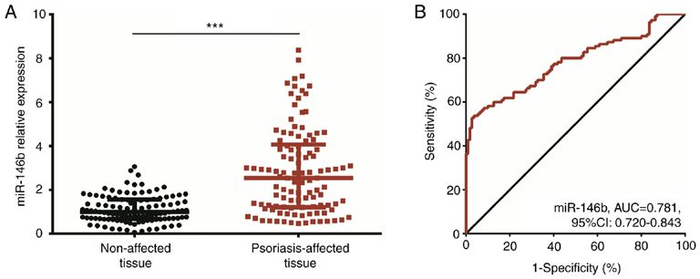
EXPERIMENTAL AND THERAPEUTIC MEDICINE 21: 296, 2021                                             3

Table I. Primer list.

Gene                                     Forward primer                                               Reverse primer

miR‑146b           5'‑ACACTCCAGCTGGGTGAGAACTGAATTCCA‑3'                               5'‑TGTCGTGGAGTCGGCAATTC‑3'
U6                 5'‑CGCTTCGGCAGCACATATACTA‑3'                                       5'‑ATGGAACGCTTCACGAATTTGC‑3'
TNF‑α              5'‑TGTTCCTCAGCCTCTTCTCCTT‑3'                                       5'‑CTCTCAGCTCCACGCCATTG‑3'
IL‑6               5'‑CTTCGGTCCAGTTGCCTTCTC‑3'                                        5'‑AGGTGAGTGGCTGTCTGTGT‑3'
IL‑17              5'‑ATTACTACAACCGATCCACCTCAC‑3'                                     5'‑CCACGGACACCAGTATCTTCTC‑3'
IL‑22              5'‑TATCACCAACCGCACCTTCAT‑3'                                        5'‑CTCATACTGACTCCGTGGAACA‑3'
GAPDH              5'‑GAGTCCACTGGCGTCTTCAC‑3'                                         5'‑ATCTTGAGGCTGTTGTCATACTTCT‑3'

TNF‑α, tumor necrosis factor α; IL, interleukin; miR, microRNA.

Table II. Characteristics of the patients (n=110).                    Correlation of miR‑146b expression in psoriatic tissue with
                                                                      disease duration, psoriasis‑affected BSA and PASI scores
Parameter                                              Value          in patients with psoriasis. The results revealed that there
                                                                      was no correlation between miR‑146b expression in psoriatic
Age (years)                                          47.4±10.8        tissue and disease duration (r= 0.058, P= 0.546; Fig. 2A) or
Sex                                                                   psoriasis‑affected BSA (r=0.160, P=0.095; Fig. 2B). However,
 Male                                                65 (59.1)        miR‑146b expression was positively correlated with the PASI
 Female                                              45 (40.9)        score (r=0.423, P
4 ZHANG et al: miR-146b IN PSORIASIS Figure 1. miR‑146b expression in psoriasis‑affected tissue and unaffected tissue. (A) miR‑146b expression in psoriasis‑affected tissue and unaffected tissue. (B) The ability of miR‑146b expression to distinguish psoriasis‑affected tissue from unaffected tissue was assessed using an ROC curve analysis. Comparisons between two individual groups were determined by Wilcoxon's rank‑sum test. The ROC curve was drawn and the AUC was calculated to determine the ability of the parameter for distinguishing psoriasis‑affected skin tissue from normal skin tissue. ***P
EXPERIMENTAL AND THERAPEUTIC MEDICINE 21: 296, 2021                                                         5

Figure 4. Association of miR‑146b expression with inflammatory cytokine levels in psoriatic tissue. Correlations between miR‑146b expression in psori‑
atic tissue and (A) TNF‑α, (B) IL‑6, (C) IL‑17 and (D) IL‑22 mRNA expression are presented. Correlations between two parameters were determined by
Spearman's correlation test. miR, microRNA; TNF, tumor necrosis factor; IL, interleukin.

at 48 h (P
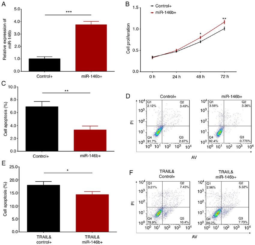
6                                                       ZHANG et al: miR-146b IN PSORIASIS

Figure 5. Impact of miR‑146b overexpression on cell proliferation and apoptosis in HaCaT cells. (A) Comparison of miR‑146b expression following
miR‑146b overexpression and control overexpression plasmid transfection in HaCaT cells. (B) Effect of miR‑146b overexpression on HaCaT cell
proliferation. (C and D) Apoptosis of HaCaT cells with miR‑146b overexpression; (C) quantified results and (D) representative flow cytometry images.
(E and F) TRAIL‑induced cell apoptosis after miR‑146b overexpression and control overexpression plasmid transfection; (E) quantified results and (F) repre‑
sentative flow cytometry images. Comparisons between two individual groups were determined using the t‑test. *P
EXPERIMENTAL AND THERAPEUTIC MEDICINE 21: 296, 2021                                             7

cytokines (including TNF‑ α, IL‑6 and IL‑17), thereby             Funding
increasing disease activity and inflammation observed in
patients with psoriasis (12,34,35). ii) miR‑146b may have         No funding was received.
activated several inflammation‑associated signaling path‑
ways (including NF‑κ B), causing a higher disease activity        Availability of data and materials
and aggravated inflammation in patients with psoriasis.
Therefore, the present study indicated that miR‑146b may          The datasets used and/or analyzed during the current study are
serve as a novel biomarker for the predication of elevated        available from the corresponding author on reasonable request.
psoriasis activity and aggravated inflammation in the
clinical setting. However, the present study had certain          Authors' contributions
limitations. First, psoriatic tissue was not easy to obtain,
which means that it may not be feasible to predict disease        XL conceived and designed the experiments. LZ and SZ
activity levels based on measurements of miR‑146b in the          performed the experiments. JW analyzed the data. SZ and
samples collected. Therefore, investigations into the predic‑     JW wrote the main manuscript text. All authors revised the
tive value of circulating miR‑146b expression are required        manuscript. All authors reviewed and approved the final
for determining psoriasis activity. In addition, the present      manuscript.
study had a single‑centered design, meaning that most of
the patients were from North China, which may result in           Ethics approval and consent to participate
selection bias. Therefore, multi‑center studies are required
for future determinations.                                        This study was approved by the Ethics Review Board of the
    Considering the malignant proliferation of keratinocytes      Affiliated Hospital of Hebei University of Engineering (Handan,
in psoriatic tissue, and to further explore the underlying        China) and all patients provided written informed consent.
mechanism of miR‑146b in the development of psoriasis,
the miR‑146b overexpression and control plasmids were             Patient consent for publication
transfected into HaCaT cells. The impact of miR‑146b
overexpression on HaCaT cell proliferation and apoptosis          Not applicable.
was subsequently evaluated. The present results revealed
that miR‑146b overexpression stimulated cell proliferation        Competing interests
and suppressed cell apoptosis, indicating that miR‑146b
increased psoriasis activity and inflammation, potentially by     The authors declare that they have no competing interests.
enhancing keratinocyte proliferation and reducing keratino‑
cyte apoptosis. To better understand the effect of miR‑146b       References
overexpression on keratinocytes under inflammatory condi‑
tions, HaCaT cell apoptosis following treatment with TRAIL         1. Parisi R, Symmons DP, Griffiths CE and Ashcroft DM;
(a protein that induces cell apoptosis) was also evaluated in         Identification and Management of Psoriasis and Associated
                                                                      ComorbidiTy (IMPACT) project team: Global epidemiology
the present study. The results demonstrated that miR‑146b             of psoriasis: A systematic review of incidence and prevalence.
overexpression inhibited TRAIL‑induced cell apoptosis.                J Invest Dermatol 133: 377‑385, 2013.
The reasons for this result may be as follows: i) miR‑146b         2. von Csiky‑Sessoms S and Lebwohl M: What's new in psoriasis.
                                                                      Dermatol Clin 37: 129‑136, 2019.
regulates multiple signaling pathways (including NF‑κ B),          3. Christophers E and van de Kerkhof PCM: Severity, heterogeneity
thereby directly accelerating keratinocyte proliferation and          and systemic inflammation in psoriasis. J Eur Acad Dermatol
inhibiting apoptosis (23,36). ii) miR‑146b mediates various           Venereol 33: 643‑647, 2019.
                                                                   4. Golińska J, Sar‑Pomian M and Rudnicka L: Dermoscopic
proteins (including TRAF6 and IRAK‑1) to promote the                  features of psoriasis of the skin, scalp and nails‑a systematic
secretion of pro‑inflammatory cytokines (including TNF‑α,             review. J Eur Acad Dermatol Venereol 33: 648‑660, 2019.
IL‑6 and IL‑17), whose secretions may indirectly stimulate         5. Hwang YJ, Jung HJ, Kim MJ, Roh NK, Jung JW, Lee YW,
keratinocyte proliferation (37). Taken together, the present          Choe YB and Ahn KJ: Serum levels of LL‑37 and inflam‑
                                                                      matory cytokines in plaque and guttate psoriasis. Mediators
study provided novel insight into the pathogenesis of psoriasis       Inflamm 2014: 268257, 2014.
and may facilitate the discovery of novel biomarkers for the       6. Menter A, St rober BE, Kaplan DH, K ivelevitch D,
prediction of psoriasis activity, as well as the discovery of         Prater EF, Stoff B, Armstrong AW, Connor C, Cordoro KM,
                                                                      Davis DMR, et al: Joint AAD‑NPF guidelines of care for the
novel treatment options.                                              management and treatment of psoriasis with biologics. J Am
    In summary, miR‑146b expression was elevated in                   Acad Dermatol 80: 1029‑1072, 2019.
psoriasis‑affected tissue when compared with that in unaf‑         7. Elmets CA, Leonardi CL, Davis DMR, Gelfand JM, Lichten J,
                                                                      Mehta NN, Armstrong AW, Connor C, Cordoro KM,
fected tissue, and its high expression was associated with            Elewski BE, et al: Joint AAD‑NPF guidelines of care for the
increased disease activity in patients with psoriasis as well         management and treatment of psoriasis with awareness and
as aggravated inflammation in psoriatic tissue. Furthermore,          attention to comorbidities. J Am Acad Dermatol 80: 1073‑1113,
miR‑146b overexpression promoted keratinocyte proliferation           2019.
                                                                   8. Guo WT and Wang Y: Dgcr8 knockout approaches to understand
and inhibited keratinocyte apoptosis in vitro.                        microRNA functions in vitro and in vivo. Cell Mol Life Sci 76:
                                                                      1697‑1711, 2019.
Acknowledgements                                                   9. Saliminejad K, Khorram Khorshid HR, Soleymani Fard S
                                                                      and Ghaffari SH: An overview of microRNAs: Biology, func‑
                                                                      tions, therapeutics, and analysis methods. J Cell Physiol 234:
Not applicable.                                                       5451‑5465, 2019.
8 ZHANG et al: miR-146b IN PSORIASIS
10. Asadzadeh Z, Mansoori B, Mohammadi A, Aghajani M, 25. Curtale G, Mirolo M, Renzi TA, Rossato M, Bazzoni F and
Haji‑Asgarzadeh K, Safarzadeh E, Mokhtarzadeh A, Duijf PHG Locati M: Negative regulation of Toll‑like receptor 4 signaling by
and Baradaran B: microRNAs in cancer stem cells: Biology, IL‑10‑dependent microRNA‑146b. Proc Natl Acad Sci USA 110:
pathways, and therapeutic opportunities. J Cell Physiol 234: 11499‑11504, 2013.
10002‑10017, 2019. 26. Echavarria R, Mayaki D, Neel JC, Harel S, Sanchez V and
11. Basso MF, Ferreira PCG, Kobayashi AK, Harmon FG, Hussain SN: Angiopoietin‑1 inhibits toll‑like receptor 4
Nepomuceno AL, Molinari HBC and Grossi‑de‑Sa MF: signalling in cultured endothelial cells: Role of miR‑146b‑5p.
MicroRNAs and new biotechnological tools for its modulation Cardiovasc Res 106: 465‑477, 2015.
and improving stress tolerance in plants. Plant Biotechnol J 17: 27. Park H, Huang X, Lu C, Cairo MS and Zhou X: MicroRNA‑146a
1482‑1500, 2019. and microRNA‑146b regulate human dendritic cell apoptosis and
12. Hou T, Liao J, Zhang C, Sun C, Li X and Wang G: Elevated cytokine production by targeting TRAF6 and IRAK1 proteins.
expression of miR‑146, miR‑139 and miR‑340 involved in regu‑ J Biol Chem 290: 2831‑2841, 2015.
lating Th1/Th2 balance with acute exposure of fine particulate 28. Croci S, Zerbini A, Boiardi L, Muratore F, Bisagni A, Nicoli D,
matter in mice. Int Immunopharmacol 54: 68‑77, 2018. Farnetti E, Pazzola G, Cimino L, Moramarco A, et al: MicroRNA
13. Tu Z, Xiong J, Xiao R, Shao L, Yang X, Zhou L, Yuan W, markers of inflammation and remodelling in temporal arteries
Wang M, Yin Q, Wu Y, et al: Loss of miR‑146b‑5p promotes from patients with giant cell arteritis. Ann Rheum Dis 75:
T cell acute lymphoblastic leukemia migration and invasion via 1527‑1533, 2016.
the IL‑17A pathway. J Cell Biochem 120: 5936‑5948, 2019. 29. Hermann H, Runnel T, Aab A, Baurecht H, Rodriguez E,
14. Kirchmeyer M, Servais FA, Hamdorf M, Nazarov PV, Ginolhac A, M a g i l n ick N, Ur ga r d E , Ša h m a t ova L , P r a n s E ,
Halder R, Vallar L, Glanemann M, Rubie C, Lammert F, et al: Maslovskaja J, et al: miR‑146b probably assists miRNA‑146a
Cytokine‑mediated modulation of the hepatic miRNome: in the suppression of keratinocyte proliferation and inflamma‑
miR‑146b‑5p is an IL‑6‑inducible miRNA with multiple targets. tory responses in psoriasis. J Invest Dermatol 137: 1945‑1954,
J Leukoc Biol 104: 987‑1002, 2018. 2017.
15. Yan M, Yang X, Shen R, Wu C, Wang H, Ye Q, Yang P, Zhang L, 30. Grine L, Dejager L, Libert C and Vandenbroucke RE: An
Chen M, Wan B, et al: miR‑146b promotes cell proliferation and inflammatory triangle in psoriasis: TNF, type I IFNs and IL‑17.
increases chemosensitivity, but attenuates cell migration and inva‑ Cytokine Growth Factor Rev 26: 25‑33, 2015.
sion via FBXL10 in ovarian cancer. Cell Death Dis 9: 1123, 2018. 31. Wang WM and Jin HZ: Interleukin‑6 in psoriasis. Zhongguo Yi
16. Yu C, Zhang L, Luo D, Yan F, Liu J, Shao S, Zhao L, Jin T, Xue Ke Xue Yuan Xue Bao 40: 284‑288, 2018 (In Chinese).
Zhao J and Gao L: MicroRNA‑146b‑3p promotes cell metastasis 32. Mease PJ: Inhibition of interleukin‑17, interleukin‑23 and the
by directly targeting NF2 in human papillary thyroid cancer. TH17 cell pathway in the treatment of psoriatic arthritis and
Thyroid 28: 1627‑1641, 2018. psoriasis. Curr Opin Rheumatol 27: 127‑133, 2015.
17. Nakasa T, Miyaki S, Okubo A, Hashimoto M, Nishida K, Ochi M 33. Hao JQ: Targeting interleukin‑22 in psoriasis. Inflammation 37:
and Asahara H: Expression of microRNA‑146 in rheumatoid 94‑99, 2014.
arthritis synovial tissue. Arthritis Rheum 58: 1284‑1292, 2008. 34. Viladomiu M, Hontecillas R, Pedragosa M, Carbo A, Hoops S,
18. Martínez‑Hernández R, Sampedro‑Núñez M, Serrano‑Somavilla A, Michalak P, Michalak K, Guerrant RL, Roche JK, Warren CA
Ramos‑Leví AM, de la Fuente H, Triviño JC, Sanz‑García A, and Bassaganya‑Riera J: Modeling the role of peroxisome
Sánchez‑Madrid F and Marazuela M: A MicroRNA signature for proliferator‑activated receptor γ and microRNA‑146 in mucosal
evaluation of risk and severity of autoimmune thyroid diseases. immune responses to Clostridium difficile. PLoS One 7: e47525,
J Clin Endocrinol Metab 103: 1139‑1150, 2018. 2012.
19. Venkatesha SH, Dudics S, Song Y, Mahurkar A and Moudgil KD: 35. Cai F, Wu F, Cao J and Chen X: MicroRNA‑146b‑3p regulates
The miRNA expression profile of experimental autoimmune the development and progression of cerebral infarction with
encephalomyelitis reveals novel potential disease biomarkers. Int diabetes through RAF1/P38MAPK/COX‑2 signaling pathway.
J Mol Sci 19: 3990, 2018. Am J Transl Res 10: 618‑628, 2018.
20. Wallace AB: The exposure treatment of burns. Lancet 1: 501‑504, 36. Weng Z, Patel AB, Vasiadi M, Therianou A and Theoharides TC:
1951. Luteolin inhibits human keratinocyte activation and decreases
21. Fredriksson T, Pettersson U: Severe psoriasis‑oral therapy with a NF‑κ B induction that is increased in psoriatic skin. PLoS One 9:
new retinoid. Dermatologica 157: 238‑244, 1978. e90739, 2014.
22. Livak KJ and Schmittgen TD: Analysis of relative gene expres‑ 37. Nestle FO, Kaplan DH and Barker J: Psoriasis. N Engl J Med 361:
sion data using real‑time quantitative PCR and the 2(‑Delta Delta 496‑509, 2009.
C(T)) method. Methods 25: 402‑408, 2001.
23. Cho S, Lee HM, Yu IS, Choi YS, Huang HY, Hashemifar SS, This work is licensed under a Creative Commons
Lin LL, Chen MC, Afanasiev ND, Khan AA, et al: Differential Attribution-NonCommercial-NoDerivatives 4.0
cell‑intrinsic regulations of germinal center B and T cells by International (CC BY-NC-ND 4.0) License.
miR‑146a and miR‑146b. Nat Commun 9: 2757, 2018.
24. Zhang Z, Dai X, Qi J, Ao Y, Yang C and Li Y: Astragalus
mongholicus (Fisch.) bge improves peripheral treg cell
immunity imbalance in the children with viral myocarditis by
reducing the levels of miR‑146b and miR‑155. Front Pediatr 6:
139, 2018.You can also read