The role of echocardiography in predicting technical problems and complications of transvenous leads extractions procedures - Authorea
←
→
Page content transcription
If your browser does not render page correctly, please read the page content below
Posted on Authorea 5 Jan 2021 | The copyright holder is the author/funder. All rights reserved. No reuse without permission. | https://doi.org/10.22541/au.160989044.41409620/v1 | This a preprint and has not been peer reviewed. Data may be preliminary.
The role of echocardiography in predicting technical problems and
complications of transvenous leads extractions procedures
Dorota Nowosielecka1 , Wojciech Jacheć2 , Anna Polewczyk3 , Lukasz Tulecki1 , Andrzej
Kleinrok1 , and Andrzej Kutarski4
1
The Pope John Paul II Province Hospital
2
Medical University of Silesia in Katowice, Faculty of Medical Sciences in Zabrze
3
Collegium Medicum of The Jan Kochanowski University
4
Medical University of Lublin
January 5, 2021
Abstract
Introduction Transesophageal echocardiography (TEE) is a useful tool in preoperative observation of patients undergoing
transvenous leads extraction (TLE) due to complications associated with implanted devices. Echocardiographic phenomena
may determine the safety of the procedure. Methods and results Data from 936 transesophageal examinations (TEE) performed
at a high volume center in patients awaiting TLE from 2015 to 2019 were assessed. TEE revealed a total of 1156 phenomena
associated with the implanted leads in 697 (64.85%) patients, including: asymptomatic masses on endocardial leads (AMEL)
(58.65%), vegetations (12,73%), fibrous tissue binding the lead to the vein or heart wall (33.76%), lead-to-lead binding sites
(18.38%), excess lead loops (19.34%), intramural penetration of the lead tip (16.13%), lead-dependent tricuspid dysfunction
(LDTD) (6.41%). Risk factors for technical difficulties during TLE in multivatiate analysis were: fibrous tissue binding the
lead to atrial wall (OR=1.738; p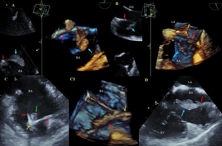
6
University of Information Technology and Management, Rzeszów, Poland
7
Department of Cardiology, Medical University, Lublin, Poland
Posted on Authorea 5 Jan 2021 | The copyright holder is the author/funder. All rights reserved. No reuse without permission. | https://doi.org/10.22541/au.160989044.41409620/v1 | This a preprint and has not been peer reviewed. Data may be preliminary.
Funding: This research did not receive any specific grant from funding agencies in the public, commercial,
or not-for-profit sectors.
Conflicts of Interest: None
Emails
Dorota Nowosielecka dornowos@wp.pl
Wojciech Jacheć wjachec@interia.pl
Anna Polewczyk annapolewczyk@wp.pl ORCID ID; https://orcid.org/0000-0002-6632-6551
Lukasz Tulecki luke27@poczta.onet.pl
Andrzej Kleinrok a.kleinrok@wp.pl
Andrzej Kutarski a_kutarski@yahoo.com
Corresponding author
Anna Polewczyk MD, PhD
Collegium Medicum of Jan Kochanowski University
19A, Aleja IX Wieków Kielc Str 25-317 Kielce, Poland
annapolewczyk@wp.pl
Phone number +48600024074
Abstract:
Introduction
Transesophageal echocardiography (TEE) is a useful tool in preoperative observation of patients undergoing
transvenous leads extraction (TLE) due to complications associated with implanted devices. Echocardiogra-
phic phenomena may determine the safety of the procedure.
Methods and results
Data from 936 transesophageal examinations (TEE) performed at a high volume center in patients awaiting
TLE from 2015 to 2019 were assessed.
TEE revealed a total of 1156 phenomena associated with the implanted leads in 697 (64.85%) patients,
including: asymptomatic masses on endocardial leads (AMEL) (58.65%), vegetations (12,73%), fibrous tissue
binding the lead to the vein or heart wall (33.76%), lead-to-lead binding sites (18.38%), excess lead loops
(19.34%), intramural penetration of the lead tip (16.13%), lead-dependent tricuspid dysfunction (LDTD)
(6.41%). Risk factors for technical difficulties during TLE in multivatiate analysis were: fibrous tissue binding
the lead to atrial wall (OR=1.738; p
Careful preoperative TEE evaluation of the consequences of extended lead implant duration (enhanced
fibrotic response) increases the probability of predicting the level of difficulty of TLE procedures, their
efficacy and risk of major complications.
Posted on Authorea 5 Jan 2021 | The copyright holder is the author/funder. All rights reserved. No reuse without permission. | https://doi.org/10.22541/au.160989044.41409620/v1 | This a preprint and has not been peer reviewed. Data may be preliminary.
Key words: transvenous leads extraction, effectiveness, complications, transesophageal echocardiography
Introduction
Transvenous lead extraction (TLE) is considered first-line strategy for the management of complications
associated with cardiac implantable electronic devices (CIED) [1,2]. Recently, due to the rising incidence
of infectious and non-infectious CIED-related complications, the number of TLE has also been increasing
[3]. According to numerous reports, the frequency of major complications of TLE ranges from 0.9 to 4.0%,
and most often there is damage to the heart or venous vessels [4-7]. Assessment of risk factors for major
complications and procedure complexity should have an impact on the selection of a suitable organizational
model of the procedure and center preferment [4-7]. The available TLE risk stratification scales most often
take into account the impact of various factors on the technical complexity of the procedure [4-7] or peri-
procedural mortality [4-7]. They are based on demographic and clinical data (patient age, gender, presence
of co-morbidities ), type of CIED system (ICD lead, number of leads) and history of pacing (age at first
implantation, number of leads designed for extraction) [4-7]. This is the first study to assess the usefulness
of new factors that may significantly affect the level of difficulty and procedure complexity as well as efficacy
and complications of TLE. These factors were identified during an echocardiographic examination of patients
selected for TLE due to CIED-related complications. Echocardiography, especially transesophageal echocar-
diography plays a key role in the evaluation of rhythm controlling devices (PM/ICD/CRT) and remains a
valuable tool for precise imaging, which is recommended by experts [8-15]. Although a number of studies
focused on the value of preoperative TEE findings (size of vegetations, presence of asymptomatic masses on
the leads), the only echocardiographic parameter discussed when estimating the procedure-related risk was
left ventricular ejection fraction (LVEF), only few studies so far have suggested that TEE can be used apart
from fluoroscopy or computerized tomography [7] to choose optimal TLE strategy [16,17], nevertheless the
effect of echocardiographic findings on procedure safety and efficacy has not been assessed.
Methods
Study group
We carried out a prospective analysis of the data from 936 preoperative TEE examinations performed at a
high volume center before transvenous lead extraction from June 2015 to October, 2019.
The extent of preoperative TEE
TEE was performed using the Philips iE33 or the GE Vivid S 70 ultrasound machine equipped with X7-2t
Live 3D or 6VT-D probes. Images were obtained before the procedure, after general anesthesia and tracheal
intubation, during preparation of the surgical field and dissection and stabilization of the leads in the device
pocket. Leads were evaluated in the mid-esophageal, inferior esophageal and modified transgastric views to
visualize the right ventricle and the tricuspid valve. In order to obtain complete visualization of the structures
(and assessment of lead/heart interaction) non-standard imaging planes were sometimes required. After the
procedure the results were entered into a computer database. We analyzed the number, location and course
of the leads: in the superior vena cava (SVC), right atrium (RA), right ventricle (RV) (taking into account
excess lead loops). We also assessed lead mobility, presence of sites at which the lead was bound to cardiac
structures, lead-to-lead binding sites and additional masses attached to the leads. An important part of the
imaging protocol was assessment of the effect of the lead on tricuspid function. Additionally, we assessed left
ventricular function (LVEF), pericardial function and possible presence of structural heart disease (atrial or
ventricular septal defects).
Definitions of echocardiographic phenomena
1. Asymptomatic masses on endocardial leads (AMEL) characterized by homogeneous echogenicity,
3smooth contour and varying degrees of organization. AMEL include components of connective tissue
(accretions), clots, masses resembling vegetations (so-called vegetation-like masses). Vegetation-like
masses may be the remnant vegetations after antibiotic treatment or (less probable) organized fibrotic
Posted on Authorea 5 Jan 2021 | The copyright holder is the author/funder. All rights reserved. No reuse without permission. | https://doi.org/10.22541/au.160989044.41409620/v1 | This a preprint and has not been peer reviewed. Data may be preliminary.
thrombi [18].
2. Hyperechoic segmental thickening of the leads defined as connective tissue overgrowth (undergoing
fibrosis, mineralization, crystallization and even ossification) [18[.
3. Bacterial vegetations: multishaped, mobile masses of inhomogeneous echogenicity attached to the leads
or/and to the neighboring anatomic structures, most frequently tricuspid leaflets. They were found only
if they were accompanied by signs of a general infection. Sometimes coexisting with AMEL (vegetation-
like masses) [18].
4. Accretion – immobile fibrous connective tissue sheath around the lead causing adherence to the endo-
cardium and vessel walls and producing images similar to segmental lead thickening but moving along
with the cardiac wall [18].
5. Excessive lead loops - result of too weak fixation of the lead during implantation or lead fracture with
break of insulation in the subclavian region [18].
6. Cardiac wall perforation by the lead: visualization of the lead tip outside the heart contour, sometimes
with fluid in the pericardial sac; placement of the lead tip close to the border of the pericardium is
referred to as penetration [18].
Transvenous leads extraction procedures
Procedures were performed in a hybrid operating room or in an operating room, using mechanical systems
such as polypropylene Byrd dilator sheaths (Cook® Medical, Leechburg, PA, USA), making use of the
oblique cutting edge of the tip to dissect leads from fibrous sheaths that immobilized the intravascular and/or
intracardiac segment of the lead [11,19]. Procedures were performed in patients under general anesthesia with
full preparation of the surgical field for cardiac surgery.
Complete procedural success, clinical success and complications of TLE were defined according to the HRS
2009 and 2017 guidelines and the 2018 EHRA expert consensus statement [1,20,21]. Complete procedural
success was defined as removal of all targeted leads and material, with the absence of any permanently
disabling complication or procedure-related death. Clinical success was achieved in patients with retention
of a small part of the lead that did not negatively affect the outcome goals of the procedure [1,20,21].
Major and minor complications were defined according to the 2018 EHRA Expert Consensus Statement on
Lead Extraction [21].
Possible technical problems during TLE include: block in lead venous entry / subclavian region, necessity
utility of Evolution / TigRail (second line tool in study center), necessity of changing of venous approach for
lead extraction (any reasons), impossible utility of lead venous entry approach – procedure (since beginning)
using another approach, need to utilise lasso-catheters or basket catheters, extracted lead break and broken
lead remnant extraction, extracted lead break and abandonment of broken lead fragment, extracted lead
fragmentation – removal in parts, lead to lead strong connection with connecting tissue scar – terrible both
leads separation, collapse / fracture of Byrd dilator, dislodgement of functional lead, reeling of ICD lead coil.
Approval of the Bioethics Committee
All patients gave their informed written consent to undergo TLE and use anonymous data from their me-
dical records, approved by the Bioethics Committee at the Regional Chamber of Physicians in Lublin no.
288/2018/KB/VII.
Statistical analysis
The Shapiro-Wilk test was used to test the normality of distribution of variables. For uniformity, all conti-
nuous variables are presented as the mean ± standard deviation. The categorical variables are presented as
number and percentage.
4The significance of difference between groups were analyzed using the unpaired “U” Mann-Whitney test
for continuous variables and the Chi2 test with Yates correction for categorical variables. The results of
analysis were considered statistically significant at a p value < 0.05. Univariate and multivariate logistic
Posted on Authorea 5 Jan 2021 | The copyright holder is the author/funder. All rights reserved. No reuse without permission. | https://doi.org/10.22541/au.160989044.41409620/v1 | This a preprint and has not been peer reviewed. Data may be preliminary.
regression analysis was used to assess the impact of echocardiographic findings on TLE complexity and
efficacy. Variables that in univariate analysis achieved statistical significance p 50%, n (%) 539 (57.585) Procedure duration 15,931 ±25.558
(sheath to sheath)
(min), mean ± SD
Diabetes mellitus 198 (21.154) Technical difficulty 233 (24.840)
(any), n (%) during TLE (any),
n (%)
Renal failure (any), 230 (24.573) Number of major 0,328 ±0.725
n (%) technical difficulty
in one patient
Charlson 4,886 ±3.764 Three or more 21 (2.239)
comorbidity index, technical problems,
mean ± SD n (%)
Indications for All 936 patients TLE procedure All 936 patients
TLE efficacy and
outcomes
LRIE with or 151 (16.132) Major complications 18 (1.923)
without pocket (any), n (%)
infection, n (%)
5Demographic CIED system
and clinical and history of
data All 936 patients pacing All 936 patients
Posted on Authorea 5 Jan 2021 | The copyright holder is the author/funder. All rights reserved. No reuse without permission. | https://doi.org/10.22541/au.160989044.41409620/v1 | This a preprint and has not been peer reviewed. Data may be preliminary.
Local (pocket) 58 (6.196) Hemopericardium, 12 (1.282)
infection (only), n n (%)
(%)
Non-infectious 727 (77.671) Tricuspid valve 6 (0.640)
indications, n (%) damage during
TLE, n (%)
CIED system and All 936 patients Rescue cardiac 12 (1.282)
history of pacing surgery, n (%)
Number of leads in 1,834 ±0.639 Lack of radiological 6 (0.640)
the system before success, n (%)
TLE, mean ± SD
Presence of 86 (9.188) Complete clinical 916 (97.863)
abandoned leads success, n (%)
before TLE, n (%)
Number of 1,837 ±0.990 Complete 917 (97.970)
procedures before procedural success,
lead extraction, n (%)
mean ± SD
The echocardiographic findings can be divided into four basic groups:
Tricuspid valve dysfunction
Tricuspid valve dysfunction not related to the route of ventricular pacing prior to TLE (18.493%)
Lead-dependent tricuspid valve dysfunction (LDTD) (6.410%)
Presence of any shadows on the leads (64.85% of patients)
1. Fibrous tissue binding the lead to the vena cava superior and right heart structures (33.761%): to the
SVC (5.98%), to the RA wall (6.94%), to the tricuspid apparatus (9.62%) and to the RV wall (11.22%)
2. Fibrous tissue binding two leads (18.38%).
3. AMEL (46.68% of patients): fibrous tissue encasing the lead (17.094%), lead thickening (29.59%),
blood clot on the lead (8.013%) vegetation-like masses (3.953%)
4. Vegetations (12.727%)
Presence of excess lead loops in the heart (19.338%)
Perforation or penetration of the lead through the cardiac wall up to the epicardium (16.132%) (Table 2)
Table 2
The most important preoperative echocardiographic findings in patients undergoing transvenous leads ex-
traction
Tricuspid valve dysfunction
(degree of regurgitation) - Average tricuspid valve
excluding patients with lead regurgitation [0-4 degree], mean
dependent TV dysfunction ±SD 1.454±0.956
Patients with severe tricuspid 162 (18.493)
regurgitation [3-4], n ( %)
6Tricuspid valve dysfunction
(degree of regurgitation) - Average tricuspid valve
excluding patients with lead regurgitation [0-4 degree], mean
Posted on Authorea 5 Jan 2021 | The copyright holder is the author/funder. All rights reserved. No reuse without permission. | https://doi.org/10.22541/au.160989044.41409620/v1 | This a preprint and has not been peer reviewed. Data may be preliminary.
dependent TV dysfunction ±SD 1.454±0.956
Lead dependent tricuspid valve Average LDTD [0-4], mean±SD 3,541±0.594
dysfunction (LDTD)
Patients with LDTD (any), n ( 60 (6.410)
%)
Patients with severe LDTD 58 (96.667)
[3-4,] n ( %)
Any shadows on the leads Patients with any shadows on 607 (64.850)
leads before TLE, n (%)
Patients with fibrous tissue Patients with fibrous tissue 236 (25.214)
binding the lead to the vena binding the lead to the vena
cava superior and heart cava superior and heart
structures, n (%) structures, n (%)
Fibrous tissue binding the lead to Fibrous tissue binding the lead to 316 (33.761)
the vena cava superior and heart the heart structures (all), n (%)
structures
Fibrous tissue binding the lead 56 (5.983)
to the SVC, n (%)
Fibrous tissue binding the lead 65 (6.944)
to the RA wall, n (%)
Fibrous tissue binding the lead to 90 (9.615)
the tricuspid apparatus, n (%)
Fibrous tissue binding the lead 105 (11.218)
to the RV wall, n (%)
Fibrous tissue binding two Fibrous tissue binding two 172 (18.377)
leads, n (%) leads, n (%)
Patients with asymptomatic Patients with asymptomatic 437 (46.688)
masses on endocardial leads masses on endocardial leads
(AMEL) (patient analysis), n (AMEL) (patient analysis), n
(%) (%)
AMEL (findings analysis) AMEL (all), n (%) 549 (58.654)
Fibrous tissue encasing the lead, 160 (29,144 / 17.094)
n ( % of all ILM / % of all pts)
Lead thickening, n ( % of all ILM 277 (50,455 / 29.594)
/ % of all pts)
Clot on the lead, n (% of all ILM 75 (13,661 / 8.013)
/ % of all pts)
Vegetation-like masses, n ( % of 37 (6,740 / 3.953)
all ILM / % of all pts)
Presence of vegetations Patients with vegetations, n (%) 119 (12.727)
Excess lead loops in the heart Patients with lead loops in the 181 (19.338)
heart (any), n (%)
Lead loops in the RA n (%) 138 (14.744)
Lead loops in the TV, n (%) 35 (3.793)
Lead loops in the RV or PA, n 28 (2.991)
(%)
7Tricuspid valve dysfunction
(degree of regurgitation) - Average tricuspid valve
excluding patients with lead regurgitation [0-4 degree], mean
Posted on Authorea 5 Jan 2021 | The copyright holder is the author/funder. All rights reserved. No reuse without permission. | https://doi.org/10.22541/au.160989044.41409620/v1 | This a preprint and has not been peer reviewed. Data may be preliminary.
dependent TV dysfunction ±SD 1.454±0.956
Perforation or penetration of Perforations, n (%) 151 (16.132)
the lead through the cardiac
wall up to the epicardium
Abbreviations :AMEL- asymptomatic masses on endocardial leads, LDTD- lead dependent tricuspid valve
dysfunction, PA- pulmonary artery SVC- superior vena cava, RA- right atrium , RV- right ventricle, TLE-
transvenous lead extraction;
Comparative analysis of the impact of several echocardiographic parameters on the course and efficacy of
TLE was presented in supplementary file.
Factors influencing the occurrence of technical difficulties – results of logistic regression analysis
Univariate regression analysis showed that the factors increasing the probability of technical difficulties
were: all types of fibrous tissue binding sites: more than 2.5-fold increase, (OR=2.649; pTechnical
Technical
Technical
Technical
Technical
Technical
Technical Major Major Major Major Maj
dif- dif- dif- dif- dif- dif- dif- com- com- com- com- com
fi- fi- fi- fi- fi- fi- fi- pli- pli- pli- pli- pli-
Posted on Authorea 5 Jan 2021 | The copyright holder is the author/funder. All rights reserved. No reuse without permission. | https://doi.org/10.22541/au.160989044.41409620/v1 | This a preprint and has not been peer reviewed. Data may be preliminary.
cul- cul- cul- cul- cul- cul- cul- ca- ca- ca- ca- ca-
ties ties ties ties ties ties ties tions tions tions tions tion
Fibrous 2,649 1.923Technical
Technical
Technical
Technical
Technical
Technical
Technical Major Major Major Major Maj
dif- dif- dif- dif- dif- dif- dif- com- com- com- com- com
fi- fi- fi- fi- fi- fi- fi- pli- pli- pli- pli- pli-
Posted on Authorea 5 Jan 2021 | The copyright holder is the author/funder. All rights reserved. No reuse without permission. | https://doi.org/10.22541/au.160989044.41409620/v1 | This a preprint and has not been peer reviewed. Data may be preliminary.
cul- cul- cul- cul- cul- cul- cul- ca- ca- ca- ca- ca-
ties ties ties ties ties ties ties tions tions tions tions tion
Fibrous 3.080 2.0446.045; p
LDTD approached the borderline of significance (OR=0.316; p=0.067) (Table 4).
6. Discussion
Posted on Authorea 5 Jan 2021 | The copyright holder is the author/funder. All rights reserved. No reuse without permission. | https://doi.org/10.22541/au.160989044.41409620/v1 | This a preprint and has not been peer reviewed. Data may be preliminary.
Transesophageal echocardiography is a very important part of patient evaluation before transvenous lead
extraction because of its ability to detect phenomena related to the presence of leads in the heart and
potentially affecting the course, safety and efficacy of the procedure [1,2]. In this study the echocardiographic
findings in patients with CIEDs were divided into four basic groups: 1.tricuspid valve dysfunction 2. presence
of any shadows on the leads before TLE 3. presence of excess lead loops in the heart and 4. perforation or
penetration of the lead through the cardiac wall up to the epicardium. From the viewpoint of the planned
transvenous lead extraction, the most important findings were the many faces of fibrosis associated with the
leads varying in location and intensity. The degree of resistance/hardness and fibrosis - the most common
enemy of the operator - appeared to determine the level of difficulty and safety of the procedure. There is a
large volume of published studies describing the presence of mobile masses attached to the leads visualized
by TTE, TEE and ICE in asymptomatic patients [22-23]. In this study additional masses on endocardial
leads (AMEL) were defined as fibrous connective tissue
(accretions), clots, vegetations-like massess. Similar to Golzio PG et al. [23], we also took into consideration
lead thickening and hyperechogenicity, frequently present (29.594%) in patients undergoing TLE (Figure 1).
Figure 1
Additional masses associated with endocardial leads detected on preprocedural TEE
1. TEE (2D and 3D, ME- bicaval) In the RA an additional mass (red arrow) attached to the lead, a
mobile mass (blue arrows) representing a bacterial vegetation
2. TEE (2D and 3 D, ME- bicaval) Segmental lead thickening (red arrow) in the atrial course with an
additional mobile mass (blue arrow) representing the connective tissue build-up (accretion, scar)
3. TEE (2D, ME- modified) In the RA cavity close to the SVC orifice an echo of two leads (red arrows)
with additional irregular masses (green arrows) at lead-to-lead binding site (yellow arrow) representing
clots. C1 – 3D imaging
4. TEE (2D, ME- modified to visualize right cardiac chambers) In the RA a mass attached to the lead
(red arrow) that may represent a pseudo vegetation (blue arrow)
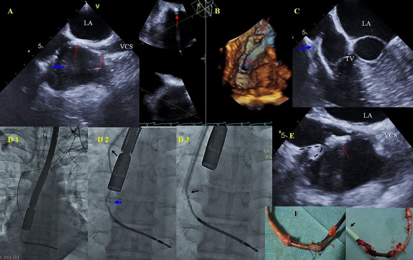
12Visualization of excess lead loops was another component of the TEE evaluation of patients before TLE.
Lead looping was usually a result of long-term contact with the myocardium, and hence a stronger adhesion
involving longer segments. In this study excess lead loops were most common in the RA (138; 14.744%
Posted on Authorea 5 Jan 2021 | The copyright holder is the author/funder. All rights reserved. No reuse without permission. | https://doi.org/10.22541/au.160989044.41409620/v1 | This a preprint and has not been peer reviewed. Data may be preliminary.
cases), and least frequent in the RV and the TPA (35; 3.793% cases). The presence of excess lead loops did
not affect the procedure-related risk, although it increased the level of complexity.
Lead loops are very well visible on fluoroscopy, but the advantage of TEE is that it permits detection of
fibrous tissue binding the lead loops to the heart walls and its possible impact on the tricuspid apparatus
(Figure 2).
Figure 2
Consequences of excess ventricular lead loops on TEE examination
1. Fluoroscopy. Ventricular lead loop in the RV cavity not affecting TV function
2. TEE (2D ME RV Inflow-Outflow modified) Ventricular lead loop forming a closed circle, with segmental
thickening
3. TEE (3D modified) The atrial lead (yellow arrow) directed towards the RAA with a well visible distal
segment and a loop formed by the ventricular lead in the RV with lead-to-lead binding site in the distal
segment (red arrow)
4. TEE (3D) Zoom in on the distal segment within the loop confirming lead-to-lead binding site (red
arrow)
5. The same as in Figure 2D
6. TEE (3D ME – bicaval) The atrial lead (yellow arrow) implanted in the RAA wall with a visible
binding site in the distal segment (blue arrow)
7. Extracted leads surrounded by the connective tissue sheath
The presence of excess lead loops in the heart is on the one side a result of suboptimal lead positioning (no
last look after the leads become lodged in the tissue), too weak tightening of the sutures, no radiological
verification of lead positioning until device replacement when the mere pulling back is already impossible.
Another echocardiographic finding i.e. fibrous tissue binding the lead to the adjacent heart and vessel
structures deserves discussion, as so far the problem has received scant attention in the research literature
[20]. In this study, fibrous tissue binding sites were recognized on inspection of the lead location and mobility
13with respect to one another and cardiac structures, looking for such signs as immediate vicinity, thickening
and lead/heart wall mobility during cardiac work (Figure 3).
Posted on Authorea 5 Jan 2021 | The copyright holder is the author/funder. All rights reserved. No reuse without permission. | https://doi.org/10.22541/au.160989044.41409620/v1 | This a preprint and has not been peer reviewed. Data may be preliminary.
Figure 3
Build-up around an ICD lead visualized on TEE and fluoroscopy and its consequences during TLE
1. TEE (2D, ME- bicaval) Segmental thickening of the ICD lead with three binding sites in the RA wall
(arrows), additionally a blue arrow points to the binding site and conductor externalization
2. TEE (2D and 3D, ME - bicaval ) The thickened ICD lead attached to the IAS (arrows)
3. TEE (2D, ME RV - Inflow) The ICD lead, hyperechoic, thickened, over the TV bound to the lateral
atrial wall at the site of externalization (blue arrow)
4. D1- Evaluation of ICD lead position and venous patency before TLE. D1 – Imaging during TLE – well
visible site of externalization (blue arrow), the tip of Byrd’s dilator marked with a black arrow. D3 –
significant pulling on the ICD lead during TLE
5. TEE (2D, bicaval) The moment of pulling on the thickened ICD lead (red arrow) seen on fluoroscopy.
D3 – significant pulling on the RA wall and the separation of pericardial layers
6. The extracted lead with multiple fragments of the connective tissue, the site of externalization and a
dilator (black arrow)
The connective tissue on the leads (scar tissue build-up around the lead, fibrous tissue binding sites, accre-
tions) is visible on TEE as lead thickening resulting in the formation of sites at which the leads are bound to
one another after being in direct contact over an extended period of time. The imaging of this phenomenon
has important implications for the course of the TLE procedure. During the extraction procedure the direct
pulling on the wall at the binding site may be too strong and cause inadvertent pulling on and uncontrolled
removal of the other lead, risking a tear of the heart wall with cardiac tamponade or hemopericardium as
the end result.
The originality of this study is that it explores the impact of TEE assessment before TLE on the course of the
procedure. Multivariate analysis showed that lead-to-lead binding sites were the strongest predictive factor
which caused a 3-fold increase in the probability of major complications during TLE. The presence of fibrous
tissue binding the lead to the atrial wall and tricuspid valve approached the borderline of significance. The
presence of binding sites in the RV wall caused a nearly 2-fold increase in the risk of technical difficulties,
thus increasing the degree of procedure complexity. The probability of technical difficulty increased also in
the presence of excess lead loops, fibrous tissue binding the lead to the RA wall and lead-to-lead binding
14sites. The presence of binding sites in the tricuspid apparatus and lead-to-lead adhesion on the borderline of
statistical significance reduced the chances of complete clinical success. The chances of procedural success
were also reduced in relation with the presence of binding sites in the SVC, RA and lead-to-lead adhesions,
Posted on Authorea 5 Jan 2021 | The copyright holder is the author/funder. All rights reserved. No reuse without permission. | https://doi.org/10.22541/au.160989044.41409620/v1 | This a preprint and has not been peer reviewed. Data may be preliminary.
whereas lead-dependent tricuspid dysfunction approached the borderline of significance.
There are numerous studies [4-7] which on the basis of demographic data (age, sex), clinical information
(indications, accompanying diseases, heart sufficiency), information about PM/ICD/CRT devices (number
and type of leads) and history of pacemaker therapy (age of leads and route of implantation) show that
initial patient assessment may identify the individuals in whom TLE may be more difficult or associated
with the occurrence of major complications. Only few studies using scoring systems provide a more precise
prediction of the level of procedure difficulty or estimate the true risk [6,7]. A review of the literature
shows that so far echocardiographic findings have not been analyzed with respect to prediction of technical
difficulties associated with TLE and complications of the procedure. Only one paper demonstrated that
low LVEF was a predictor of major complications [6], another paper documented an eventful postoperative
course in patients with right ventricular dysfunction [4]. The evidence from another study suggests that
information from CT examination may be useful for estimating procedure difficulty [8]. Yet another study
implies that accurate Doppler blood flow measurements in the SVC may identify patients with significant
lead fibrosis requiring powered sheaths for successful removal. Although numerous papers have emphasized
the role of the connective tissue (scar tissue binding the lead to the SVC and heart wall) in estimating
procedure complexity and its complications [3,6,7], to the best of our knowledge we are the first to use the
information about the degree of connective tissue build-up to predict technical difficulties and risk of major
complications associated with TLE.
When developing a risk calculator for prediction of complications (SAFeTy TLE) [7] we found out that
lead-to-lead binding site was an extremely important prognostic factor, however other information (S = sum
of lead dwell times, A = anemia, Fe = female, T == treatment (previous procedures), Y = young patients)
appeared more significant in multivariate analysis. We are of the opinion that all forms of connective tissue
response (scar tissue binding the lead to the vein and heart structures, lead-to-lead adhesion) are extremely
significant factors that increase procedure complexity and its radiological efficacy, however they do not
necessarily translate into major complications at an experienced high volume center. Nevertheless TEE
before TLE should become a tool that provides additional information about procedure-related risk.
7. Limitations
This is a single-center, observational, prospective study. TLE was performed using mechanical systems
without laser energy. Comparison of diagnostic sensitivity of TEE and ICE was not the aim of the study.
8. Conclusions
Enhanced inflammatory response in the form of connective tissue sheaths that surround the leads and their
cardiac and vascular binding sites very significantly affect the degree of TLE complexity, its efficacy defined as
clinical, procedural and radiological success and the occurrence of major complications. Careful preoperative
TEE evaluation of the consequences of extended lead implant duration (enhanced fibrotic response) by
means of preoperative transesophageal echocardiography increases the probability of predicting the level of
TLE complexity, its efficacy defined by clinical, procedural and radiological success and the risk of major
complications associated with TLE.
Acknowledgements: We would like to thank all doctors participated in transvenous leads extraction
procedures
Funding: This research did not receive any specific grant from funding agencies in the public, commercial,
or not-for-profit sectors.
Disclosures: Authors declared none conflict of interest
References
151. Kusumoto FM, Schoenfeld MH, Wilkoff BL, Berul I, Birgersdotter-Green UM, Carrillo R, et al. 2017
HRS expert consensus statement on cardiovascular implantable electronic device lead management and
extraction. Heart Rhythm. 2017;14:e503-e551
Posted on Authorea 5 Jan 2021 | The copyright holder is the author/funder. All rights reserved. No reuse without permission. | https://doi.org/10.22541/au.160989044.41409620/v1 | This a preprint and has not been peer reviewed. Data may be preliminary.
2. Bongiorni MG, Kennergren C, Butter C, Deharo JC, Kutarski A, Rinaldi CA, et al. The European
Lead Extraction ConTRolled (ELECTRa) study: a European Heart Rhythm Association (EHRA)
Registry of Transvenous Lead Extraction Outcomes. Eur Heart J. 2017;38:2995-3005.
3. Poole JE, Gleva MJ, Mela T, Chung MK, Uslan DZ, Borge R, et al. Complication rates associated with
pacemaker or implantable cardioverter-defibrillator generator replacements and upgrade procedures:
results from the REPLACE registry. Circulation. 2010; 19;122:1553-1561.
4. Agarwal SK, Kamireddy S, Nemec J, Voigt A, Saba S. Predictors of complications of endovascular
chronic lead extractions from pacemakers and defibrillators: a single-operator experience. J Cardiovasc
Electrophysiol. 2009;20:171-175.
5. Nowosielecka D, Jacheć W, Polewczyk A, Tulecki L, Tomków K, Stefańczyk P, et al. Transesophageal
echocardiography as a monitoring tool during transvenous lead extraction – does it improve procedure
effectiveness? J Clin Med. 2020; 8:1382
6. Segreti L, Giannotti Santoro M, Di Cori A, Zucchelli G, Viani S, De Lucia R, et al. Utility of risk scores
to predict adverse events in cardiac lead extraction. Expert Rev Cardiovasc Ther. 2018;16:695-705
7. Jacheć W, Polewczyk A, Polewczyk M, Tomasik A, Kutarski A. Transvenous Lead Extraction SAFeTY
Score for Risk Stratification and Proper Patient Selection for Removal Procedures Using Mechanical
Tools. J Clin Med. 2020;9 pii: E361. doi: 10.3390/jcm9020361.
8. Lewis RK, Pokorney SD, Greenfield RA, Hranitzky PM, Hegland DD, Schroder JN, et al. Preprocedu-
ral ECG-gated computer tomography for prevention of complications during lead extraction. Pacing
Clin Electrophysiol 2014;37:1297-1305.
9. Lo R, D’Anca M, Cohen T, Kerwin T. Incidence and prognosis of pacemaker lead-associated masses:
a study of 1,569 transesophageal echocardiograms. J Invasive Cardiol 2006;18:599–601.
10. Downey BC, Juselius WE, Pandian NG , Mark Estes III NA, Link MS. Incidence and significance
of pacemaker and implantable cardioverter-defibrillator lead masses discovered during transesophageal
echocardiography. Pacing Clin Electrophysiol 2011;34:679–683.
11. Hilberath JN, Burrage PS, Shernan SK, Varelmann DJ, Wilusz K, Fox JA, et al. Rescue transoe-
sophageal echocardiography for refractory haemodynamic instability during transvenous lead extrac-
tion. Eur Heart J Cardiovasc Imaging. 2014;15:926-932.
12. Oestreich BA, Ahlgren B, Seres T, Zipse MM, Tompkins C, Varosy PD, et al. Use of Transesophageal
Echocardiography to Improve the Safety of Transvenous Lead Extraction. JACC Clin Electrophysiol.
2015;1:442-448.
13. Bongiorni MG, Di Cori A, Soldati E, Zucchelli G, Arena G, Segreti L, et al. Intracardiac echocar-
diography in patients with pacing and defibrillating leads: A Feasibility Study. Echocardiography
2008;25:632-638.
14. Narducci ML, Pelargonio G, Russo E, Marinaccio L, Di Monaco A, Perna F, et al. Usefulness of
intracardiac echocardiography for the diagnosis of cardiovascular implantable electronic device-related
endocarditis. J Am Coll Cardiol. 2013;61:1398–1405.
15. Dalal A, Asirvatham SJ, Chandrasekaran K, Seward JB, Tajik T. Intracardiac echocardiography in
the detection of pacemaker lead endocarditis. J Am Soc Echocardiogr 2002;15:1027–1028.
16. Nowosielecka D, Polewczyk A, Jacheć W, Tulecki L, Tomków K, Stefańczyk P, et al. A new approach
to the continuous monitoring of transvenous lead extraction using transesophageal echocardiography-
analysis of 936 procedures. Echocardiography. 2020;37:601-611.
17. Strachinaru M, Kievit CM, Yap SC, Hirsch A, Geleijnse ML, Szili-Torok T. Multiplane/3D trans-
esophageal echocardiography monitoring to improve the safety and outcome of complex transvenous
lead extractions. Echocardiography. 2019;36:980-986.
18. Nowosielecka D, Polewczyk A, Jacheć W, Tulecki L, Kleinrok A, Kutarski A. Echocardiographic find-
ings in patients with cardiac implantable electronic devices- analysis of factors predisposing to lead-
associated changes. Clin Physiol Funct Imaging. 2020;18. Online ahead of print.
1619. Kutarski A, Czajkowski M, Pietura R, Obszanski B, Polewczyk A, Jachec W, et al. Effectiveness,
safety, and long-term outcomes of non-powered mechanical sheaths for transvenous lead extraction.
Europace. 2018;20:1324-1333
Posted on Authorea 5 Jan 2021 | The copyright holder is the author/funder. All rights reserved. No reuse without permission. | https://doi.org/10.22541/au.160989044.41409620/v1 | This a preprint and has not been peer reviewed. Data may be preliminary.
20. Wilkoff BL, Love CJ, Byrd CL, Bongiorni MG, Carrillo RG, Crossley GH 3rd, et al. Transvenous Lead
Extraction: Heart Rhythm Society Expert Consensus on Facilities, Training, Indications, and Patient
Management. Heart Rhythm, 2009; 6: 1085-1104.
21. Bongiorni MG, Burri H, Deharo JC, Starck C, Kennergren C, Saghy L, et al. 2018 EHRA ex-
pert consensus statement on lead extraction: recommendations on definitions, endpoints, research
trial design, and data collection requirements for clinical scientific studies and registries: endorsed by
APHRS/HRS/LAHRS. Europace. 2018 1;20:1217
22. Bongiorni MG, Soldati E, Zucchelli G, Di Cori A, Segreti L, De Lucia R, et al. Transvenous removal
of pacing and implantable cardiac defibrillating leads using single sheath mechanical dilatation and
multiple venous approaches: high success rate and safety in more than 2000 leads. Eur Heart J.
2008;29:2886-1893
23. Golzio PG, Errigo D, Peyracchia M, Gallo E, Frea S, Castagno D, et al. Prevalence and prognosis
of lead masses in patients with cardiac implantable electronic devices without infection. J Cardiovasc
Med. 2019,20:372–378
17You can also read