Alleviating Paravertebral Muscle Spasm after Radiofrequency Ablation Treatment of Hypersensitive Basivertebral and Sinuvertebral Nerves for ...
←
→
Page content transcription
If your browser does not render page correctly, please read the page content below
Pain Physician 2021; 24:E883-E892 • ISSN 2150-1149

      Prospective Study

                    Alleviating Paravertebral Muscle Spasm
                    after Radiofrequency Ablation Treatment of
                    Hypersensitive Basivertebral and Sinuvertebral
                    Nerves for Chronic Discogenic Back Pain
                             Ji Yeon Kim, MD1, Hyeun Sung Kim, MD, PhD1, Pang Hung Wu, MBBS, FRCS2, and
                             Il-Tae Jang MD, PhD1

      From: 1Nanoori Gangnam            Background: Paraspinal muscle spasm caused by pain from a lumbar degenerative disc is frequently
  Hospital, Seoul, Spine Surgery,       investigated in patients with low back pain. Radiofrequency ablation (RFA) surgery could alleviate
  Korea, Rep. of South; 2National
       University Health System,        paraspinal muscle spasms.
          JurongHealth Campus,
 Orthopaedic Surgery, Singapore         Objectives: We performed RFA surgery on the high-intensity zone (HIZ) and hypersensitive
                                        sinuvertebral and basivertebral nerves to evaluate its outcome. The paravertebral muscle cross-sectional
      Address Correspondence:           area (CSA) was measured on magnetic resonance imaging (MRI) before and after surgery to evaluate
    Hyeun Sung Kim, MD, PhD
                                        the effect of RFA surgery on the paravertebral muscle.
   Department of Neurosurgery,
    Nanoori Gangnam Hospital
    731 Eonju-ro, Gangnam-gu,           Study Design: Prospective cohort study.
            Seoul 06048, Korea
                       E-mail:          Setting: A single spine surgery center.
    neurospinekim@gmail.com
                                        Methods: A comparative study was performed on 2 different uniportal spinal endoscopic surgery
Disclaimer: There was no external       groups; 23 patients who underwent RFA surgery for chronic discogenic back pain and 45 patients
funding in the preparation of this
                                        who underwent posterior decompression surgery for lumbar spinal stenosis with 12 months of follow-
                     manuscript.
                                        up. Paravertebral muscle cross-sectional area, Schiza grade, Modic type, and HIZ size were measured
 Conflict of interest: Each author      on pre- and post-operative MRI. An endoscopic video review was performed to evaluate the presence
       certifies that he or she, or a   of intraoperative twitching and grade the degree of epidural neovascularization and adhesion. Visual
member of his or her immediate          analog scale VAS, modified Oswestry Disability Index, ODI and MacNab’s criteria were evaluated for
        family, has no commercial
   association (i.e., consultancies,    outcome measures.
 stock ownership, equity interest,
  patent/licensing arrangements,        Results: Intraoperative endoscopic video evaluation showed neovascularization and adhesion
etc.) that might pose a conflict of     adjacent to the disc and pedicle. In the RFA surgery group, there were 7 patients (30.43%) with grade
   interest in connection with the      2 and 16 (69.57%) with grade 3 neovascularization; intraoperative twitching was observed in 19 out
            submitted manuscript.
                                        of 23 patients (82.61%). After performing an RFA on the sinuvertebral and basivertebral nerves for the
            Manuscript received:        treatment of discogenic back pain, the results showed significant improvement in pain and disability
                      10-21-2020        scores. The mean CSA of the paraspinal muscle in the RFA surgery group was significantly increased
    Revised manuscript received:        after surgery at the L4–L5 and L5–S1 levels (L4–L5: 3901 ± 1096.7 mm² to 4167 ± 1052.1 mm², P =
                     03-02-2021
                                        0.000; L5-S1: 3059 ± 968.5 mm² to 3323 ± 1046.2 mm², P = 0.000) compared to preoperative CSA.
        Accepted for publication:
                      03-19-2021
                                        Limitations: This study was limited by its small sample size.
           Free full manuscript:
  www.painphysicianjournal.com          Conclusion: Hypersensitive sinuvertebral and basivertebral nerves are strongly associated with
                                        epidural neovascularization with adhesion and the pathological pain pathway in degenerative disc
                                        disease. Epidural neovascularization with adhesion reflects aberrant neurological connections, which
                                        are associated with reflex inhibitory mechanisms of the multifidus muscle, which induces spasm.
                                        RFA treatment of the region of epidural neovascularization with adhesion effectively treated chronic
                                        discogenic back pain and could induce paraspinal muscle spasm release.

                                        Key words: Discogenic back pain, high-intensity zone, sinuvertebral nerve, basivertebral nerve,
                                        radiofrequency ablation, multifidus muscle

                                        Pain Physician 2021: 24:E883-E892

                                                    www.painphysicianjournal.com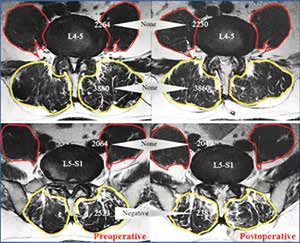
Pain Physician: September/October 2021 24:E883-E892

C        hronic low back pain (CLBP) is one of the most
         disabling conditions globally and is associated
         with tremendous socioeconomic and health
care consequences (1). In the normal intervertebral
disc (IVD), there is no nerve structure in the inner layer
                                                             surgery group met the following inclusion criteria: 1)
                                                             interlaminar approach at the L4–L5 or L5–S1 levels; 2)
                                                             Schiza grade A spinal stenosis or none; 3) HIZ in the
                                                             posterior annulus fibrosis on T2-weighted MRI; 4) disc
                                                             degeneration on MRI; 5) no nerve root compressive
of the fibrous ring (2) and no significant difference in     lesions, such as disc protrusion or degenerative bulg-
nerve density distribution between the outer fibrous         ing disc; 6) severe LBP with less leg pain or without
ring and endplate (3). In degenerative conditions,           leg pain for more than 6 months. We also applied the
inflammatory pathways involving secreted cytokines           following exclusion criteria: 1) predominant nonme-
induce nerve fibers and granulation tissue to grow in        chanical leg pain and neurogenic claudication; 2) MRI
the fissure of a degenerated IVD, leading to discogenic      showing prolapsed uncontained disc or significant
back pain (4,5). Previous reports have suggested that        facet arthropathy; 3) necessity of discectomy or re-
the high-intensity zone (HIZ) forms because annular          moval of any disc fragment for neural compression
tears cause injured disc material to accumulate and          during endoscopic spine surgery.
cause further degenerative changes within the IVD (6-             The patients included in the endoscopic posterior
9). However, a recent systematic review found evidence       decompression surgery group met the following inclu-
that an HIZ is a risk factor for CLBP (10-12).               sion criteria: 1) Schiza grade B–D; 2) severe leg pain
     In the present prospective study, we performed          with neurogenic intermittent claudication; 3) one
endoscopic radiofrequency ablation (RFA) surgery on          symptomatic lesion at the L4–L5 or L5–S1 level. The fol-
the HIZ with the surrounding sinuvertebral nerve (SVN)       lowing exclusion criteria were applied to both surgery
and basivertebral nerve (BVN) to treat discogenic low        groups: 1) previous spinal surgery, traumatic fracture,
back pain (LBP). We then evaluated the short- and            infection, or tumor; 2) postoperative epidural or muscle
long-term effects of this procedure on the patient’s         hematoma on postoperative MRI; 3) inability to mea-
score, MacNab’s criteria, and disability score. A recent     sure the CSA of the paravertebral muscle because of
study observed “Kim’s twitching” of the buttock and          severe sarcopenia.
paraspinal muscle during RFA (13). Therefore, we                  The Ethics Committee of Nanoori Hospital ap-
measured the cross-sectional area (CSA) of the para-         proved this study. Informed consent was obtained from
vertebral muscle on magnetic resonance imaging (MRI)         all patients.
before and after surgery to evaluate the effect of RFA
on the paravertebral muscle and find the neurological        Surgical Procedures
connections between the SVN, BVN, and paravertebral              Patients were classified into the following 2 cat-
muscles.                                                     egories according to the type of disease and the endo-
                                                             scopic surgical procedures conducted:
Methods
                                                             RFA via Interlaminar Endoscopic Lumbar
Study Patients                                               Discectomy (RF-IELD)
     This was a prospective study involving 23 patients          This procedure was performed under seated re-
who underwent uniportal spinal endoscopic RFA                gional epidural anesthesia. The patient was placed
surgery due to chronic discogenic back pain and 45           prone. The approach side was decided based on either
patients who underwent uniportal spinal endoscopic           symptom severity or, in the case of mid-axial back
posterior decompression surgery due to lumbar spinal         pain, the location of the HIZ. Under intraoperative
stenosis. The procedures were performed by a single          fluoroscopy, the endoscope was docked in the junc-
professional senior spine surgeon at a single center dur-    tion of the ipsilateral facet joint and IVD line. The
ing the calendar year 2018. All patients were followed       endoscope had a 30° viewing angle, a 7.3 mm outer
for at least 12 months. We set the posterior decompres-      diameter, and a 4.7 mm diameter working channel.
sion group as a comparative group because pain relief        The working channel was pushed into the spinal canal
could be obtained by posterior neural decompression          away from the neural elements toward the disc space.
without RFA surgery or discectomy, which may influ-          A bipolar radiofrequency electrocoagulator was used
ence the paraspinal muscle.                                  to ablate the hypersensitive basivertebral nerve around
     The patients included in the endoscopic RFA             the suprapedicular area and sinuvertebral nerve be-

E884                                                                                      www.painphysicianjournal.com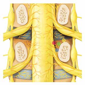
Paravertebral Muscle Changes after Lumbar Endoscopic RFA Treatment

tween the posterior annulus and posterior longitudinal                a)
ligament, which was covered with epidural neovascu-
larization and adhesions (Fig. 1) (13). The procedure
was performed unilaterally on the symptomatic side,
and RFA was also performed on the annulus containing
the HIZ lesion. The procedure was performed with the
working channel protecting the neural elements from
possible damage.

Lumbar Endoscopic Unilateral Laminotomy For
Bilateral Decompression (LE-ULBD)
     This procedure was performed under regional
epidural or general anesthesia. The patient was
placed prone. The endoscopic system has a working
cannula with a 13.7 mm outer diameter and a 10.2
mm inner diameter, and a 15º view angle. The endo-
scope was docked on the junction of the ipsilateral
facet joint and IVD line. We drilled just enough lamina
to safely remove the thickened flavum and sufficiently                b)
decompress the neural tissues. Next, we confirmed the
neovascularization grade, but RFA was not performed
in this group.

Data Collection

Clinical Data Collection
     Information on patient characteristics such as age,
gender, symptom duration, and surgery indication were
collected; the nature of the surgery and the level of
operation were also documented. For each patient, the
following information was collected before and after
surgery (one week, 3 month, and final follow-ups) by
attending physicians: visual analog scale (VAS) scores
for LBP and leg pain, modified Oswestry Disability score
(ODI), and MacNab’s criteria for evaluating patient’s
pain response and disability.
                                                                Fig. 1. Depiction of a coronal mid-pedicle cut of the
MRI Acquisition                                                 lumbar spine. A) Epidural neovascularization and
     One junior professional neurosurgeon analyzed              adhesions (black arrows) are in the intervertebral and
and measured the MRI parameters. Images were ob-                parapedicular areas, in which the sinuvertebral nerve and
                                                                basivertebral nerve are located, respectively. The high-
tained using a GE Signa 1.5-T HDxT MRI Machine (GE
                                                                intensity zone (white arrow) is in the intervertebral disc
Healthcare, Milwaukee, WI). All measurements were               with pathological neuroticized sinuvertebral nerve. B)
performed using an Infinitt PACS M6 Version (Infinitt           Kim’s twitching points. A: Middle parapedicular point,
Healthcare Co., Seoul, Republic of Korea). The images           B: upper parapedicular point (most common site), C:
were obtained using a fast spin-echo sequence with an           suprapedicular point, D: vertebral point.
echo train length of 28, a bandwidth of 25 Hz, a rep-
etition time of 3,000 milliseconds, an echo time of 110       before surgery and within 3 days after surgery without
milliseconds, a field of view of 160 × 160 mm, a number       a drain.
of excitations of 2, a slice thickness of 3 mm, and a slice        Before surgery, we observed the types of Modic
gap of 0.5 mm. MRI was performed within one week              changes and Schiza grade (14) for the degree of spinal

www.painphysicianjournal.com                                                                                            E885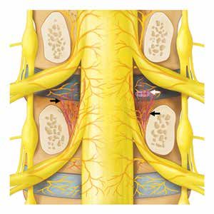
Pain Physician: September/October 2021 24:E883-E892

stenosis. The transverse diameter and CSA of the HIZ                mize the change in value, the CSA was obtained from
were measured on axial T2-weighted MRI before and                   2 consecutive transverse images at the same position
after surgery by pixel counting in the RF-IELD group.               (mid and upper disc level), and the average value was
Transverse images were acquired obliquely at each                   obtained.
disc level. Paravertebral muscle CSA was assessed by                     Surgical procedures were performed within the
carefully outlining the muscle mass on the T2-weight-               sublaminar space from the caudal to the cranial direc-
ed axial images, excluding fat and/or fibrous tissue                tion, so the paraspinal muscle at the mid and upper disc
external to the muscle fascia. Next, the CSA was au-                levels was not directly violated. Furthermore, patients
tomatically measured by pixel counting (Figs. 2A,3B).               with postoperative muscle or epidural hematoma were
One study of CSA reported significant modifications                 excluded. The paravertebral muscle was classified into
when the slice orientation was modified (15). To mini-              2 sections: the paraspinal muscle (multifidus + erector

 a)                                                                  b)

 c)                                                                  d)

   Fig. 2. Illustrations of measurement. The cross-sectional area (CSA) of the paraspinal muscle group (multifidus and erector
   spinae muscles) and psoas muscles at the L4–L5 and L5–S1 mid disc level (left: preoperative, right: postoperative). The
   pattern of CSA change was represented as “Positive,” “None,” and “Negative.” A) RF-IELD group: psoas muscle was
   categorized as “None,” but the paraspinal muscle was “Positive” at both levels. The high-intensity zone was located at the L5–
   S1 left paracentral area (blue arrow), but disappeared after radiofrequency ablation treatment (yellow arrow). B) LE-ULBD
   group: psoas muscle at both levels and paraspinal muscle at the L4–L5 level were categorized as “None”; the paraspinal
   muscle at the L5–S1 level showed “Negative.” C) Mean CSA of the paraspinal muscle. A P-value < 0.05 was considered to
   indicate significant difference. D) Mean CSA of the psoas muscle.

E886                                                                                                  www.painphysicianjournal.com
Paravertebral Muscle Changes after Lumbar Endoscopic RFA Treatment
spinae) and the psoas muscle, and the CSA was ob-
tained from each section. Although value modification
was minimized, it could not be excluded entirely, so
differences between +100 and -100 mm² were defined
as no change (“None”). When the CSA increased by
more than 100 mm², it was described as “Positive”;
when it decreased more than 100 mm², it was defined
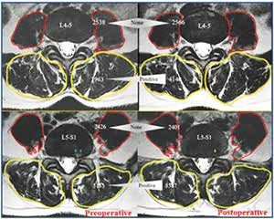
as “Negative” (Fig. 3).
Intraoperative Findings
Two experienced neurosurgeons reviewed the
endoscopic videos to evaluate intraoperative twitching
and to grade the degree of neovascularization and ad- Fig. 3. The pattern of paravertebral (paraspinal + psoas)
hesion. In mismatched cases, the results were adjusted muscle cross-sectional area (CSA) change was described
to reflect agreements after repeated assessments. We as a percentage within each group. When the CSA
increased by more than 100 mm², it was “Positive”; when
used a novel degenerative spinal neovascularization
it decreased by more than 100 mm², it was “Negative”;
grading described in a previous study (13), as follows: when it was between +100 mm² and -100 mm², it was
Grade 1, normal vascularization; Grade 2, neovascular- categorized as “None”.
ization without adhesion; Grade 3, neovascularization
with adhesion. Neovascularization and adhesion were
observed in the epidural space between the thecal sac, group and 45 patients in the LE-ULBD group. The mean
traversing and exiting nerve roots, posterior longitudi- age was 47.1 ± 12.5 years (range: 19–76 years) in the RE-
nal ligament, annulus, and vertebral bodies (Fig. 1A). IELD group and 59.8 ± 13.8 years (range: 33–81 years) in
The bipolar radiofrequency probe was applied to the the LE-ULBD group. The mean symptom duration was
medial suprapedicular area (Fig. 1B); twitching of the 20.1 ± 7.5 months in the RF-IELD group and 9.5 ± 2.6
buttock and paraspinal region were then observed, months in LE-ULBD group. The mean follow-up dura-
which typically starts when the BVN is coagulated; a tion was 15.5 ± 3.4 months in the RF-IELD group and
previous study termed this iatrogenic twitching “Kim’s 16.6 ± 4.1 months in the LE-ULBD group. The treated
twitching” (13). Once the BVN was completely ablated, spinal levels in the RF-IELD group were L4–L5 (n = 10)
Kim’s twitching stopped, even when the radiofrequency and L5–S1 (n = 13); in the group LE-ULBD, they were
was applied to the same area. Kim’s twitching was also L4–L5 (n = 42) and L5–S1 (n = 3; Table 1).
observed when the SVN was coagulated around the Preoperative MRI showed Modic change in 12 out
HIZ in the intervertebral disc (Fig. 1A). During regional of 23 patients in the RF-IELD group (52.12%) and in
epidural anesthesia, pain sensation was decreased, but 14 out of 45 patients in the LE-ULBD group (31.11%).
motor function was not, so no problems were encoun- There was a higher proportion of patients with Modic
tered when evaluating muscle twitching. change in the RF-IELD group (Table 1). All patients in
the RF-IELD group showed Schiza grade A, which means
Statistical Analysis no or minor stenosis. In the LE-ULBD group, there were
Statistical analyses were performed using PASW Sta- 7 patients with Grade B (moderate stenosis), 23 with
tistics ver. 18.0 (SPSS Inc., Chicago, IL, USA). The paired t- Grade C (severe stenosis), and 15 with Grade D (ex-
test was used to compare pre- and postoperative VAS, ODI, treme stenosis) (Table 1).
and MacNab’s outcome criteria to assess the outcomes as Intraoperative endoscopic videos showed neovas-
well as HIZ size, disc height, and paravertebral muscle CSA cularization and adhesion in the epidural space adja-
to evaluate the morphological changes. Independent tt- cent to the disc and pedicle; there were 7 patients with
test was used to compare outcomes and measured values Grade 2 and 16 with Grade 3 in the RF-IELD group; in
between the 2 independent groups. A P-value < 0.05 was the LE-ULBD group, there were 8 with Grade 1, 23 with
considered to indicate significant difference. Grade 2, and 10 with Grade 3. Kim’s twitching was ob-
served in 19 out of the 23 patients in the RF-IELD group
Results (Table 1) when the BVN is coagulated at the parape-
We included a total of 23 patients in the RF-IELD dicular point and when the SVN is coagulated around
www.painphysicianjournal.com E887Pain Physician: September/October 2021 24:E883-E892
Table 1. Background data and clinical results of the 2 groups.
Variable RF-IELD (n = 23) LE-ULBD (n = 45) P-value
Gender (men/women) 9/14 20/25 N/A
Age (years, mean/range) 47.1 ± 12.5 (19~76) 59.8 ± 13.8 (33~81) N/A
Symptom duration (months) 20.1 ± 7.5 9.5 ± 2.6 N/A
Follow-up duration (months) 15.5 ± 3.4 16.6 ± 4.1 N/A
Treated level (n / %) N/A
L4–L5 10 (48) 42 (93)
L5–S1 13 (52) 3 (7)
Modic type (n) N/A
Type 1 6 7
Type 2 5 7
Type 3 1 0
Total (n/%) 12 (52) 14 (31) N/A
Schiza grade (n/%) N/A
A 23 (100) 0
B 0 7 (16)
C 0 23 (51)
D 0 15 (33)
Neovascularization grade (n/%) N/A
Grade 1 0 8 (18)
Grade 2 7 (30) 27 (60)
Grade 3 16 (70) 10 (22)
Kim’s twitching (n/%) N/A
Negative 4 (17)
Positive 19 (83)
HIZ size (range)
Transverse diameter
6.25 ± 3.05 (2 ~ 12), 1.65 ± 1.90 (0 ~ 9.64) P = 0.000*
(mm, before and after) (Range)
CSA (mm², Pre and Post) (Range) 13.43 ± 10.63 (3.4 ~ 35.4), 1.84 ± 4.28(0 ~ 15.33) P = 0.000*
7.73 ± 1.29, 3.22 ± 0.63 (P = 0.000*),
VAS (before surgery, 1-week, 7.22 ± 1.28, 2.17 ± 0.64 (P = 0.000*), 1.65 ± 0.56 (P =
2.4 ± 0.74 (P = 0.000*), 2.2 ± 0.75 (P P = 0.929
3-month, and final follow-ups) 0.000*), 1.65 ± 0.56 (P = 0.000*)
= 0.000*)
73.2 ± 10.89, 29.3 ± 2.96 (P =
ODI (before surgery, 1-week, 68.3 ± 10.56, 25.3 ± 2.98 (P = 0.000*), 22.3 ± 2.38 (P =
0.000*), 25.6 ± 2.44 (P = 0.000*), P = 0.444
3-month, and final follow-ups) 0.000*), 21.4 ± 2.60 (P = 0.000*)
24.4 ± 2.56 (P = 0.000*)
MacNab’s criteria (n) N/A
Fair 0 3
Good 11 33
Excellent 12 9
Descriptive details of outcome measurement for preoperative MRI (Modic type, disc height, Schiza grade), intraoperative findings (neovascu-
larization grade, Kim’s twitching), pain measurement (VAS, ODI before surgery and at one week, 3 month and final follow-ups, MacNab’s cri-
teria). Independent t-test is used to compare the measured values between the 2 independent groups. Values are presented as mean ± standard
deviation. *P < 0.05 was considered to indicate significant difference. N/A; not applicable. HIZ; high-intensity zone.
E888 www.painphysicianjournal.comParavertebral Muscle Changes after Lumbar Endoscopic RFA Treatment
the HIZ in the intervertebral disc (Fig. 1). The transverse a)
length and CSA of the HIZ were measured before and
after surgery in the RF-IELD group. The mean value of
the transverse length was changed from 6.25 ± 3.05
mm to 1.65 ± 1.90 mm, and the CSA was changed from
13.43 ± 10.63 mm² to 1.84 ± 4.28 mm². There was a sta-
tistically significant decrease in both parameter values
(P = 0.00) (Table 1).
We measured VAS score (mean and range) be-
fore surgery and at the one week, 3 month, and final
follow-ups, with the following results: 7.22 (5–9), 2.17
(1–4), 1.65 (1–3), and 1.65 (1–3), respectively, in the RF-
IELD group and 7.73 (6–9), 3.22 (2–4), 2.40 (1–4), and
2.20 (1–4), respectively, in the LE-ULBD group (Table 1). b)
There was a significant improvement at the one week,
3 month, and final follow-ups in all surgical groups (P
= 0.000 in all cases and in both groups) (Fig. 4A). How-
ever, there was no significant difference between the 2
surgical groups (P = 0.929).
The ODI scores (mean and range) were measured
before surgery and at the one week, 3 month, and final
follow-ups, with the following results: 68.30 (48–84),
25.30 (20–34), 22.30 (18–26), and 21.40 (16–24) in the
RF-IELD group and 73.18 (56–86), 29.33 (22–42), 25.60
(18–42), and 24.40 (16–38) in the LE-ULBD group (Table
1). There was a statistical improvement at the one week,
Fig. 4. (Upper)(A) The mean visual analog score (VAS)
3 month, and final follow-ups in both surgical groups (P
and (Lower)(B) Oswestry Disability Index (ODI) were
= 0.000 in all cases and in both groups) (Fig. 4B). There measured before surgery and at the one week, 3 month, and
was no significant difference between the 2 groups (P = final follow-ups. All P-values < 0.05 were considered to
0.444). MacNab’s criteria showed excellent outcomes in indicate significant difference.
12 cases and good outcomes in 11 cases in the RF-IELD
group, as well as excellent outcomes in 9 cases, good at the L4–L5 level, while there were 15 (65%) “Posi-
outcomes in 33 cases, and fair outcomes in 3 cases in the tive” changes and 8 (35%) “None” changes out of 23
LE-ULBD group (Table 1). Table 2 and Figs. 2 and 3 detail patients at the L5–S1 level (Table 2) (Fig. 3). In the LE-
the mean CSA and pattern of CSA change in the paraspi- ULBD group, significantly more patients had changed
nal and psoas muscles. The mean CSA at each level was to “Negative” (L4–L5: 44%, L5–S1: 44%) or “None”
calculated as the sum of the bilateral measured values. (L4–L5: 20%, L5–S1: 40%) than to “Positive” (L4–5:
The mean CSA of the paraspinal muscle in the RF- 36%, L5–S1: 16%) (Fig. 3). The mean CSA of the psoas
IELD group was significantly increased after surgery at muscle showed an inconsistent direction of increase
both the L4–L5 and L5–S1 levels (L4–L5: 3901 ± 1096.7 and decrease at the L4–5 and L5–S1 levels in both sur-
mm² to 4167 ± 1052.1 mm²; P = 0.000, L5–S1: 3059 ± gical groups, unlike the paraspinal muscle (Fig. 2D).
968.5 mm² to 3323 ± 1046.2 mm²; P = 0.000). However, The pattern of psoas CSA change was not affected by
in the LE-ULBD group, it was significantly decreased surgery regardless of surgical type, because more than
after surgery at the L5–S1 level (2533 ± 820.8 mm² to half of the patients were classified as “None” (L4–5:
2383 ± 781.3 mm²; P = 0.001), but not at the L4–L5 lev- 56%, L5–S1: 78% in the RF-IELD group; L4–L5: 58%,
el (3880 ± 750.4 mm² to 3860 ± 738.2 mm²; P = 0.067) L5–S1: 68% in the LE-ULBD) (Fig. 3).
(Fig. 2C). The pattern of CSA change in the paraspinal
muscle was strictly measured. In the RF-IELD group,
Discussion
there were 16 (70%) “Positive” changes, 4 (17%) Several treatment strategies are effective in degen-
“None” changes, and 3 (13%) “Negative” changes erative disc disease, including RFA of the BVN through
www.painphysicianjournal.com E889Pain Physician: September/October 2021 24:E883-E892
Table 2. Paraspinal muscle and psoas muscle changes of the 2 groups.
Mean CSA (mm²) Pattern of CSA change (n)
Muscle Procedure Level
Pre-op. Post-op. P-value Negative None Positive
L4–L5 3901 ± 1096.7 4167 ± 1052.1 P = 0.000* 3 4 16
RF-IELD
L5–S1 3059 ± 968.5 3323 ± 1046.2 P = 0.000* 0 8 15
(n = 23)
Total 3220 ± 1284.9 3366 ± 1352.4 P = 0.000*
Paraspinal
L4–L5 3880 ± 750.4 3860 ± 738.2 P = 0.067 20 9 16
LE-ULBD
L5–S1 2533 ± 820.8 2383 ± 781.3 P = 0.000* 20 18 7
(n = 45)
Total 3072 ± 1138.6 3045 ± 1145.8 P = 0.238
L4–L5 2538 ± 1081.9 2566 ± 1126.5 P = 0.271 4 13 6
RF-IELD
L5–S1 2418 ± 893.0 2413 ± 919.3 P = 0.814 2 18 3
(n = 23)
Total 2738 ± 985.1 2868 ± 985.1 P = 0.553
Psoas
L4–L5 2264 ± 851.3 2230 ± 867.5 P = 0.287 12 26 7
LE-ULBD
L5–S1 2064 ± 778.5 2049 ± 816.3 P = 0.463 7 31 7
(n = 45)
Total 2299 ± 833.5 2216 ± 816.3 P = 0.788
The mean cross-sectional area (CSA) and pattern of CSA change of the paraspinal muscle and psoas muscle at the L4–L5 and L5–S1 levels for the
RF-IELD and LE-ULBD groups. When CSA increased by more than 100 mm², it was defined as “Positive”; when it decreased by more than 100
mm², it was defined as “Negative”; when it was between +100 mm² and -100 mm², it was defined as no change (“None”). Values are presented as
mean ± standard deviation. *P < 0.05 was considered to indicate significant difference.
intraosseous probe insertion (16). The sinuvertebral hypersensitive nerves and favorable clinical outcomes
nerve is involved in nociceptive transmission in de- after RFA surgery. Furthermore, neovascularization
generative IVD. The ascending branch, which becomes grade is probably a crucial inferred sign, because nei-
intraosseous near the pedicle and plays a vital role in ther the SVN nor the BVN are usually seen, even with
endplate nociceptive transmission, is termed the BVN endoscopic magnified vision (Fig. 1A).
(13) (Fig. 1B). In the present study, we performed an The mean CSA of the paraspinal muscle in the RF-
RFA on the SVN and BVN for discogenic back pain in IELD group was significantly increased after surgery,
young patients; the results showed significant improve- and more than half of the patients had a “Positive”
ment in pain and disability scores (Table 1). CSA value at all levels (Figs. 2C, 3). This result could
Changes in HIZ size were measured using trans- mean that paraspinal muscle spasm was alleviated after
verse diameter and CSA; we sought to confirm that RFA in more than half of the patients, even though no
postoperative HIZ change was associated with symp- needle electromyography was carried out. The leading
tom improvement. Of the 23 patients in the RF-IELD causes of the paraspinal muscle changes, pain reduc-
group, the HIZ was eliminated in 17 after RFA on the tion, or secondary RFA effect were unclear. Both re-
annulus containing the HIZ lesion. Nonetheless, we duced pain after decompression surgery and pain inhi-
found no clear correlation between elimination of bition by RFA could alleviate paraspinal muscle spasm.
the HIZ and the degree of symptom improvement. To Therefore, we formed the LE-ULBD surgical group as a
further investigate whether such a correlation occurs, a control to evaluate the paraspinal muscle response to
prospective study should be carried out with a control the RFA procedure.
group of patients suffering from discogenic back pain However, in the LE-ULBD group, the number of
without HIZ. “Positive” changes was much less than in the RF-IELD
Patients included in the present study who had group. Instead, there were more “Negative” changes
symptomatic HIZ and discogenic back pain showed a (Fig. 3), probably because the paraspinal muscle react-
high association with Kim’s twitching (19 out of 23) and ed and was changed by postoperative pain in patients
a high grade of neovascularization (Grade 3 in 16 out with negative changes. Therefore, even though muscle
of 23 patients); they also showed significant improve- stimulation does occur due to postoperative pain, the
ments in VAS, ODI, and MacNab’s criteria after RFA increased CSA values in the RF-IELD group must have
surgery. Therefore, high neovascularization grade and been meaningful.
Kim’s twitching are closely correlated with changed The psoas muscle at the L4–L5 and L5–S1 levels was
E890 www.painphysicianjournal.comParavertebral Muscle Changes after Lumbar Endoscopic RFA Treatment
unaffected by the RFA or decompression surgery—al- though value modification was minimized in this way,
most all patients were categorized as “None”. Similar it could not be excluded entirely. Third, the study did
results of the psoas CSA in patients with LBP have oc- not show changes in the entire lumbar paravertebral
curred previously (17,18), perhaps because the psoas muscle, because only the L4–L5 and L5-S1 levels were
major is innervated by direct branches of the anterior included. A larger prospective trial with 3D muscle re-
rami of the lumbar plexus at the L1–L3 levels. construction using specific software (Muscl’ X or custom
Several etiologies of multifidus muscle atrophy in software) should be performed to measure the entire
patients with LBP have been reported, including disuse muscle volume. Fourth, the changed muscle observed
atrophy, reflex inhibition, and dorsal ramus syndrome after surgery may not have been permanent.
(19-21). Postoperative paraspinal muscle change is likely Despite these limitations, the present study was
more correlated with the medial branches of the poste- meaningful as a first step to find an aberrant pathway
rior primary ramus and its reflex inhibitory mechanism between the hypersensitive SVN, BVN, and paraspinal
to the multifidus than with other paraspinal muscles. muscles, especially the multifidus.
In the present study, we observed Kim’s twitching;
this indicated a close connection between the SVN (in-
Conclusion
cluding the BVN) and the paraspinal muscles innervated Hypersensitive SVN and BVN are strongly associ-
by the posterior ramus of the spinal nerve. When RFA is ated with epidural neovascularization with adhesion
applied to the hypersensitive SVN and BVN, branches of and the pathological pain pathway in degenerative
the primary ventral ramus stimulate aberrant connec- disc disease. Epidural neovascularization with adhesion
tions with the traversing nerve or exiting nerve root. reflects the aberrant neurological connections, which
A strong impulse then propagates along the posterior are associated with reflex inhibitory mechanisms of the
ramus of the spinal nerve and knock down its inhibitory multifidus muscle, which induces the spasm. RFA treat-
mechanism. Eventually, the multifidus alleviated. This ment of epidural neovascularization with adhesion
mechanism may be the main cause of postoperative effectively treated chronic discogenic back pain and
paraspinal (multifidus + erector spinae) CSA increase. could alleviate paraspinal muscle spasm.
The results of the present study suggest that multifidus
muscle spasms would be alleviated if the reflex inhibi- Acknowledgments
tory mechanism were disconnected or diminished by Hyeun-Sung Kim and Ji Yeon Kim contributed
RFA. equally to this work as first authors. We thank Keong
The present study has several limitations. First, Rae Kim who contributed to the statistical analysis
the study populations were not homogeneous in age, of the data and Jae Eun Park for coordinating the
gender, pathology, or number of patients, so the re- scientific research. We would like to thank Editage
sults may not have represented the actual difference (www.editage.co.kr) for English language editing.
between these 2 patient groups. Second, to reduce
modification to the minimum, the CSA was assessed by Ethics Statement
carefully outlining the muscle mass, excluding fat and This study was approved by the Ethics Committee of
fibrous tissue external to the muscle fascia, and mea- Nanoori Hospital. Informed consent was obtained from
suring the average value from 2 consecutive slices. Al- the patients. IRB approval number: NR-IRB 2020-018.
References
1. Simon J, McAuliffe M, Shamim F, B, Blumbergs P, Fraser R. ISSLS 5. Garcia-Cosamalon J, del Valle ME,
Vuong N, Tahaei A. Discogenic low back prize winner: The innervation of the Calavia MG, et al. Intervertebral disc,
pain. Phys Med Rehabil Clin N Am 2014; intervertebral disc: A quantitative sensory nerves and neurotrophins: Who
25:305-317. analysis. Spine (Phila Pa 1976) 2003; is who in discogenic pain? J Anat 2010;
2. Roberts S, Evans EH, Kletsas D, Jaffray 28:2570-2576. 217:1-15.
DC, Eisenstein SM. Senescence in 4. Ohtori S, Miyagi M, Inoue G. Sensory 6. Chen JY, Ding Y, Lv RY, et al. Correlation
human intervertebral discs. Eur Spine J nerve ingrowth, cytokines, and between MR imaging and discography
2006; 15:S312-316. instability of discogenic low back pain: A with provocative concordant pain in
3. Fagan A, Moore R, Vernon Roberts review. Spine Surg Relat Res 2018; 2:11-17. patients with low back pain. Clin J Pain
www.painphysicianjournal.com E891Pain Physician: September/October 2021 24:E883-E892
2011; 27:125-130. A, et al. What low back pain is and why stenosis based on the morphology of
7. Lam KS, Carlin D, Mulholland RC. we need to pay attention. Lancet 2018; the dural sac on magnetic resonance
Lumbar disc high-intensity zone: The 391:2356-2367. images. 2018 27:1146-1156). Eur Spine J
value and significance of provocative 13. Kim HS, Wu PH, Jang IT. Lumbar 2019; 28:2223.
discography in the determination of degenerative disease part 1: Anatomy 17. Danneels LA, Vanderstraeten GG,
the discogenic pain source. Eur Spine J and pathophysiology of intervertebral Cambier DC, Witvrouw EE, De Cuyper
2000; 9:36-41. discogenic pain and radiofrequency HJ. CT imaging of trunk muscles in
8. Carragee EJ, Paragioudakis SJ, Khurana ablation of basivertebral and chronic low back pain patients and
S. 2000 Volvo Award winner in clinical sinuvertebral nerve treatment for healthy control subjects. Eur Spine J
studies: Lumbar high-intensity zone chronic discogenic back pain: A 2000;9:266-272.
and discography in subjects without prospective case series and review of 18. Cooper RG, St Clair Forbes W, Jayson
low back problems. Spine (Phila Pa 1976) literature. Int J Mol Sci 2020; 21:1483. MI. Radiographic demonstration of
2000; 25:2987-2992. 14. Schizas C, Theumann N, Burn A, et paraspinal muscle wasting in patients
9. Park KW, Song KS, Chung JY, et al. al. Qualitative grading of severity with chronic low back pain. Br J
High-intensity zone on L-spine MRI: of lumbar spinal stenosis based on Rheumatol 1992; 31:389-394.
Clinical relevance and association with the morphology of the dural sac on 19. Hultman G, Nordin M, Saraste
trauma history. Asian Spine J 2007; magnetic resonance images. Spine H, Ohlsèn H. Body composition,
1:38-42. (Phila Pa 1976) 2010; 35:1919-1924. endurance, strength, cross-sectional
10. Teraguchi M, Yim R, Cheung JP, 15. Henderson L, Kulik G, Richarme D, area, and density of MM erector spinae
Samartzis D. The association of high- Theumann N, Schizas C. Is spinal in men with and without low back pain.
intensity zones on MRI and low back stenosis assessment dependent on J Spinal Disord 1993; 6:114-123.
pain: A systematic review. Scoliosis slice orientation? A magnetic resonance 20. Hodges P, Holm AK, Hansson T,
Spinal Disord 2018; 13:22. imaging study. Eur Spine J 2012; Holm S. Rapid atrophy of the lumbar
21:S760-S764. multifidus follows experimental disc or
11. Brinjikji W, Diehn FE, Jarvik JG, et al.
MRI findings of disc degeneration 16. Li Y, Feng X, Tan J, Peng B. Letter to nerve root injury. Spine (Phila Pa 1976)
are more prevalent in adults with the Editor concerning “Intraosseous 2006; 31:2926-2933. doi:10.1097/01.
low back pain than in asymptomatic basivertebral nerve ablation for the brs.0000248453.51165.0b
controls: A systematic review and meta- treatment of chronic low back pain: A 21. Kader DF, Wardlaw D, Smith FW.
analysis. AJNR Am J Neuroradiol 2015; prospective randomized double-blind Correlation between the MRI changes
36:2394-2399. sham-controlled multi-center study” in the lumbar multifidus muscles and
by Fischgrund JS, et al. (Qualitative leg pain. Clin Radiol 2000; 55:145-149.
12. Hartvigsen J, Hancock MJ, Kongsted
grading of severity of lumbar spinal
E892 www.painphysicianjournal.comYou can also read