Oblique Lateral Interbody Fusion versus Transforaminal Lumbar Interbody Fusion in Degenerative Lumbar Spondylolisthesis: A Single-Center ...
←
→
Page content transcription
If your browser does not render page correctly, please read the page content below
Hindawi
BioMed Research International
Volume 2021, Article ID 6693446, 14 pages
https://doi.org/10.1155/2021/6693446

Research Article
Oblique Lateral Interbody Fusion versus Transforaminal Lumbar
Interbody Fusion in Degenerative Lumbar Spondylolisthesis: A
Single-Center Retrospective Comparative Study

          Xing Du, Yuxiao She, Yunsheng Ou , Yong Zhu, Wei Luo, and Dianming Jiang
          Department of Orthopedics, The First Affiliated Hospital of Chongqing Medical University, 400016 Chongqing, China

          Correspondence should be addressed to Yunsheng Ou; ouyunsheng2001@163.com

          Received 30 December 2020; Revised 18 February 2021; Accepted 15 March 2021; Published 22 March 2021

          Academic Editor: Ying-Qi Zhang

          Copyright © 2021 Xing Du et al. This is an open access article distributed under the Creative Commons Attribution License, which
          permits unrestricted use, distribution, and reproduction in any medium, provided the original work is properly cited.

          Objective. To compare the efficacy of oblique lateral interbody fusion (OLIF) and transforaminal lumbar interbody fusion (TLIF) in
          single-level degenerative lumbar spondylolisthesis (DLS). Methods. A retrospective analysis of patients who underwent single-level
          DLS surgery in our department from 2015 to 2018 was performed. According to the surgical method, the enrolled patients were
          divided into two groups, namely, the OLIF group who underwent OLIF combined with percutaneous pedicle screw fixation
          (PPSF) and the TLIF group. Clinical outcomes included operation time, operation blood loss, postoperative drainage, hospital
          stay, visual analog scale (VAS) score, Oswestry disability index (ODI), and complications, and imaging outcomes included
          upper vertebral slip, intervertebral space height (ISH), intervertebral foramen height (IFH), intervertebral space angle (ISA),
          lumbar lordosis (LL), and bone fusion rate. All outcomes were recorded and analyzed. Results. A total of 65 patients were finally
          included, and there were 28 patients and 37 patients in the OLIF group and the TLIF group, respectively. The OLIF group
          showed shorter operation time, less blood loss, less postoperative drainage, and shorter hospital stay than the TLIF group
          (P < 0:05). The ISH, IFH, ISA, and LL were all larger in the OLIF group at postoperative and last follow-up (P < 0:05), but the
          degree of upper vertebral slip was found no difference between the two groups (P > 0:05). The bone graft fusion rate of OLIF
          group and TLIF group at 3 months, 6 months, and last follow-up was 78.57%, 92.86%, and 100% and 70.27%, 86.49%, and
          97.30%, respectively, and no significant differences were found (P > 0:05). Compared with the TLIF group, the OLIF group
          showed a superior improvement in VAS and ODI at 1 month, 3 months, and 6 months postoperative (P < 0:05), but no
          differences were found at 12 months postoperative and the last follow-up (P > 0:05). There was no significant difference in
          complications between the two groups, with 4 patients and 6 patients in the OLIF group and TLIF group, respectively (P > 0:05
          ). Conclusions. Compared with TLIF, OLIF showed the advantages of less surgical invasion, better decompression effect, and
          faster postoperative recovery in single-level DLS surgery.

1. Introduction                                                         conservative treatment for more than 3 months, surgery
                                                                        should be considered [3].
Lumbar spondylolisthesis is defined as the forward slip of the               The surgical methods of DLS mainly include anterior and
upper vertebrae relative to the lower. There are many causes            posterior surgery [4]. In anterior lumbar interbody fusion
of lumbar spondylolisthesis, including degeneration, trauma,            (ALIF), the discectomy and spondylolisthesis reduction can
dysplasia, and pathology, among which degenerative lumbar               be performed under direct vision, and the correction and
spondylolisthesis (DLS) is the most common [1]. It is                   maintain of intervertebral space height and lumbar lordosis
reported that the incidence of DLS is about 5% to 7% and                (LL) can be also achieved by implanting a large cage [5].
often causes low back pain, lower limb pain, or weakness,               But ALIF has a high risk of abdominal vascular or viscera
and even cauda equina syndrome in severe cases [2]. The                 injury, as well as the rate of postoperative complications
treatment of DLS mainly includes conservative and surgical              [6]. In recent years, transforaminal lumbar interbody fusion
treatment. For patients with no obvious efficacy after regular            (TLIF) has been the most commonly used posterior surgery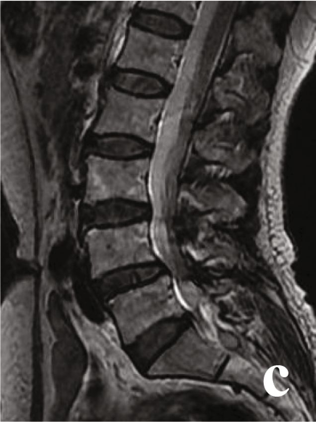
2                                                                                                BioMed Research International

for DLS [7]. By resecting the facet joints, loosening surround-    the psoas and abdominal aorta, as well as the extent of upper
ing fibrous tissue, and implanting pedicle screws and inter-        vertebral slip, spinal canal stenosis, and nerve root compres-
vertebral cage, TLIF can obtain a good spondylolisthesis           sion. Surgery was performed when basic diseases such as dia-
reduction effect and maintain satisfactory spinal stability         betes, coronary heart disease, and high blood pressure were
[8]. However, patients undergoing TLIF often suffer from            under control.
chronic back pain after surgery, which may be due to the
resection of paravertebral muscle and facet joints [9]. In addi-   2.3. Surgical Procedure. The choice of surgical method was
tion, surgeons need to open the spinal canal in TLIF surgery,      mainly based on the following principles: OLIF combined
and the intraoperative nerve stimulation may also cause            PPSF was mainly used for patients whose preoperative MRI
numbness or weakness of the lower limbs after surgery              or CT showed an appropriate operative window between
[10]. Thus, more and more surgeons have been seeking min-          the psoas and abdominal aorta. If preoperative MRI or CT
imally invasive surgical methods for DLS.                          showed no operative window between the psoas and abdom-
    Oblique lateral interbody fusion (OLIF) is a minimally         inal aorta or the operative window was narrow, TLIF should
invasive anterior retroperitoneal approach surgery, which          be considered.
has been very popular in recent years. In OLIF surgery, the
surgeon enters the retroperitoneal space through blunt sepa-       2.3.1. OLIF Group. Place the patient in a lateral supine posi-
ration, pulls the psoas muscle backward, reaches the opera-        tion after general anaesthesia and use C-arm X-rays to iden-
tive segment or intervertebral space through the anatomical        tify the surgical level. Then cut a 4 cm incision in the outer
space between the abdominal aorta and the psoas muscle,            abdominal area, separate the layers of abdominal muscles,
and performs decompression and fusion procedures [11].             and push away the extraperitoneal fat with fingers. Find the
OLIF was reported with satisfactory efficacy in lumbar               front edge of the psoas with a Cobb periosteal stripper, push
degenerative diseases in cohort studies [12, 13]; however,         back the psoas and place an OLIF retractor. After exposing
no study compared its efficacy with TLIF surgery in DLS.             the vertebra, insert a positioning needle and confirm the sur-
Our previous study conclude that OLIF combined with per-           gical level by C-arm X-rays and place different extenders to
cutaneous pedicle screw fixation (PPSF) had less surgical           extend the channel to 22 mm. Then, remove the interverte-
trauma and faster pain relief and rapid lumbar function            bral disc completely and implant a cage (Medtronic, USA)
recovery in lumbar tuberculosis [14]. Thus, we really won-         filled with granular bone, which was derived from allogeneic
dered whether OLIF may or may not be extrapolated to the           bone (Gold Bone Way, China). Not any bone fusion promot-
DLS surgery.                                                       ing substance was used in bone graft materials. Then, place a
    Therefore, we conducted this retrospective study to com-       drainage tube and close the incision layer by layer. Adjust the
pare the efficacy of OLIF and TLIF in single-level DLS, in           patient to a prone position and do posterior internal fixation
order to provide evidence for the application of OLIF in           with percutaneous pedicle screw instrumentation (IRENE,
DLS patients.                                                      China). Finally, use a C-arm X-ray to confirm the good posi-
                                                                   tion of posterior fixation and close the posterior incision.
2. Materials and Methods
                                                                   2.3.2. TLIF Group. Place the patient in a prone position after
The ethical approval of this study was obtained from the           general anaesthesia. Make a posterior median incision after
Ethics Committee of the First Affiliated Hospital of Chong-          identification of the surgical level by C-arm X-rays. Accord-
qing Medical University (No. 2020-049), and all the partici-       ing to preoperative clinical features, the side with lower limb
pants gave the informed consent before taking part. This           symptoms was defined as the decompression side. Strip the
study had been registered in the Chinese Clinical Trial Regis-     sacrospinous muscle of the decompression side, expose the
try (ChiCTR2000039446). This study was reported according          lamina and facet joints of the surgical level, and then implant
to the STROCSS criteria [15].                                      the pedicle screws. For the contralateral side, expose the facet
                                                                   joints and implant the pedicle screws via the Wiltes
2.1. Patients. A retrospective analysis of medical records of      approach. Then, resect part of facet joints and lamina of the
DLS patients hospitalized in our hospital from 2015 to 2018        decompression side and remove the intervertebral disc
was conducted.                                                     completely and implant a cage (Guona, China) filled with
    Inclusion criteria: (1) single-level DLS (L2/3-L4/5). (2)      granular bone through the intervertebral foramen. The used
Mild symptomatic DLS (Meyerding Grade: I or II). (3) Age           granular bone was derived from the lamina, spinous process,
 > 18 years. (3) Underwent OLIF combined with PPSF (OLIF           and facet articular process. However, in most cases, the autol-
group) or TLIF (TLIF group). (4) More than 12 months of            ogous bone volume was not enough, so they were often
follow-up time. (5) Clinical and imaging data were                 mixed with some allogeneic granular bone (Gold Bone
completed.                                                         Way, China). Not any bone fusion promoting substance
    Exclusion criteria: (1) a previous lumbar spinal surgery       was used in bone graft materials. Then, correct the spondylo-
history. (2) Recurrent DLS after surgical treatment. (3) DLS       listhesis by properly pulling up and pressurizing the posterior
with severe cardiovascular disease or malignant tumor, etc.        screw system (IRENE, China) and obtain an appropriate LL
                                                                   by using a prebending rod. After the confirmation of correc-
2.2. Preoperative Management. X-rays, CT, and MRI were             tion by C-arm X-rays, the surgical wound was rinsed and
taken in all patients to assess the surgical window between        hemostasis was carefully performed. The posterior screw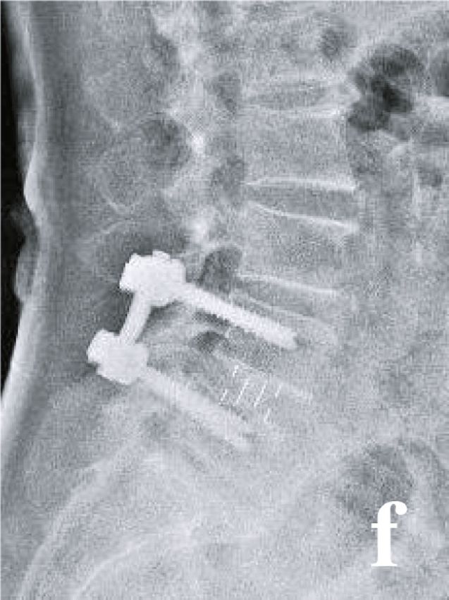
BioMed Research International                                                                                                    3

system was then properly pulled up and pressurized in order
to correct the spondylolisthesis and obtain an appropriate LL,
and a C-arm X-ray was used to confirm the correction. The
surgical wound was rinsed, and hemostasis was carefully per-
formed. Finally, place a drainage tube and close the incision
layer by layer.

2.4. Postoperative Management. In the first 3 days after sur-
gery, antibiotics were used to prevent infection. When post-
operative drainage was less than 40 ml/d, the drainage tube
was removed. An X-ray examination of the lumbar spine
was taken after extubation. After discharge, a modeled rigid
lumbar brace was applied continuously for 3 months.
Patients were requested to wear the lumbar braces every
day when getting out of bed, moving, or sitting. Their family
members were asked to supervise the brace wearing, and the
medical team conducted telephone follow-up once a week to
timely evaluate the patient’s compliance. Follow-up of X-
rays, CT, and MRI (if necessary) was conducted for 1, 3, 6,
and 12 months after surgery.

2.5. Outcomes. Clinical outcomes: (1) operation time, opera-
tion blood loss, postoperative drainage, and hospital stay.
(2) Visual analog scale (VAS) score and Oswestry disability        Figure 1: Diagram of the measurement of ISH (red line), ITH
index (ODI) at 1, 3, 6, and 12 months postoperative and            (green line), ISA (blue line), and LL (yellow line).
the last follow-up. (3) Complications.
    Imaging outcomes: (1) the degree of upper vertebral            3. Results
slip: the ratio of the slip distance of the upper vertebrae
to the length of the upper endplate of the lower vertebrae.        According to the inclusion and exclusion criteria, our study
(2) Intervertebral space height (ISH): the mean of anterior        finally included 65 patients, and there were 28 patients and
and posterior ISH. (3) Intervertebral space foramen (IFH):         37 patients in the OLIF group and the TLIF group, respec-
the distance between the lower margin of the superior              tively. There were no significant differences in age
pedicle and vertebral body connection and the upper mar-           (P = 0:641), gender (P = 0:683), body mass index (BMI)
gin of the inferior pedicle and vertebral body connection.         (P = 0:591), ASA grade (P = 0:779), operative level
(4) Intervertebral space angle (ISA): the angle between            (P = 0:890), bone mineral density (BMD) (P = 0:101), and
the upper and lower endplate of the intervertebral space.          follow-up time (P = 0:282) between the two groups.
(5) Lumbar lordosis (LL): the angle between the upper              (Table 1).
endplate of the L1 vertebral body and the upper endplate               The OLIF group showed shorter operation time
of the S1 vertebral body. The measurement methods of               (P < 0:001), less operation blood loss (P < 0:001), less postop-
ISH, IFH, ISA, and LL were shown in Figure 1. (6) Bone             erative drainage (P < 0:001), and shorter hospital stay
graft fusion: according to Bridwell et al.’s study [16], bone      (P < 0:001) than the PLIF group. (Figure 2).
graft fusion was divided into four levels. Grade I: fused              No significant differences were found in preoperative
with remodeling and trabeculae. Grade II: graft intact,            ISH, IFH, ISA, and LL between the two groups (P = 0:508,
not fully remodeled and incorporated though; no                    0.649, 0.231, and 0.522, respectively). However, the ISH,
lucencies. Grade III: graft intact, but a definite lucency at       IFH, ISA, and LL at postoperative and the last follow-up were
the top or bottom of the graft. Grade IV: definitely not            significantly larger in the OLIF group than those in the TLIF
fused with resorption of bone graft and with collapse.             group (postoperative: P < 0:001, 0.002,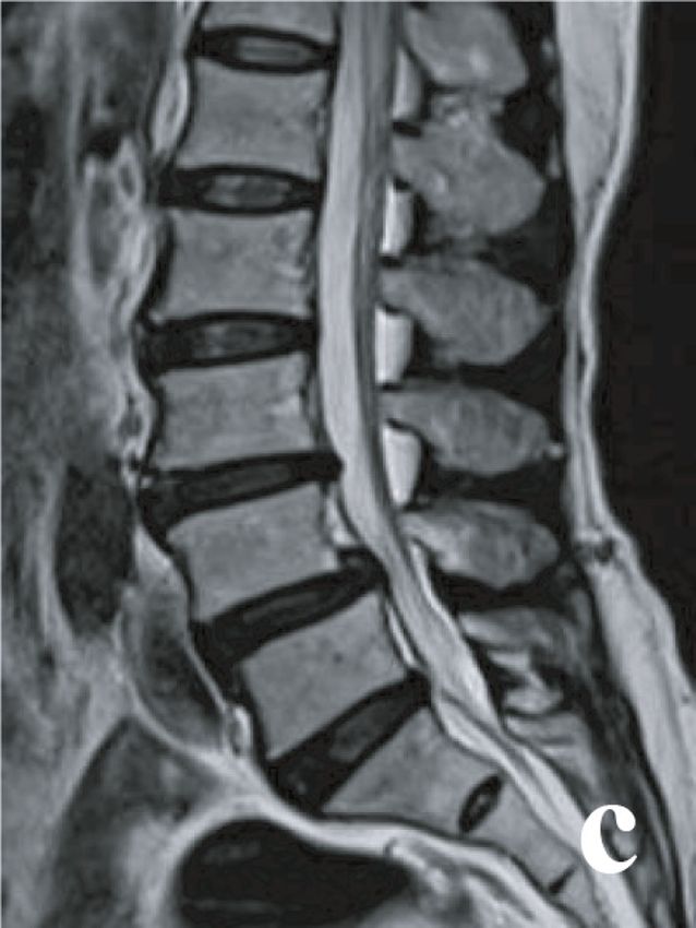
4                                                                                                                                                    BioMed Research International

                             Table 1: Comparison of preoperative clinical features between the two groups.

Clinical features                                                        TLIF group (N = 37)                                             OLIF group (N = 28)               P value
Age (year), mean ± SD                                                         52:8 ± 7:1                                                      53:6 ± 6:4                    0.641
Gender (n), Male/Female                                                         23/14                                                           16/12                       0.683
BMI (kg/m2), mean ± SD                                                        22:5 ± 2:3                                                      22:8 ± 2:1                    0.591
ASA grade (n)                                                                                                                                                               0.779
  I                                                                                24                                                               17
  II                                                                               10                                                                9
  III                                                                              3                                                                2
Operation level (n)                                                                                                                                                         0.890
  L3/4                                                                          10                                                               8
  L4/5                                                                          27                                                              21
BMD (T score), mean ± SD                                                     −2:3 ± 1:0                                                      −1:9 ± 0:9                     0.101
Follow-up time (month), mean ± SD                                             22:1 ± 7:0                                                      20:3 ± 6:1                    0.282

                                                           150                                                         200

                                                                                           Operation blood loss (ml)

                                                                              ⁎                                        150
                             Operation time (min)

                                                           100
                                                                                                                                                ⁎
                                                                                                                       100

                                                            50
                                                                                                                        50

                                                             0                                                              0
                                                                 TLIF       OLIF                                                 TLIF         OLIF
                                                                   (a)                                                             (b)
                                                           200                                                         10

                                                                                                                       8
                             Postoperative drainage (ml)

                                                           150
                                                                                                                                               ⁎
                                                                                           Hospital stay (d)

                                                                              ⁎                                        6
                                                           100
                                                                                                                        4

                                                            50
                                                                                                                       2

                                                             0                                                         0
                                                                 TLIF       OLIF                                                TLIF         OLIF
                                                                   (c)                                                            (d)

Figure 2: Comparison of operation time (a), operation blood loss (b), postoperative drainage (c), and hospital stay (d) between the two
groups. (∗ Compared with TLIF group, P < 0:05).

the two groups (P = 0:451, 0.412, and 0.389, respectively)                                           postoperative, and last follow-up (VAS score: P = 0:760,
(Figure 3).                                                                                          0.064, and 0.408, respectively; ODI: P = 0:604, 0.088, and
    No significant differences were found in VAS score and                                             0.216, respectively). However, the OLIF group showed a bet-
ODI between the two groups at preoperative, 12 months                                                ter improvement in VAS score and ODI at 1 month, 3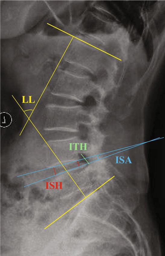
BioMed Research International 5
15 20 #⁎
#⁎ # & &⁎
# &⁎ 15
10 &
ISH (mm)
IFH (mm)
10
5
5
0 0
Preoperative
Postoperative
Last follow-up
Preoperative
Postoperative
Last follow-up
TLIF TLIF
OLIF OLIF
(a) (b)
20 #⁎ 60
#⁎
# # & &⁎
15 &⁎
40
&
10
20
5
0 0
Preoperative
Postoperative
Last follow-up
Preoperative
Postoperative
Last follow-up
TLIF TLIF
OLIF OLIF
(c) (d)
40 110
Upper vertebral slip (%)
Bone graft fusion (%)
100
30
90
20
80
# #
10
70
0 60
3 months 6 months Last follow-up
Preoperative
Postoperative
Last follow-up
OLIF
TLIF
TLIF
OLIF
(e) (f)
Figure 3: Comparison of ISH (a), ITH (b), ISA (c), LL (d), degree of upper vertebral slip (e), and bone graft fusion rate (f) between the two
groups at different follow-up time. (∗ Compared with TLIF group, P < 0:05; &compared with preoperative, P < 0:05; #compared with
postoperative, P < 0:05).
months, and 6 months postoperative than the TLIF group Four patients with complications were found in the OLIF
(VAS score: P < 0:001,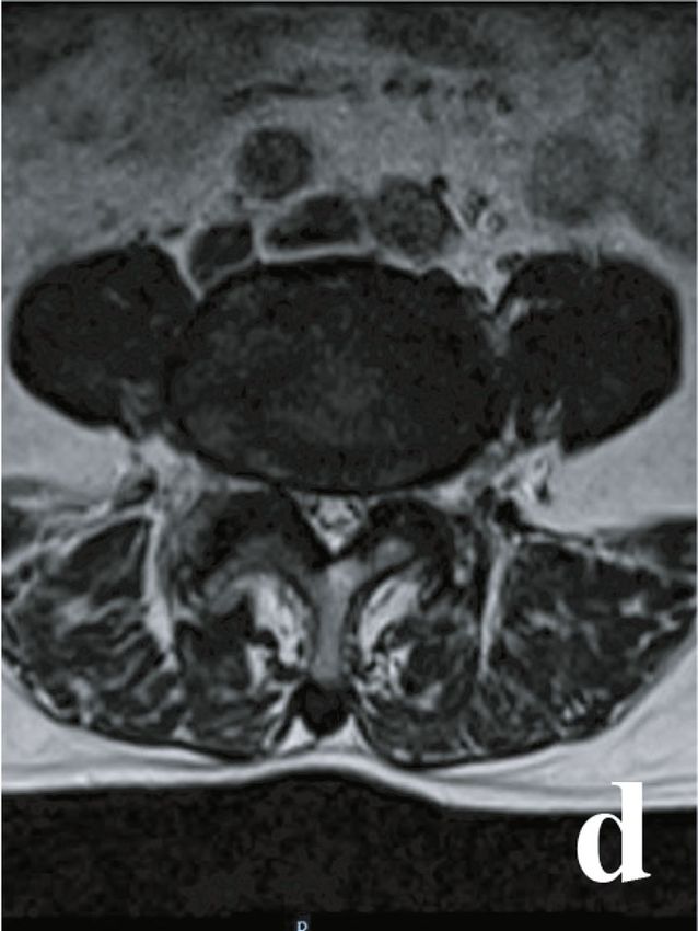
6 BioMed Research International
10 80
8
60
VAS score ⁎ ⁎
ODI (%)
6 ⁎
40 ⁎
4 ⁎
⁎
20
2
0 Time 0 Time
Preoperative
1 month
3 months
6 months
Preoperative
12 months
Last follow-up
1 month
3 months
6 months
12 months
Last follow-up
OLIF OLIF
TLIF TLIF
(a) (b)
Figure 4: Comparison of VAS score (a) and ODI (b) between the two groups at different follow-up time. (∗ Compared with TLIF group, P
< 0:05).
transient sympathetic injury. While 6 patients with compli- OLIF by comparing the changes of imaging parameters
cations were found in the TLIF group, including 3 patients before and after surgery [21–23]. In our study, it was found
of dural tear, 1 patient of transient thigh pain and/or numb- that the ISH, IFH, and ISA for postoperative and last
ness, 1 patient of transient ankle dorsiflexion weakness, and 1 follow-up were significantly larger in the OLIF group than
patient of postoperative incision infection. There was no sig- the TLIF group. The reasons may be as follows: (1) in
nificant difference in the complications rate (14.3% vs. OLIF surgery, a large cage with a degree of inclination
16.2%) between the two groups (P = 0:446). Finally, all com- angle was implanted into the intervertebral space, while
plications were cured after treatment. in TLIF surgery, because of the narrow operating space,
only a small cage, almost without an inclination angle,
3.1. Typical Cases. Typical cases were shown in Figures 5 and could be implanted through the intervertebral foramen
6. [24]. (2) In TILF surgery, only one side of the paraverteb-
ral muscle and the facet joints was removed while the con-
4. Discussion tralateral side was usually preserved, so the intervertebral
space may not be effectively extended, especially for
In this study, TLIF showed longer operation time and more patients with severe facet joints degeneration or even facet
operation blood loss than OLIF. This result was similar to a joints fusion [25]. LL is an important imaging index to
previous study. The possible reasons were as follows: (1) in evaluate the efficacy of DLS surgery. It was confirmed that
TLIF surgery, one side of paravertebral muscle was peeled LL was closely related to postoperative lumbar back pain,
off, and part of facet joints and lamina was also resected and effective correction and maintenance of LL were of
[17], while neither was done in OLIF surgery. (2) In OLIF great significance to relieve lumbar back pain and improve
surgery, surgeons could directly reach the operative interver- lumbar function [26]. In this study, both the postoperative
tebral space and did the discectomy under direct vision [18], and last follow-up LL were found significantly larger in the
while in TLIF surgery, discectomy could not be performed OLIF group than the TLIF group. This was closely related
under direct vision, especially for the contralateral side of to the effective correction and maintenance of ISH, IFH,
the decompression side [19]. (3) In OLIF surgery, the spinal and ISA in the OLIF group [27]. However, no differences
canal was not opened, so the risk of nerve injury was low, in upper vertebral slip at preoperative, postoperative, and
while TLIF surgery required opening the intervertebral last follow-up were found between the two groups. This
foramina, which had a risk of nerve injury, so surgeons paid may be owing to the following reasons: (1) in TLIF sur-
more attention to the separation and the protection of the gery, the posterior pedicle system could effectively correct
nerve roots [20]. The less postoperative drainage and shorter spondylolisthesis by its strong pulling force [28]. (2) Both
hospital stay of the OLIF group may also be related to the of the two groups achieved a higher rate of bone graft
larger surgical damage of TLIF surgery. In addition, the pro- fusion during the follow-up, and vertebral spondylolisth-
longed postoperative bedtime caused by the risk of spinal esis would not deteriorate once bone graft fusion was
instability due to the intraoperative dissection of paraverteb- achieved. Although OLIF showed a better decompression
ral muscle and facet joints may also be the reasons for the efficacy than TLIF in this study, it is still necessary to
longer hospital stay of the TLIF group. extend the follow-up time to confirm this conclusion in
OLIF is an indirect decompression surgery, and many future research, as spinal canal remodeling during long-
studies are aimed at evaluating the decompression effect of term follow-up after surgery had been proved [29].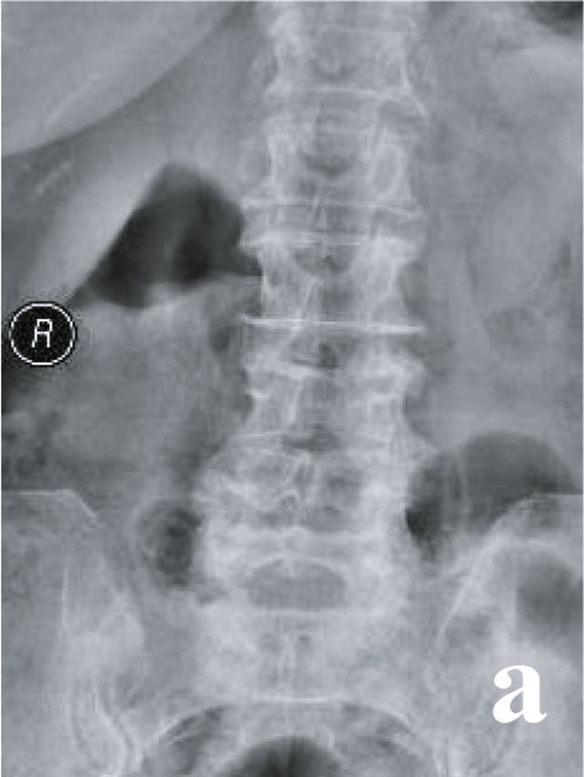
BioMed Research International                                      7

                                (a)                          (b)

                                (c)                          (d)

                                      Figure 5: Continued.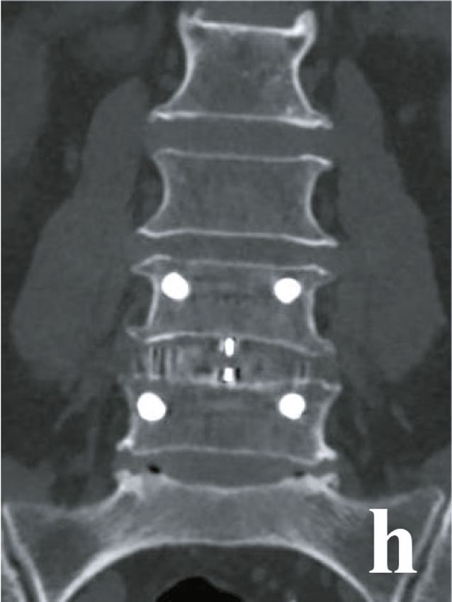
8                                      BioMed Research International

    (e)                          (f)

    (g)                          (h)

          Figure 5: Continued.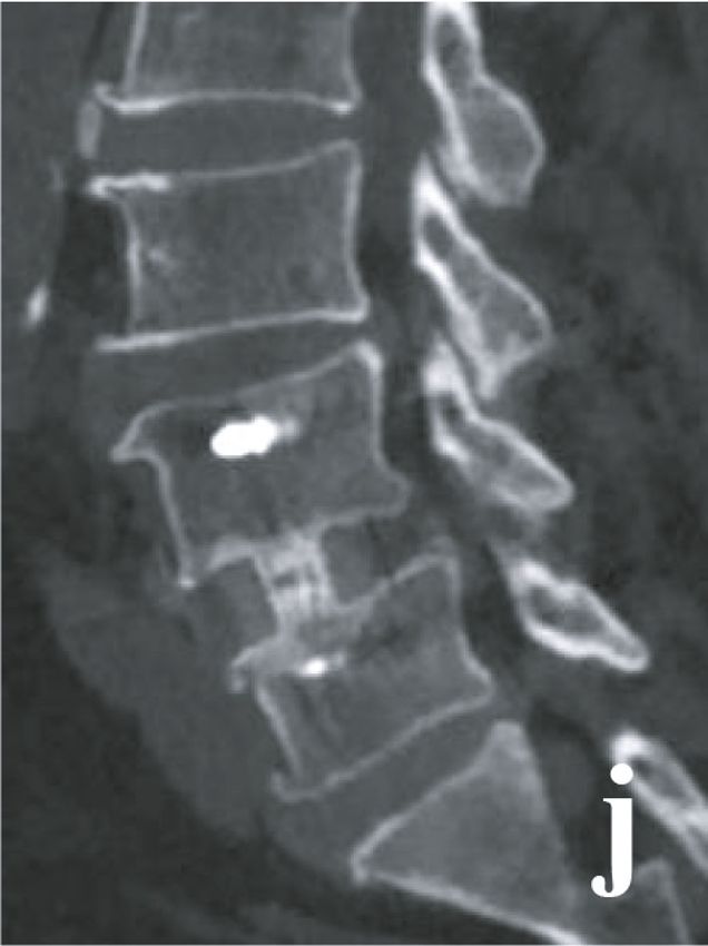
BioMed Research International 9
(i) (j)
(k) (l)
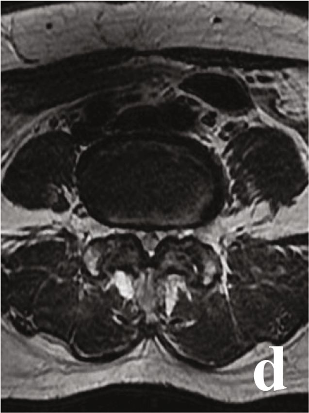
Figure 5: A 49-year-old male with L4/5 DLS in TLIF group. (a)–(d) Preoperative X-ray and MRI. (e) and (f) Postoperative X-ray. (g)–(j) CT
at 6 months postoperative. (k) and (l) X-ray at 18 months postoperative.
In the present study, the OLIF group and TLIF group Postoperative pain relief and lumbar function recovery
showed similar bone graft fusion rate. We thought the rea- were the main focus of both the surgeons and patients. In this
sons may be as follows: (1) in both groups, the intervertebral study, it was found that the short-term (in 12 months) post-
discs were completely removed, and the cartilage endplate operative pain and lumbar functional in the OLIF group were
was also completely scraped. This would create a suitable superior to the TLIF group, but there were no significant dif-
good graft bed for fusion [30]. (2) Granular bone graft was ferences in long-term follow-up. The reasons may be as fol-
used in both groups, and our previous study had showed that lows: (1) less damage to paraspinal muscles and facet joints
granular bone graft had a high fusion rate [31]. (3) Both was found in the OLIF group [33], so low back pain and lum-
groups used a posterior pedicle screw system to create a sta- bar function were better than the TLIF group in the short-
ble local mechanical environment, which was also beneficial term follow-up. (2) OLIF surgery did not open the spinal
for bone graft fusion [32]. canal and had little stimulation to nerve roots [34]. (3) For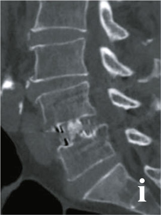
10                                      BioMed Research International

     (a)                          (b)

     (c)                          (d)

           Figure 6: Continued.
BioMed Research International 11
(e) (f)
(g) (h)
Figure 6: Continued.12 BioMed Research International
(i) (j)
(k) (l)
Figure 6: A 50-year-old female with L4/5 DLS in the OLIF group. (a)–(d) Preoperative X-ray and MRI. (e) and (f) Postoperative X-ray. (g)–(j)
CT at 5 months postoperative. (k) and (l) X-ray at 20 months postoperative.
most patients in both groups, bone graft fusion had achieved, and susceptible to being irritated or injured [36, 37]. The
and the paravertebral muscles had also been recovered at 1 main complications of TLIF were nerve root stimulation or
year postoperative. The above results were also the reasons injury and dural tear [38]. However, we found no significant
for shorter hospital stays in the OLIF group. Although difference in complication rate between the two groups. This
stand-alone OLIF was reported with less injury to paraver- result also indicated the safety of the two surgical methods for
tebral muscles [33], PPSF was used in the OLIF group in DLS.
our study, because preoperative BMD revealed that most In our opinion, the indications of OLIF combined PPSF
patients had osteopenia or even osteoporosis. for DLS were as follows: (1) severe back pain and/or leg pain
Segmental artery injury, transient thigh numbness were with poor response to regular conservative treatment of 3
the common complications in the OLIF group [35], because months. (2) Progressive exacerbation of instability or spon-
the lumbar plexus, lumbar sympathetic trunk, and segmental dylolisthesis. (3) Single-segmental DLS (L2/3-L4/5) with
artery are all located laterally in front of the lumbar vertebrae mild degree of spondylolisthesis (Grade I or II). (5) AnBioMed Research International 13
appropriate operative window was shown between the psoas ogy Innovation and Application Development Project of
and abdominal aorta on preoperative MRI or CT [39]. Chongqing (cstc2020jscx-msxmX0219).
This study also has some limitations. First, it is a retro-
spective study with a small sample size and short follow-up
period. Second, different experiences in OLIF/TLIF surgery References
may also cause bias.
[1] M. Bydon, M. A. Alvi, and A. Goyal, “Degenerative lumbar
5. Conclusion spondylolisthesis: definition, natural history, conservative
management, and surgical treatment,” Neurosurgery Clinics
Compared with TLIF, OLIF showed the advantages of less of North America, vol. 30, no. 3, pp. 299–304, 2019.
surgical invasion, better decompression effect, and faster [2] Y. X. J. Wang, Z. Káplár, M. Deng, and J. C. S. Leung, “Lumbar
postoperative recovery in single-level DLS surgery. degenerative spondylolisthesis epidemiology: a systematic
review with a focus on gender-specific and age-specific preva-
lence,” Journal of Orthopaedic Translation, vol. 11, pp. 39–52,
Abbreviations 2017.
DLS: Degenerative lumbar spondylolisthesis [3] M. Karsy and E. F. Bisson, “Surgical versus nonsurgical treat-
ALIF: Anterior lumbar interbody fusion ment of lumbar spondylolisthesis,” Neurosurgery Clinics of
TLIF: Transforaminal lumbar interbody fusion North America, vol. 30, no. 3, pp. 333–340, 2019.
OLIF: Oblique lateral interbody fusion [4] W. R. Spiker, V. Goz, and D. S. Brodke, “Lumbar interbody
PPSF: Percutaneous pedicle screw fixation fusions for degenerative spondylolisthesis: review of tech-
niques, indications, and outcomes,” Global Spine Journal,
VAS: Visual analog scale
vol. 9, no. 1, pp. 77–84, 2018.
ODI: Oswestry disability index
ISH: Intervertebral space height [5] J. Y. Cho, T. S. Goh, S. M. Son, D. S. Kim, and J. S. Lee, “Com-
parison of anterior approach and posterior approach to instru-
IFH: Intervertebral foramen height
mented interbody fusion for spondylolisthesis: a meta-
ISA: Intervertebral space angle analysis,” World Neurosurgery, vol. 129, pp. e286–e293, 2019.
LL: Lumbar lordosis
[6] B. Kapustka, G. Kiwic, P. Chodakowski et al., “Anterior lum-
SD: Standard deviation
bar interbody fusion (ALIF): biometrical results and own expe-
BMI: Body mass index riences,” Neurosurgical Review, vol. 43, no. 2, pp. 687–693,
BMD: Bone mineral density. 2020.
[7] A. K. Chan, V. Sharma, L. C. Robinson, and P. V. Mumma-
Data Availability neni, “Summary of guidelines for the treatment of lumbar
spondylolisthesis,” Neurosurgery Clinics of North America,
The clinical data in this study is available from the corre- vol. 30, no. 3, pp. 353–364, 2019.
sponding author on reasonable request.
[8] C. Wipplinger, C. Melcher, R. N. Hernandez et al., “"One and a
half" minimally invasive transforaminal lumbar interbody
Additional Points fusion: single level transforaminal lumbar interbody fusion
with adjacent segment unilateral laminotomy for bilateral
Research Registration. This study had been registered in Chi- decompression for spondylolisthesis with bisegmental steno-
nese Clinical Trial Registry (Number: ChiCTR2000039446, sis,” J Spine Surg, vol. 4, no. 4, pp. 780–786, 2018.
http://www.chictr.org.cn/showproj.aspx?proj=63238). [9] S. H. Min, M. H. Kim, J. B. Seo, J. Y. Lee, and D. H. Lee, “The
quantitative analysis of back muscle degeneration after poste-
Conflicts of Interest rior lumbar fusion: comparison of minimally invasive and
conventional open surgery,” Asian Spine J, vol. 3, no. 2,
The authors have no conflicts of interest to disclose in rela- pp. 89–95, 2009.
tion to this article. [10] V. A. Mehta, M. J. McGirt, G. L. Garcés Ambrossi et al.,
“Trans-foraminal versus posterior lumbar interbody fusion:
Authors’ Contributions comparison of surgical morbidity,” Neurological Research,
vol. 33, no. 1, pp. 38–42, 2013.
Conception and design were done by Xing Du and Yunsheng [11] J. X. J. Li, K. Phan, and R. Mobbs, “Oblique lumbar interbody
Ou; data analysis and interpretation were done by Xing Du, fusion: technical aspects, operative outcomes, and complica-
Yuxiao She, and Yong Zhu; data collection and management tions,” World Neurosurgery, vol. 98, pp. 113–123, 2017.
were done by Xing Du, Yuxiao She, Wei Luo, and Dianming [12] J. Li, D. Zhang, Y. Shen, and X. Qi, “Lumbar degenerative dis-
Jiang; manuscript writing and critical revisions were done by ease after oblique lateral interbody fusion: sagittal spinopelvic
all authors; overall responsibility was done by Xing Du and alignment and its impact on low back pain,” Journal of Ortho-
Yunsheng Ou. paedic Surgery and Research, vol. 15, no. 1, p. 326, 2020.
[13] S. Y. Chang, Y. Nam, J. Lee, B. S. Chang, C. K. Lee, and H. Kim,
Acknowledgments “Impact of preoperative diagnosis on clinical outcomes of obli-
que lateral interbody fusion for lumbar degenerative disease in
This work was supported by the Natural Science Foundation a single-institution prospective cohort,” Orthopaedic Surgery,
of Chongqing (cstc2019jcyj-msxmX0358) and the Technol- vol. 11, no. 1, pp. 66–74, 2019.14 BioMed Research International
[14] X. Du, Y. S. Ou, Y. Zhu, W. Luo, G. Y. Jiang, and D. M. Jiang, ters and clinical outcomes after oblique lumbar interbody
“Oblique lateral interbody fusion combined percutaneous ped- fusion,” World Neurosurgery, vol. 133, pp. e156–e164, 2020.
icle screw fixation in the surgical treatment of single-segment [28] Z. X. Wu, C. Zhan, G. Cui et al., “Stress distribution on the
lumbar tuberculosis: a single- center retrospective comparative screws in posterior lumbar fusion of isthmic spondylolisthesis
study,” International Journal of Surgery, vol. 83, pp. 39–46, with 2- or 3-vertebra fixation techniques: a biomechanical
2020. cadaveric study,” The Journal of Surgical Research, vol. 176,
[15] R. Agha, A. Abdall-Razak, E. Crossley et al., “STROCSS 2019 no. 1, pp. 95–101, 2012.
guideline: strengthening the reporting of cohort studies in sur- [29] S. Ohtori, S. Orita, K. Yamauchi et al., “Change of lumbar liga-
gery,” International Journal of Surgery, vol. 72, pp. 156–165, mentum flavum after indirect decompression using anterior
2019. lumbar interbody fusion,” Asian Spine J, vol. 11, no. 1,
[16] K. H. Bridwell, L. G. Lenke, K. W. McEnery, C. Baldus, and pp. 105–112, 2017.
K. Blanke, “Anterior fresh frozen structural allografts in the [30] T. Steffen, A. Tsantrizos, and M. Aebi, “Effect of implant
thoracic and lumbar spine,” Spine, vol. 20, no. 12, pp. 1410– design and endplate preparation on the compressive strength
1418, 1995. of interbody fusion constructs,” Spine (Phila Pa 1976),
[17] C. J. Fu, W. C. Chen, M. L. Lu, C. H. Cheng, and C. C. Niu, vol. 25, no. 9, pp. 1077–1084, 2000.
“Comparison of paraspinal muscle degeneration and decom- [31] X. Du, Y. S. Ou, S. Xu, B. He, W. Luo, and D. M. Jiang, “Com-
pression effect between conventional open and minimal inva- parison of three different bone graft methods for single seg-
sive approaches for posterior lumbar spine surgery,” Scientific ment lumbar tuberculosis: a retrospective single-center
Reports, vol. 10, no. 1, p. 14635, 2020. cohort study,” International Journal of Surgery, vol. 79,
[18] S. Ohtori, S. Orita, K. Yamauchi et al., “Mini-open anterior ret- pp. 95–102, 2020.
roperitoneal lumbar interbody fusion: oblique lateral Inter- [32] Y. Y. Hsieh, F. Y. Tsuang, Y. J. Kuo, C. H. Chen, C. J. Chiang,
body fusion for lumbar spinal degeneration disease,” Yonsei and C. L. Lin, “Biomechanical analysis of single-level inter-
Medical Journal, vol. 56, no. 4, pp. 1051–1059, 2015. body fusion with different internal fixation rod materials: a
[19] X. Cheng, K. Zhang, X. Sun et al., “Clinical and radiographic finite element analysis,” BMC Musculoskeletal Disorders,
outcomes of bilateral decompression via a unilateral approach vol. 21, no. 1, p. 100, 2020.
with transforaminal lumbar interbody fusion for degenerative [33] W. He, Y. S. Da He, Y. Xing et al., “Quantitative analysis of
lumbar spondylolisthesis with stenosis,” The Spine Journal, paraspinal muscle atrophy after oblique lateral interbody
vol. 17, no. 8, pp. 1127–1133, 2017. fusion alone vs. combined with percutaneous pedicle screw fix-
[20] N. E. Epstein, “More nerve root injuries occur with minimally ation in patients with spondylolisthesis,” BMC Musculoskeletal
invasive lumbar surgery: let's tell someone,” Surgical Neurology Disorders, vol. 21, no. 1, p. 30, 2020.
International, vol. 7, Suppl 3, pp. S96–S101, 2016. [34] R. J. Mobbs, K. Phan, G. Malham, K. Seex, and P. J. Rao, “Lum-
[21] W. Limthongkul, T. Tanasansomboon, W. Yingsakmongkol, bar interbody fusion: techniques, indications and comparison
T. Tanaviriyachai, K. Radcliff, and W. Singhatanadgige, “Indi- of interbody fusion options including PLIF, TLIF, MI-TLIF,
rect decompression effect to central canal and ligamentum fla- OLIF/ATP, LLIF and ALIF,” Journal Spine Surgery, vol. 1,
vum after extreme lateral lumbar interbody fusion and oblique no. 1, pp. 2–18, 2015.
lumbar interbody fusion,” Spine (Phila Pa 1976), vol. 45, [35] Z. Y. Zeng, Z. W. Xu, D. W. He et al., “Complications and pre-
no. 17, pp. E1077–E1084, 2020. vention strategies of oblique lateral interbody fusion tech-
[22] M. Wu, J. Li, M. Zhang et al., “Efficacy and radiographic anal- nique,” Orthopaedic Surgery, vol. 10, no. 2, pp. 98–106, 2018.
ysis of oblique lumbar interbody fusion for degenerative lum- [36] H. Wang, Y. Zhang, X. Ma, X. Xia, F. Lu, and J. Jiang, “Radio-
bar spondylolisthesis,” Journal of Orthopaedic Surgery and graphic study of lumbar sympathetic trunk in oblique lateral
Research, vol. 14, no. 1, p. 399, 2019. interbody fusion surgery,” World Neurosurgery, vol. 116,
[23] J. Sato, S. Ohtori, S. Orita et al., “Radiographic evaluation of pp. e380–e385, 2018.
indirect decompression of mini-open anterior retroperitoneal [37] S. Orita, K. Inage, T. Sainoh et al., “Lower lumbar segmental
lumbar interbody fusion: oblique lateral interbody fusion for arteries can intersect over the intervertebral disc in the oblique
degenerated lumbar spondylolisthesis,” European Spine Jour- lateral interbody fusion approach with a risk for arterial injury:
nal, vol. 26, no. 3, pp. 671–678, 2017. radiological analysis of lumbar segmental arteries by using
[24] C. Sun, H. Wang, J. Jiang, F. Lu, X. Ma, and X. Xia, “Length of magnetic resonance imaging,” Spine (Phila Pa 1976), vol. 42,
lumbar interbody cage using radiological measurements of no. 3, pp. 135–142, 2017.
Chinese endplates and the apophyseal ring,” World Neurosur- [38] B. Garg and N. Mehta, “Minimally invasive transforaminal
gery, vol. 116, pp. e1204–e1213, 2018. lumbar interbody fusion (MI-TLIF): a review of indications,
[25] P. A. Robertson, W. A. Armstrong, D. L. Woods, and J. J. Raw- technique, results and complications,” J Clin Orthop Trauma,
linson, “Lordosis recreation in transforaminal and posterior vol. 10, Suppl 1, pp. S156–S162, 2019.
lumbar interbody fusion: a cadaveric study of the influence [39] L. Liu, Y. Liang, H. Zhang et al., “Imaging anatomical research
of surgical bone resection and cage angle,” Spine (Phila Pa on the operative windows of oblique lumbar interbody fusion,”
1976), vol. 43, no. 22, pp. E1350–E1357, 2018. PLoS One, vol. 11, no. 9, article e0163452, 2016.
[26] C. Y. Chuang, M. Y. Liaw, L. Y. Wang et al., “Spino-pelvic
alignment, balance, and functional disability in patients with
low-grade degenerative lumbar spondylolisthesis,” Journal of
Rehabilitation Medicine, vol. 50, no. 10, pp. 898–907, 2018.
[27] L. Xiao, Q. Zhao, X. Sun, C. Liu, Y. Zhang, and H. Xu, “Rela-
tionship between alterations of spinal/pelvic sagittal parame-You can also read