The intraoperative use of a calliper predicts leg length and offset after total hip arthroplasty. Component subsidence influences the leg length
←
→
Page content transcription
If your browser does not render page correctly, please read the page content below
Fansur et al. Journal of Orthopaedic Surgery and Research                (2021) 16:424
https://doi.org/10.1186/s13018-021-02559-3

 RESEARCH ARTICLE                                                                                                                                    Open Access

The intraoperative use of a calliper predicts
leg length and offset after total hip
arthroplasty. Component subsidence
influences the leg length
Maliha Fansur1, Nagib A. Yurdi2 and Reinhard Stoewe3*

  Abstract
  Background: The purpose of total hip arthroplasty (THA) post-surgery and proper physiotherapy is positive
  recovery for the patient. Consideration is given to hip replacement biomechanics by ensuring no discrepancies in
  limb length (LL) and a stable prosthesis. Therefore, the patient must have proper preoperative planning and
  communication and a clear understanding of what to expect.
  Methods: A prospective series of 59 THA operated by a single surgeon via Hardinge approach was studied, using
  an intraoperative calliper (CAL) to predict the change of LL and offset. We compared the results of the
  intraoperative changes before and after THA implantation with the reference of these values on anteroposterior x-
  ray pelvis. The importance of leg length balance and a good offset restoration is questioned, and the effect of
  component subsidence on leg length is considered.
  Results: The average preoperative leg length discrepancy was −6.0 mm, postoperatively +3.6 mm. There was a
  strong correlation between the CAL measurements and the values on the x-ray (LL, r=0.873, p
Fansur et al. Journal of Orthopaedic Surgery and Research   (2021) 16:424                                                 Page 2 of 8

uncertainty about the ideal postoperative leg length and               Table 1 Patient demographic characteristics and procedure
offset after THA. Recently, also as response to patient’s              data
dissatisfaction and threatening litigation [2], there have             Age mean                                            51 (17–74)
been an increasing number of studies to suggest im-                    Gender (M/F)                                        30/24
provements concerning this aspect by using intraopera-                 Operated hip (L/R)                                  30/29
tive tests, calibration gauges, and other devices to
                                                                       BMI mean                                            30 (20–47)
improve functional outcome and reduce possible com-
                                                                       Discharge post-OP day mean                          4.8 (1–10)
plications. One difficulty when approaching this problem
is the interference with other phenomena observed in                   Primary THA/revision THA                            56/3
THA surgery, such as hip instability, gait disorders, in-              Femoral component (uncemented/cemented)             55/4
fection, fractures, and subsidence of prosthetic                       Acetabular component (uncemented/cemented)          57/2
components.
   While the use of an intraoperative calibration gauge is
widespread in some countries, e.g., Norway, there are                    Preoperatively, all patients were examined clinically. In
other countries where it is hardly used, e.g., Germany                 case of suspected LLD, an examination of the patient
and the UAE. Therefore, the authors decided it was                     standing, using lift blocks under the short leg, and visu-
valuable to present the results from this study suggesting             ally examining the level pelvis was completed. In this
the value of an intraoperative calibration gauge to make               study, the gold standard and reference to measure pre-
THA more predictable. The hypothesis of this study is                  operative LLD was the anterior-posterior (AP) weight-
that the use of an intraoperative calliper can predict the             bearing x-ray of the pelvis [3]. Other radiological exami-
postoperative leg length and offset, and therefore im-                 nations to further examine LLD preoperatively, such as
prove the outcome of THA operations. The first section                 x-ray long-film standing, scanograms, computerized
presents the results related to postoperative leg length               digital radiographs, and CT [4, 5], were utilized in excep-
and offset using an intraoperative calliper (CAL). The                 tional circumstances on some patients. The results of
values from anterior-posterior (AP) x-ray of the pelvis                these additional examinations performed were not in-
with a calibration ball are used as a reference. Utilizing             cluded in this study.
this measurement method, the target zone for intraoper-
ative leg length measurement is discussed with respect                 Surgical technique
to other interfering factors.                                          All surgeries were performed via a direct lateral Hard-
                                                                       inge approach, patient in lateral decubitus. All prosthesis
                                                                       was distributed by DePuy Synthes. The CAL was sup-
Materials and methods
                                                                       plied by Smith & Nephew, Memphis, TN, USA. Its
This study was completed at Healthpoint Hospital in the
                                                                       proper use and possible mistakes while using it are de-
city of Abu Dhabi, UAE. Healthpoint Hospital is a semi-
                                                                       scribed well in the literature [6, 7]. The reference pin for
public hospital in the capital of the UAE functioning as
                                                                       the use of CAL was positioned into the pelvis in line
a local hospital and a reference hospital for the whole
                                                                       with the skin incision close to the iliac crest through an
country. This clinical study was registered and autho-
                                                                       additional 3-mm skin incision. Before the dislocation of
rized by the institutional research ethics committee
                                                                       the hip, the most lateral point of the greater trochanter
REC016.
                                                                       was marked with diathermy on the bone and the sur-
                                                                       rounding soft tissue. After attaching the free arm of
Method                                                                 CAL to the reference pin, the tip was gently led onto the
This is a retrospective study of prospectively collected               mark on the greater trochanter without creating any ten-
data where 54 patients and 59 hips were reviewed. All                  sion in the CAL. Figure 1 shows the calliper set in place
THA operations were performed by the senior author                     before the dislocation of the hip operating a right hip
RS between 2018 and 2020. All patients signed a consent                patient lateral supine.
form to participate in this study. All patients with stable              In this position, the first set of data was recorded and
and reproducible hip conditions on the side to be oper-                documented. The second set of data was recorded like-
ated, including primary and revision surgery, were in-                 wise when the trial implants were in place. If there was a
cluded in this study. Patients with acute proximal                     substantial aberration of this data from expected values,
femoral fractures and dislocated dysplasia hips were ex-               the reason, such as cup malpositioning or severe varus/
cluded. In a reflection of the population diversity of the             valgus malalignment of the stem, was found and cor-
UAE, the clinical group came from 21 different national                rected. At this stage, and at the end of the operation be-
backgrounds. The patient demographic characteristics                   fore wound closure, also different clinical tests were
are shown in Table 1.                                                  done to check the stability of the hip and to look for any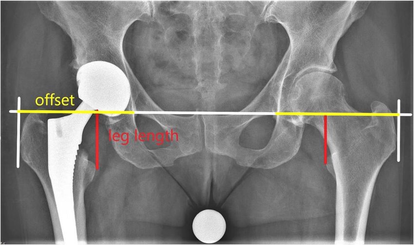
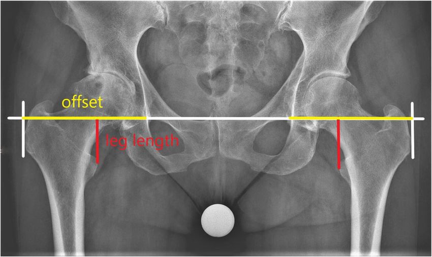
Fansur et al. Journal of Orthopaedic Surgery and Research         (2021) 16:424                                                  Page 3 of 8

 Fig. 1 The calliper set in place before the dislocation of the hip operating a right hip patient lateral supine

possible impingement, the priority being to achieve a                          closely related to global offset, which by definition is the
stable hip [8]. Based on the second set of data, the final                     sum of the femoral offset and the lateralization of the
implants were chosen (neck length, standard or high off-                       hip joint center of rotation (acetabular offset) [9, 10].
set of the stem, stem size) and set in place. The third set
of data was the values after the last reposition of the hip
with the final components in place. To avoid deviation                         Radiological analysis
due to positioning, this third set of data was taken with                      To avoid any bias, the radiological data were collected,
a particular focus on maintaining the operative limb in                        examined, and evaluated completely independent by au-
the same position while taking the first set concerning                        thor MF. For each patient, a set of two radiological ex-
flexion and abduction. This process is also commonly                           aminations, which were all performed at the same
described in the literature [7]. The first and third set of                    department using the same x-ray machine, was evalu-
data were used in this study. The change of leg length                         ated. An anterior-posterior (AP) weight-bearing x-ray of
can be calculated immediately. This is not the case for                        the pelvis with patella facing forward was taken. Using
the femoral offset, which per definition is the distance                       the Woolson method [3], leg length was measured by
from the center of rotation of the femoral head to a line                      drawing a line from the interteardrop line to the apex of
bisecting the long axis of the femur [9]. We were aware                        the trochanter minor. The offset, which means global
that by using CAL we measured a change in a value                              offset, was assimilated by a line, parallel to the

 Fig. 2 X-ray pelvis pre-OP
Fansur et al. Journal of Orthopaedic Surgery and Research (2021) 16:424 Page 4 of 8
interteardrop line, from the bottom of the teardrop to No control group was available as CAL is used in all
the height of the most lateral point of the trochanter THA operations by the senior author.
major (Fig. 2).
According to this preoperative x-ray, a rough estima- Results
tion of the required change in leg length and offset was Fifty-four patients and 59 hips with complete data sets
calculated comparing the situation with the contralateral were included for analysis. The mean age of the patients
hip. At this point, also other possible pathologies, e.g., a was 51 years (17–74), with 30 out of 54 patients being
future operation on the contralateral side, were consid- male. Fifty-six out of 59 operations were primary THA’s,
ered. At our institution, we do not perform simultaneous and 3 were revisions. The indication for operation was
bilateral THA. The perception of the likelihood of a sec- degenerative osteoarthritis in 32 cases, rheumatoid arth-
ond operation on the contralateral side and the conse- ritis 4 cases, hip dysplasia (Crowe I to III) 6 cases, and
quences on the LLD of the second operation influenced stage 4 non-traumatic avascular caput necrosis 10 cases.
our target of LLD of the first operation. Four patients were operated because of posttraumatic
Postoperatively, all patients were encouraged to be avascular necrosis. The three revisions were performed
mobilized with full weight-bearing as tolerated. due to polyethylene wear, aseptic loosening of a cemen-
Dependent upon early mobilization and the patient’s ted acetabular component, and undercoverage of acetab-
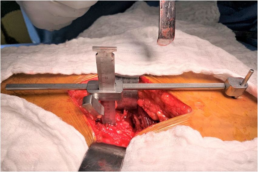
pain level, a second x-ray of the pelvis was taken on the ular component, each. The BMI of the patients was
third postoperative day using the same technique (Fig. mean 30 (20–47). The operational time was mean 127
3). A calibration ball of 25 mm was used for both exami- min (78–211). When operated, 11 of the patients had
nations. It was placed between the patient’s legs as close already a THA on the contralateral side. The day of dis-
to the focal point of the x-ray beam as practically charge was postoperatively mean day 4.8 (1–10). The
possible. use of the CAL increased the operational time by about
4 min. There were no intraoperative or postoperative
Statistics complications reported due to the use of the CAL, espe-
Mean, median, range, and standard deviation were cal- cially no wound healing problems of the additional small
culated for the various measurement parameters. Data proximal incision. On these 59 THA operations, the fol-
was analyzed with the SPSS V22.0 statistical software lowing complications were observed: on five patients,
(IBM Corp., USA). Pearson correlation testing was per- there was intraoperatively a suspicion of a proximal fem-
formed to compare the radiographic results with the in- oral calcar fracture which was treated by a cerclage. Two
traoperative measurements of the change in leg length patients had more than 3° varus/valgus malalignment of
and the change in offset. This study hypothesized that the femoral stem. One patient suffered an undisplaced
these two values correlate, and herewith, the use of CAL pelvic fracture intraoperatively and was treated conser-
predicts the postoperative leg length and offset. A correl- vatively without any further consequences. One patient
ation value of r ≥ 0.5 was found to be a strong correl- had directly postoperatively an inlay displacement of the
ation. A p < 0.05 was considered to be statistically ceramic acetabular inlay; one patient had an undercover-
significant. age of the acetabular component. Both of these patients
Fig. 3 X-ray pelvis post-OPFansur et al. Journal of Orthopaedic Surgery and Research (2021) 16:424 Page 5 of 8 were taken for revision. Six patients had blood transfu- in measurements [15]. As mentioned, additional clinical sion postoperatively. tests to check stability and impingement were done in- The preoperative x-ray measurements show an LLD traoperatively. There were no patients where it was ne- mean of −6.0 mm (−29 to 25), while the postoperative x- cessary to change the neck length or other prosthesis ray measurements show an LLD mean of 3.6 mm (−7 to parts to prioritize stability over achieving the scheduled 43). Of the operated 59 hips, 43 had an LLD up to 5 limb length and offset, abandoning the calliper input. In mm, 54 up to 10 mm, 57 up to 15 mm, and 2 over 20 agreement with Barbier et al., the calliper seems to bet- mm, directly after surgery. Comparing the pre- and ter predict the leg length than the offset [12]. Our study post-operative x-ray showed an increase in leg length on also confirms that good results using the calliper can be the operated side with a mean of 10.1 mm (−3 to 26), achieved with other surgical approaches as we used a while intraoperatively on the calliper, a change of mean different approach than Barbier et al. or Enke et al. [11, 10.0 mm (−3 to 27) was observed. This shows a high 12]. positive correlation of r = 0.873, p
Fansur et al. Journal of Orthopaedic Surgery and Research   (2021) 16:424                                                Page 6 of 8

experience with it. It is not cost-prohibitive in relation             available literature, we still have challenges with a proper
to instrument supply and has reliable international avail-             interpretation of these results in relation to the benefit
ability. The literature and the results of our study show              for the patient. We cannot even define a clear target
convincingly that the use of an intraoperative calibration             zone for postoperative offset. In the clinical setup, any
gauge improves significantly the postoperative results                 LLD seems to be more relevant than any discrepancy in
concerning LLD [6, 11, 12].                                            offset. Therefore, when intraoperatively a situation arises
   Within the literature, other authors have expressed                 where a mismatch in LL and offset has to be compro-
doubts that this is also the case concerning the offset                mised, the LL should be made accurate. Further studies
[12]. One apparent reason is the fact that the calliper                will be required to clarify this situation.
measurements are related to a change in “total offset”                    Postoperative migration and subsidence of both com-
and not in “femoral offset,” which seems to be more im-                ponents, the acetabular and the femoral component, are
portant for the functional outcome after THA [5, 19].                  known and well-studied phenomena after THA surgery.
We compared the postoperative total offset with the pre-               The Corail stem, used for 57 of our 59 operated hips, is
operative offset of the same hip. In the literature, there             uncemented and fully hydroxyapatite coated. According
is disagreement about which value should be used as ref-               to Selvaratnam et al., most of the subsidence of this stem
erence when comparing postoperative offset after THA.                  occurs within the first 6 weeks after the operation [27].
Some authors use as reference the preoperative offset of               In literature, the amount of postoperative subsidence of
the operated hip [5, 20], some use the offset of the                   the stem and the percentage of patients concerned with
contralateral not operated hip [19], while others use ab-              it greatly differ. Faisal et al. report that the uncemented
solute values as 42–48 mm [21].                                        collarless Corail stem can be used safely for all patient
   Similar to some studies, we chose to measure the                    groups, even the elderly, and almost no subsidence oc-
change of offset on digital radiographs of the pelvis [11,             curs [28]. Using the same stem, Ries et al. found a mean
12, 20]. We consider this a major weakness of this study.              subsidence of 3.1 mm after a mean follow-up of 7
Reproducibility is poor, with a mean error of about 9.7                months [29]. Other authors report that about 30% of the
mm, which means 22% assuming an offset of 45 mm,                       stems subside more than 3 mm within the first 6 weeks
when using x-ray for measuring offset [22]. While CT                   [27, 30]. It might be considered that the use of a collared
seems to be the golden standard to measure offset [23],                stem is the answer to avoid stem subsidence. However,
already a software upgrade of the digital x-ray gives                  in clinical series, only about 39% of collars have primary
more precise results Ein-Bild-Röntgen-Analyse [5]. Due                 contact to the bone [31]. In a large series, it is shown
to software incompatibility with our existing PACS sys-                that the mean subsidence of uncemented stems is 2.9
tem, this upgrade was not used for this study.                         mm (0–20.4 mm) whereas the addition of a collar leads
   Although interest in postoperative offset after THA is              to a lesser degree of subsidence but does not avoid it
increasing, it is still unconfirmed that differences in off-           [29]. A collarless uncemented stem respects the press-fit
set lead to severe complications. In a meta-analysis                   principle and, therefore, should lead to better bony
study, De Fine et al. did not report any difference in                 osseointegration of the prosthesis and less aseptic loos-
bearing surface wear, implant loosening, or dislocation                ening. It should be mentioned that even a cemented
rate when looking at THA patients with different fem-                  femoral stem, the Exeter stem, has a subsidence of 1.42
oral offsets using hard-bearing surfaces [24]. A study                 mm (0.43–3.91) within the first 2 years and afterwards
from the New Zealand registry does not show any differ-                0.08 mm/year [32].
ence in the revision rate related to different offsets due                The Pinnacle cup, used for 57 of our 59 operated hips,
to different designs of uncemented femoral stems [21].                 is uncemented, hemispheric, and fully hydroxyapatite
Studies investigating clinical results after THA report                coated. According to Dammerer et al., within the first 2
the growing tendency that patients with decreased fem-                 years, this cup shows a mean total migration of 1.42 mm
oral offset are more Trendelenburg positive and have a                 (0.1–6.3) [33]. Another study using a similar cup shows
worse outcome on the Oxford Hip Score (OHS) than                       an even higher migration within the first 2 years postop-
patients with a restored or increased offset [5, 10, 25,               eratively for patients being operated for rheumatoid
26]. This might emphasize the importance of restoring                  arthritis 2.62 mm (0.55–8.22) than for osteoarthritis 1.44
the abductor lever arm to at least a certain length [19].              mm (0.1–5.62) [34]. Nieuwenhuijse et al. report that
Summarizing these results, in case of indecision rather                even the cemented Exeter cup shows a mean cranial mi-
an increase in offset might be recommended [5, 10, 19].                gration of 0.94 mm within the first 2 years [35].
   In our study, we saw a strong correlation between the                  Until now, all studies evaluating leg length after THA
results of our intraoperatively used calibration gauge                 are based on the target that, directly postoperatively, the
with the measurements on the x-ray pelvis concerning                   patient should have the same leg length as the contralat-
change of offset r = 0.542, p
Fansur et al. Journal of Orthopaedic Surgery and Research            (2021) 16:424                                                                   Page 7 of 8

same way. Even though the leg length postoperatively                             Acknowledgements
might be precisely the same, the patient often has a feel-                       We would like to thank Enas Al Dalalsheh for the productive and rewarding
                                                                                 discussions, and Diane Presly and Caroline Moos for their editorial support.
ing immediately after the operation that the operated ex-
tremity is “too long”. This may be due to postoperative                          Authors’ contributions
swelling and pain and usually disappears within the first                        MF study design, collecting data, statistical analysis, writing and revision of
                                                                                 the manuscript; NY revision of the manuscript; RS mentor, initiation of the
weeks. Another study reveals that even at the long view,
                                                                                 study, study design, performing the operations, collecting data, revision of
64% of patients after THA perceive an LLD despite                                the manuscript. The authors read and approved the final manuscript.
radiologically this is not apparent, setting the threshold
for LLD at ≥ 5 mm [1]. The list of states that can lead to                       Funding
                                                                                 Herewith, the authors declare that they did not receive any funding related
a perception of LLD is long: spine pathology, pelvic obli-                       to this study. The funding of the publication fee is guaranteed by the
quity, LLD concerning status post THA, neurological                              authors.
impairment, knee malalignment [37], and other discrep-
ancies below the hip, leading to a very complex situ-                            Availability of data and materials
                                                                                 Herewith, the authors confirm that all data and materials, used for this study,
ation. According to the authors, there is an additional                          are available on request.
time-dependent factor to be considered: the migration of
the prosthesis components, which contributes to a                                Declarations
change in perception of LLD. Summarizing the studies
                                                                                 Ethics approval and consent to participate
named above, patients can easily expect a change in LLD                          This study was registered and approved by Healthpoint Research and Ethics
of 5 mm or more sometime after surgery due to subsid-                            committee (REC016), and in accordance with the Abu Dhabi Health
ence of the stem and the cup. Subsidence of prosthesis                           Department standards, rules, and regulations. All patient education and
                                                                                 informed consent have been performed in accordance with the ethical
components after THA is common. A certain amount of                              standards and policy related to research site requirements. All patients
subsidence and long-term shortening of the extremity                             signed a consent form to participate in this study.
should not be evaluated as a complication. It should be
                                                                                 Consent for publication
seen as the prosthesis’ natural process when embedded                            Herewith, the authors confirm their consent that their manuscript is
in a living environment. In our opinion, the orthopedic                          published in the Journal of Orthopaedic Surgery and Research.
surgeon, the patients, and legal institutions have to con-
sider these factors when evaluating the success of a                             Competing interests
                                                                                 The authors declare that they have no competing interests.
THA operation. This must be reflected upon intensively
in pre- and postoperative counseling and be part of the                          Author details
                                                                                 1
surgical consent of the THA patient.                                              Department of Radiology, Lincoln County Hospital, Lincoln LN2 5QY, UK.
                                                                                 2
                                                                                  Department of Orthopedics, Healthpoint Hospital, Abu Dhabi, UAE.
                                                                                 3
                                                                                  Department of Orthopedics, Sønderborg Hospital, 6400 Sønderborg,
                                                                                 Denmark.
Conclusion
The technique using an intraoperative calibration gauge                          Received: 12 May 2021 Accepted: 15 June 2021

to predict postoperative leg length and total offset is safe
and does not lead to any complications. It improves the                          References
postoperative results concerning LLD, and therefore, we                          1. Wylde V, Whitehouse SL, Taylor AH, Pattison GT, Bannister GC, Blom AW.
                                                                                     Prevalence and functional impact of patient-perceived leg length
recommend that the tool be used routinely for all THA                                discrepancy after hip replacement. Int Orthop. 2009;33(4):905–9. https://doi.
operations. This study was done to describe the tech-                                org/10.1007/s00264-008-0563-6.
nique and to establish its use. Further comparative con-                         2. Bokshan SL, Ruttiman RJ, DePasse JM, Eltorai AEM, Rubin LE, Palumbo MA,
                                                                                     et al. Reported litigation associated with primary hip and knee arthroplasty.
trolled studies will be needed to confirm the advantage                              J Arthroplasty. 2017;32(12):3573–7.e1. https://doi.org/10.1016/j.arth.2017.07.
of this technique. The intraoperative gauge gives also                               001.
good estimation of postoperative offset, but its clinical                        3. Woolson ST, Hartford JM, Sawyer A. Results of a method of leg-length
                                                                                     equalization for patients undergoing primary total hip replacement. J
and anatomical relevance is not clear and should be sub-                             Arthroplasty. 1999;14(2):159–64. https://doi.org/10.1016/s0883-5403(99)9011
ject to further studies. We suggest a target of +5 mm of                             9-5.
direct postoperative LLD after consideration of the fre-                         4. Gurney B. Leg length discrepancy. Gait Posture. 2002;15(2):195–206. https://
                                                                                     doi.org/10.1016/s0966-6362(01)00148-5.
quency and consequences of subsidence of the pros-                               5. Clement ND, S Patrick-Patel R, MacDonald D, Breusch SJ. Total hip
thesis components of THA patients.                                                   replacement: increasing femoral offset improves functional outcome. Arch
                                                                                     Orthop Trauma Surg. 2016;136(9):1317–23. https://doi.org/10.1007/s00402-01
                                                                                     6-2527-4.
Abbreviations                                                                    6. Bourne RB, Rorabeck CH. Soft tissue balancing: the hip. J Arthroplasty. 2002;
BMI: Body mass index; CAL: Calliper; CT : Computed tomography; F: Female;            17(4 Suppl 1):17–22. https://doi.org/10.1054/arth.2002.33263.
L: Left; LL: Leg length; LLD: Leg length discrepancy; M: Male; OHS: Oxford hip   7. Kawamura H, Watanabe Y, Nishino T, Mishima H. Effects of lower limb and
score; OP : Operation; PACS: Picture Archiving and Communications Systems;           pelvic pin positions on leg length and offset measurement errors in
R: Right; THA: Total hip arthroplasty; TN: Tennessee; UAE: United Arabic             experimental total hip arthroplasty. J Orthop Surg Res. 2021;16(1):193.
Emirates; USA: United States of America                                              https://doi.org/10.1186/s13018-021-02347-z.
Fansur et al. Journal of Orthopaedic Surgery and Research (2021) 16:424 Page 8 of 8
8. Sculco PK, Cottino U, Abdel MP, Sierra RJ. Avoiding hip instability and limb 28. Faisal M, Thomas G, Young SK. Subsidence of the Corail femoral component
length discrepancy after total hip arthroplasty. Orthop Clin North Am. 2016; in the elderly. A retrospective radiological review. Hip Int. 2011;21(3):325–9.
47(2):327–34. https://doi.org/10.1016/j.ocl.2015.09.006. https://doi.org/10.5301/HIP.2011.8409.
9. Lecerf G, Fessy MH, Philippot R, Massin P, Giraud F, Flecher X, et al. Femoral 29. Ries C, Boese CK, Dietrich F, Miehlke W, Heisel C. Femoral stem subsidence
offset: anatomical concept, definition, assessment, implications for in cementless total hip arthroplasty: a retrospective single-centre study. Int
preoperative templating and hip arthroplasty. Orthop Traumatol Surg Res. Orthop. 2019;43(2):307–14. https://doi.org/10.1007/s00264-018-4020-x.
2009;95(3):210–9. https://doi.org/10.1016/j.otsr.2009.03.010. 30. Rattanaprichavej P, Laoruengthana A, Chotanaphuti T, Khuangsirikul S,
10. Shapira J, Chen SL, Rosinsky PJ, Maldonado DR, Meghpara M, Lall AC, et al. Phreethanutt C, Pongpirul K. Subsidence of hydroxyapatite-coated femoral
The effect of postoperative femoral offset on outcomes after hip stem in Dorr type C proximal femoral morphology. J Arthroplasty. 2019;
arthroplasty: a systematic review. J Orthop. 2020;22:5–11. https://doi.org/10.1 34(9):2011–5. https://doi.org/10.1016/j.arth.2019.05.017.
016/j.jor.2020.03.034 Erratum in: J Orthop. 2020 Dec 14;23:273. 31. Meding JB, Ritter MA, Keating EM, Faris PM. Comparison of collared and
11. Enke O, Levy YD, Bruce WJ. Accuracy of leg length and femoral offset collarless femoral components in primary uncemented total hip
restoration after total hip arthroplasty with the utilisation of an arthroplasty. J Arthroplasty. 1997;12(3):273–80. https://doi.org/10.1016/s0883-
intraoperative calibration gauge. Hip Int. 2020;30(3):296–302. https://doi. 5403(97)90023-1.
org/10.1177/1120700019836383. 32. Nieuwenhuijse MJ, Valstar ER, Kaptein BL, Nelissen RG. The Exeter femoral
12. Barbier O, Ollat D, Versier G. Interest of an intraoperative limb-length and stem continues to migrate during its first decade after implantation: 10-12
offset measurement device in total hip arthroplasty. Orthop Traumatol Surg years of follow-up with radiostereometric analysis (RSA). Acta Orthop. 2012;
Res. 2012;98(4):398–404. https://doi.org/10.1016/j.otsr.2012.02.004. 83(2):129–34. https://doi.org/10.3109/17453674.2012.672093.
13. Desai AS, Dramis A, Board TN. Leg length discrepancy after total hip 33. Dammerer D, Ruzicka A, Blum P, Putzer D, Liebsch M, Lair J, et al. Two-year
arthroplasty: a review of literature. Curr Rev Musculoskelet Med. 2013;6(4): radiologic assessment of the Pinnacle cup-a migration analysis with EBRA.
336–41. https://doi.org/10.1007/s12178-013-9180-0. Arch Orthop Trauma Surg. 2021;141(1):149–54. https://doi.org/10.1007/s004
14. Wayne N, Stoewe R. Primary total hip arthroplasty: a comparison of the 02-020-03648-4.
lateral Hardinge approach to an anterior mini-invasive approach. Orthop 34. Moon JK, Jung JW, Kim Y, Yang JH, Park YS, Kim YH. Acetabular cup
Rev (Pavia). 2009;1(2):e27. https://doi.org/10.4081/or.2009.e27. migration after primary cementless total hip arthroplasty in rheumatoid
15. Heaver C, St Mart JP, Nightingale P, Sinha A, Davis ET. Measuring limb arthritis and its influencing factors: a comparative study with osteoarthritic
length discrepancy using pelvic radiographs: the most reproducible hip. Int Orthop. 2020;44(6):1047–53. https://doi.org/10.1007/s00264-020-04
method. Hip Int. 2013;23(4):391–4. https://doi.org/10.5301/hipint.5000042. 502-3.
16. Gurney B, Mermier C, Robergs R, Gibson A, Rivero D. Effects of limb-length 35. Nieuwenhuijse MJ, Valstar ER, Kaptein BL, Nelissen RG. Good diagnostic
discrepancy on gait economy and lower-extremity muscle activity in older performance of early migration as a predictor of late aseptic loosening of
adults. J Bone Joint Surg Am. 2001;83(6):907–15. https://doi.org/10.2106/ acetabular cups: results from ten years of follow-up with roentgen
00004623-200106000-00013. stereophotogrammetric analysis (RSA). J Bone Joint Surg Am. 2012;94(10):
17. Farrell CM, Springer BD, Haidukewych GJ, Morrey BF. Motor nerve palsy 874–80. https://doi.org/10.2106/JBJS.K.00305.
following primary total hip arthroplasty. J Bone Joint Surg Am. 2005;87(12): 36. Sykes A, Hill J, Orr J, Humphreys P, Rooney A, Morrow E, et al. Patients'
2619–25. https://doi.org/10.2106/JBJS.C.01564. perception of leg length discrepancy post total hip arthroplasty. Hip Int.
18. Reikerås O, Haaland JE, Lereim P. Femoral shortening in total hip 2015;25(5):452–6. https://doi.org/10.5301/hipint.5000276.
arthroplasty for high developmental dysplasia of the hip. Clin Orthop Relat 37. Fang C, McAlpine K, Gustin M, Niu R, Freccero D, Gordon M, et al. Limb
Res. 2010;468(7):1949–55. https://doi.org/10.1007/s11999-009-1218-7. lengthening after primary total knee arthroplasty: customized patient-
19. Bjørdal F, Bjørgul K. The role of femoral offset and abductor lever arm in specific instrumentation does not affect expected limb lengthening. Adv
total hip arthroplasty. J Orthop Traumatol. 2015;16(4):325–30. https://doi. Orthop. 2021;2021 Feb 22:5573319–6. https://doi.org/10.1155/2021/5573319.
org/10.1007/s10195-015-0358-7.
20. Herman KA, Highcock AJ, Moorehead JD, Scott SJ. A comparison of leg Publisher’s Note
length and femoral offset discrepancies in hip resurfacing, large head Springer Nature remains neutral with regard to jurisdictional claims in
metal-on- metal and conventional total hip replacement: a case series. J published maps and institutional affiliations.
Orthop Surg Res. 2011;6(1):65. https://doi.org/10.1186/1749-799X-6-65.
21. Wyatt MC, Kieser DC, Kemp MA, McHugh G, Frampton CMA, Hooper GJ.
Does the femoral offset affect replacements? The results from a National
Joint Registry. Hip Int. 2019;29(3):289–98. https://doi.org/10.1177/112070001
8780318.
22. Lechler P, Frink M, Gulati A, Murray D, Renkawitz T, Bücking B, et al. The
influence of hip rotation on femoral offset in plain radiographs. Acta
Orthop. 2014;85(4):389–95. https://doi.org/10.3109/17453674.2014.931196.
23. Pasquier G, Ducharne G, Ali ES, Giraud F, Mouttet A, Durante E. Total hip
arthroplasty offset measurement: is C T scan the most accurate option?
Orthop Traumatol Surg Res. 2010;96(4):367–75. https://doi.org/10.1016/j.
otsr.2010.02.006.
24. De Fine M, Romagnoli M, Toscano A, Bondi A, Nanni M, Zaffagnini S. Is
there a role for femoral offset restoration during total hip arthroplasty? A
systematic review. Orthop Traumatol Surg Res. 2017;103(3):349–55. https://
doi.org/10.1016/j.otsr.2016.12.013.
25. Asayama I, Naito M, Fujisawa M, Kambe T. Relationship between
radiographic measurements of reconstructed hip joint position and the
Trendelenburg sign. J Arthroplasty. 2002;17(6):747–51. https://doi.org/10.1
054/arth.2002.33552.
26. Judge A, Arden NK, Batra RN, Thomas G, Beard D, Javaid MK, et al. The
association of patient characteristics and surgical variables on symptoms of
pain and function over 5 years following primary hip-replacement surgery: a
prospective cohort study. BMJ Open. 2013;3(3):e002453. https://doi.org/1
0.1136/bmjopen-2012-002453.
27. Selvaratnam V, Shetty V, Sahni V. Subsidence in collarless Corail hip
replacement. Open Orthop J. 2015;9(1):194–7. https://doi.org/10.2174/1
874325001509010194.You can also read