Infrared thermography for assessment of thoracic paravertebral block: a prospective observational study - BMC Anesthesiology
←
→
Page content transcription
If your browser does not render page correctly, please read the page content below
Zhang et al. BMC Anesthesiology     (2021) 21:168
https://doi.org/10.1186/s12871-021-01389-4

 RESEARCH                                                                                                                                          Open Access

Infrared thermography for assessment of
thoracic paravertebral block: a prospective
observational study
Shuang Zhang1†, Yong Liu1†, Xiaohu Liu2, Tianzhu Liu1, Pengcheng Li2 and Wei Mei1*

  Abstract
  Background: There was no “gold standard” to assess the success or failure of thoracic paravertebral block (TPVB).
  Measurement of skin temperature with infrared thermography (IT) would be a reliable method to evaluate the
  effectiveness of regional blocks. This study aimed to explore the feasibility of using skin temperature difference (Td)
  determined by IT between the blocked and unblocked side to predict the spread of TPVB.
  Methods: Sixty-one patients undergoing elective unilateral breast or thoracoscopic surgery were enrolled in this
  prospective observational study. TPVB was performed at T4 and T5 under real-time ultrasound guidance with 10 mL
  of 0.4% ropivacaine for each patient, respectively. Td between the blocked and unblocked side were measured with
  IT from T2 to T10 at the anterior chest wall before TPVB and 5 min, 10 min, 15 min and 20 min after TPVB. Pinprick
  test was performed at 20 min after TPVB. Successful TPVB was defined as no sensation to pinprick in 3 or more
  adjacent dermatomes corresponding to the site of injection at 20 min after TPVB. Td was compared to pinprick test
  for evaluating its effectiveness in predicting the success of TPVB. The sensitivity, specificity, and cut-off value of Td
  for predicting successful TPVB were determined by receiver operator characteristic (ROC) curve analysis.
  Results: Compared with the baseline value before block, Td from T2 to T10 were significantly increased at each
  time point in successful blocks. In failed blocks, Td was not increased in any dermatome. The increase of Td at T4-
  T7 was more than 1 °C 20 min after successful TPVB. Fifteen minutes after block, Td increase at T4 had the greatest
  potential to predict block success. The area under the ROC curve was 0.960 at a cut-off value of 0.63 °C with a
  sensitivity of 83.3% and a specificity of 100.0%.
  Conclusions: This study suggested that the increase of Td at T4 dermatome determined by IT between the
  blocked and unblocked side is an early, quantitative, and reliable predictor of successful TPVB.
  Trial registration: Clinical trial registration: NCT04078347.
  Keywords: Pain management, Skin temperature, Sensory blocked extent, Thoracic paravertebral block

* Correspondence: wmei@hust.edu.cn
†
 Shuang Zhang and Yong Liu contributed equally to this work.
1
 Department of Anesthesiology and Pain Medicine, Tongji Hospital, Tongji
Medical College, Huazhong University of Science and Technology, Wuhan,
China
Full list of author information is available at the end of the article

                                       © The Author(s). 2021 Open Access This article is licensed under a Creative Commons Attribution 4.0 International License,
                                       which permits use, sharing, adaptation, distribution and reproduction in any medium or format, as long as you give
                                       appropriate credit to the original author(s) and the source, provide a link to the Creative Commons licence, and indicate if
                                       changes were made. The images or other third party material in this article are included in the article's Creative Commons
                                       licence, unless indicated otherwise in a credit line to the material. If material is not included in the article's Creative Commons
                                       licence and your intended use is not permitted by statutory regulation or exceeds the permitted use, you will need to obtain
                                       permission directly from the copyright holder. To view a copy of this licence, visit http://creativecommons.org/licenses/by/4.0/.
                                       The Creative Commons Public Domain Dedication waiver (http://creativecommons.org/publicdomain/zero/1.0/) applies to the
                                       data made available in this article, unless otherwise stated in a credit line to the data.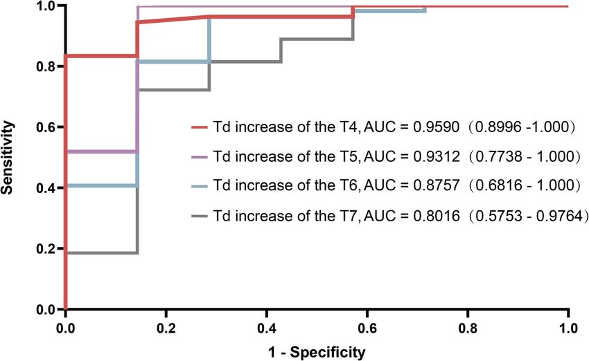
Zhang et al. BMC Anesthesiology    (2021) 21:168                                                                  Page 2 of 8

Introduction                                                    Study intervention
Thoracic paravertebral block (TPVB) produces ipsilateral        TPVB was performed in the induction room. Room
somatic and sympathetic blockade in multiple contigu-           temperature was maintained a constant 24 ± 0.5 °C.
ous thoracic dermatomes. It is a widely used analgesic          Intravenous access was established on arrival at the
technique for thoracic, chest wall, breast, urologic,           block room. A standard monitoring with electrocardiog-
abdominal or orthopedic surgery [1–5]. One characteris-         raphy, non-invasive blood pressure, and pulse oximetry
tic of TPVB is the unpredictability of local anesthetic         was applied to patients. Each patient lay supine and all
spreading in the paravertebral space [6–8]. It is very          clothing were removed from the upper body. The pa-
important to assess the spread of TPVB to ensure the            tients were allowed to acclimatize for 10 min.
expected analgesic effects.
   Many methods, including pinprick test, cold test,
                                                                Ultrasound-guided TPVB
pupillary dilation reflex and analgesia nociception index,
                                                                Ultrasound-guided TPVB was performed by one experi-
have been used to assess the outcome of TPVB [3, 9–11].
                                                                enced anesthetist with a low frequency (2 ~ 5 MHz)
None of these methods has been proven to be an optimal
                                                                curved array transducer (SonoSite M-Turbo; SonoSite
one. It is a generally accepted notion that skin temperature
                                                                Inc., Bothell, WA, USA). Patients were placed in the lat-
will increase after successful regional anesthesia because of
                                                                eral position with the side to be operated upwards.
sympathetic blockade. This type of temperature change can
                                                                Using aseptic precautions, the T4 and T5 paravertebral
be detected by infrared thermography. Infrared thermog-
                                                                space was located by counting from the 12th rib to the
raphy has been successfully applied in predicting the
                                                                4th rib. TPVB was performed at the T4 paravertebral
effectiveness of various regional blocks including upper and
                                                                space first. The transducer was placed at an oblique
lower extremity block, epidural and spinal anesthesia [12].
                                                                transverse position along the long axis of the rib and
In addition, clinical applications of thermal image are
                                                                tilted until the transverse process, the internal intercostal
spreading and range from regional anesthesia to kidney
                                                                membrane and the pleura were visualized. After infiltra-
transplantation [13]. However, its usefulness in TPVB has
                                                                tion with 2 ml of 1% lidocaine, a 22-gauge, 120-mm
not been determined.
                                                                stimuplex needle (Stimuplex® D; B. Braun; Melsungen;
   The goal of this study was to determine whether skin
                                                                Germany) was advanced from lateral to medial with in-
temperature difference (Td) determined by IT between
                                                                plane technique under real-time ultrasound guidance.
the blocked and unblocked side can predict the spread
                                                                Once the needle passed through the internal intercostal
of TPVB.
                                                                membrane, 10 ml of 0.4% ropivacaine was injected.
                                                                Using the same technique, another 10 ml of 0.4% ropiva-
Methods
                                                                caine was injected at the T5 paravertebral space.
Study design
This prospective observational study was approved by
the Ethical Committee of Tongji Hospital, China (num-           Infrared thermography
ber TJ-IRB20190424) and was registered at clinicaltrials.-      During the test, the patient’s chest wall was exposed in
gov (NCT04078347) on September 6, 2019. Written                 air. The rest of the body was covered with a blanket, and
informed consent was obtained from all subjects. The            forced air warming device was used to ensure comfort
reporting in the current manuscript follows the recom-          for the patients. The skin temperature of the patient’s
mendations in the STROBE guideline.                             anterior chest wall was accessed continuously by
                                                                computer-assisted infrared thermal cameras (Image for-
Inclusion and exclusion criteria                                mat: (640 × 480) IR pixel, Recording and storage of IR
The patients who listed to undergo elective, unilateral         frames rates with up to 240 Hz, Thermal resolution: up
major breast surgery or thoracoscopic surgery were              to 0.02 K, Measurement accuracy: +/− 1%) (VarioCAM®,
screened for inclusion. The inclusion criteria were             HD Research600, InfraTec, Germany). Infrared imaging
American Society of Anesthesiologists physical status           was taken before TPVB (t = 0) to provide a baseline
class I-II, and patients undergo elective surgery with          value. Then thermographic images were repeated at 5
TPVB for perioperative analgesia. Exclusion criteria were       min intervals until 20 min post the completion of TPVB
patient refusal, skin infection at the site of needle inser-    (t = 5, t = 10, t = 15 and t = 20). Temperature data were
tion, younger than 18 years, body mass index>35 kg/m2,          stored for off-line analysis and analyzed by the self-
significant thoracic kyphoscoliosis, preoperative use of        contained system (IRBIS® 3 plus, InfraTec, GmbH,
vasodilatory drugs, coagulopathy, preoperative use of           Germany). Skin temperature of each dermatome ranged
analgesic medications, history of previous thoracic or          from T2 to T10 was measured in the representative rect-
breast surgery, allergy to local anesthetics, and periph-       angle (Fig. 1A). The rectangles were placed on the
eral neuropathy.                                                photographed chest wall on a vertical, mid-clavicular
Zhang et al. BMC Anesthesiology (2021) 21:168 Page 3 of 8
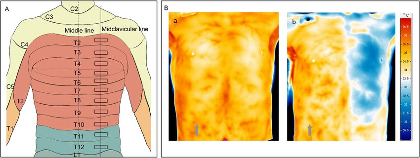
Fig. 1 A Anterior view of thoracic segments diagram, showing the representative rectangle areas (RAs) measured by infrared thermography. B
thermographic image of a 41-yr-old male patient. (a) thermographic image before thoracic paravertebral block; (b) thermographic image at 15
min after thoracic paravertebral block. Grey arrow indicated the blocked side
line. The other side, which was not blocked, was as in the failed group. While considering the dropout rate
control. (presumably 20%), the sample size was finally deter-
Temperature difference (Td) was defined as the differ- mined to be 65 subjects.
ence of skin temperature between the blocked side and
the unblocked side at a certain dermatome. Td was cal- Statistical analysis
culated at each measurement time point for each derma- Statistical analyses were performed using SPSS 22.0
tome. A characteristic infrared thermographic image (IBM Corp., New York, NY, USA). As a diagnostic test,
before and after the block was shown in Fig. 1B. ROC curves were constructed to determine the sensitiv-
ity, specificity, and cut-off values of Td for predicting
Block assessment by pinprick test
Pinprick test was evaluated at t = 20 immediately after Table 1 Patient characteristics. Data are expressed as the mean
infrared thermographic imaging. Pinprick sensation was (SD) or number of patients (%) in each group
assessed using a 22-gauge short bevel needle from T2 to Successful TPVB Failed TPVB
T10 at midclavicular line bilaterally. Pinprick response Simple size, n 54 7
was recorded quantitatively as 1 (sensation) or 0 (no Sociodemographic Charactertics
sensation/numb).
Sex, n (%)
Successful block Male 21 (40.4) 5 (57.1)
Successful block was defined as the pinprick score was 0 Female 33 (61.1) 2 (42.9)
in 3 or more adjacent dermatomes corresponding to the Mean Age (SD) in years 55 (10) 49 (16)
site of injection at 20 min after block [14, 15]. Otherwise, Mean BMI (SD) in kg/m2 23 (3) 24 (1)
it was defined as a failed block. Patients were transferred Surgical Charactertics
to operating room 30 min after TPVB. All patients re-
Block side, n (%)
ceived general anesthesia. Patient controlled analgesia
with sufentanil was provided for all patients following Left 21 (38.9) 3 (42.9)
operation. Right 33 (61.1) 4 (57.1)
ASA status (I ~ II), n (%)
Sample size estimation ASA I 23 (42.6) 3 (42.9)
The sample size was calculated using MedCalc Software ASA II 31 (57.4) 4 (57.1)
version 15.2 (MedCalc Software, Ostend, Belgium). We
Surgery, n (%)
hypothesized that the area under the receiver operator
characteristic (ROC) curve was 0.8 with 0.5 for null hy- Mastectomy 7 (13.0) 1 (14.3)
pothesis value. The incidence of the failed block was es- Mastectomy + ALND 4 (7.4) 1 (14.3)
timated to be 14% on the basis of our previous pilot Lung lobectomy 36 (66.7) 3 (42.6)
study. Setting a significance level of 0.05 and the type 2 Lung wedge resection 9 (16.7) 2 (28.6)
error of 0.2. The minimum required sample size was 49 Abbreviations: SD standard deviation, BMI Body mass index, ASA American
with 42 patients in the successful group and 7 patients Society of AnesthesiologistsZhang et al. BMC Anesthesiology (2021) 21:168 Page 4 of 8 successful block. The optimal cut-off point was calcu- Results lated by ROC curves with the maximal Youden index From October 2019 to August 2020, a total of 65 pa- value (sensitivity+specificity-1). The area under the curve tients were assessed for eligibility to participate in this and the 95% confidence interval (CI) were reported as study. Two patients failed to provide the written in- well. formed consent. Two patients were excluded by exclu- Continuous variables were displayed as means (stand- sion criteria. Finally, 61 patients were included. ard deviation) or medians (interquartile range [IQR] As determined by pinprick test, successful block was [25–75]), and discrete variables are expressed as num- achieved in 54 patients. There were no differences in bers (n). The normally distributed data after Kolmogo- terms of demographic characteristics between patients rov–Smirnov test were compared using the independent with successful block and patients with failed block sample t-test, non–normal distributed data were ana- (Table 1). lyzed using the Mann-Whitney U test. Categorical data Sensory block spread from T2 to T10. The number of were compared by χ2 test or Fisher’s exact. A P value of patients with loss of pinprick sensation for each derma- < 0.05 was considered statistically significant. tome was shown in Fig. 2A. The median dermatomes Fig. 2 A Number of patients with loss of pinprick sensation at 20 min after thoracic paravertebral block. B Density distribution for upper and lower level of loss of pinprick sensation at 20 min after thoracic paravertebral block. Median values are shown as black lines
Zhang et al. BMC Anesthesiology      (2021) 21:168                                                                                    Page 5 of 8

with loss of pinprick sensation were 5 (4–7) in the suc-                    ROC curves were constructed for Td increase at 15
cessful blocks. The median upper level was T3 (T2–T3)                     min after block to predict successful block (Supplemen-
and lower level was T7 (T6–T8) (Fig. 2B).                                 tary Table 1 and Fig. 4). The area under the ROC curve
  Tds were similar between successful and failed blocks                   (AUC) of T4 was 0.960 (95% CI: 0.8996–1.000) with the
at each dermatome at time zero (t = 0). In the successful                 cut-off point value of 0.63 °C, showing the greatest po-
blocks, Td increased rapidly from 5 min to 20 min after                   tential to predict successful block (Fig. 5). The sensitivity
block (t = 5, t = 10, t = 15 and t = 20) (P < 0.01, respect-              and specificity were 83.3 and 100.0%, respectively.
ively). Td did not increase (t = 5, t = 10, t = 15 and t = 20)              There were no significant differences in hemodynamic
at any dermatome in the failed blocks (P > 0.05, respect-                 parameters (mean arterial pressure and heart rate) be-
ively). In addition, Td was higher at each time point after               tween successful and failed blocks.
block (t = 5, t = 10, t = 15 and t = 20) in the successful
blocks than that in the failed blocks (P < 0.05, respect-                 Discussion
ively). The increase of Td at T4–T7 were more than 1 °C                   The results of our study showed that Td increase could
at t = 20 in the successful blocks (Fig. 3).                              be an early, quantitative, and reliable indicator of

 Fig. 3 Temperature (Td) values of the thoracic dermatome (T2-T10) in patients who were performed thoracic paravertebral blocks (TPVB). *P <
 0.05 compared with failed TPVB at each time point.!P < 0.01 compared with the baseline value
Zhang et al. BMC Anesthesiology        (2021) 21:168                                                                                         Page 6 of 8

 Fig. 4 Receiver operator characteristic (ROC) curve of Td increase at 15 min after thoracic paravertebral block. Td increase was calculated as Td at
 each time point after paravertebral block minus Td at baseline. Td: difference of skin temperature between the blocked and the unblocked side
 at a certain dermatome. AUC, area under the curve

successful TPVB. The occurrence of temperature                                blocks, sciatic nerve blocks, spinal and epidural
increase secondary to regional anesthesia is a well-                          anesthesia [12, 16]. A previous study found ipsilateral
recognized phenomenon. This type of temperature in-                           warming after TPVB [17]. However, its usefulness in
crease can be noninvasively and accurately detected by                        predicting the success of TPVB needs to be determined.
IT. Some studies have investigated the possibility of in-                     The thoracic paravertebral place contains the intercostal
frared thermography to determine the success or failure                       nerve and the sympathetic trunk. Successful TPVB can
of peripheral nerve blocks, such as brachial plexus (arm)                     reliably block both the intercostal nerve and sympathetic

 Fig. 5 Td increase at T4 in patients with successful and failed TPVB. A cut-off value of 0.63 °C at 15 min after the block is marked. Horizontal lines
 represent medians, boxes represent quartiles, and whiskers represent ranges. Td: difference of skin temperature between the blocked and the
 unblocked side at a certain dermatome. TPVB: thoracic paravertebral block
Zhang et al. BMC Anesthesiology   (2021) 21:168                                                                                   Page 7 of 8

nerve. Blockade of small unmyelinated sympathetic                block because the onset time of ropivacaine in some pa-
nerve fibers with local anesthetics causes vasodilatation,       tients is more than 20 min [25]. Secondly, we didn’t
an increase in blood flow and an increase in local               measure core temperatures which could influence skin
temperature [18, 19]. In reality, the chest wall                 temperature after TPVB. Thirdly, we didn’t use loss of
temperature will change over time because the differ-            sensation to surgical stimulus as the standard of success-
ence between the skin and ambient temperature. It is             ful TPVB. Instead of surgical stimulus, we use pinprick
difficult to predict the effectiveness of TPVB by the            sensation to evaluate the effectiveness of TPVB. In
absolute skin temperature values of the blocked side.            addition, the post-operative pain was not measured in
We use Td between the blocked and unblocked side to              present study. Lastly, we have not recorded video during
predict the success or failure of TPVB. The results of           the temperature changing after TVBP in the anterior
our study showed that Td in the successful blocks in-            chest wall.
creased significantly as early as 5 min after TPVB. ROC
analysis showed that the highest area under the ROC              Conclusions
curve (AUC) values were achieved at T4 level 15 min              Whether skin temperature difference between the
after TPVB. The AUC was 0.960 with a sensitivity of              blocked and unblocked side can predict the outcome of
83.3% and a specificity of 100.0%. It suggests the credit-       thoracic paravertebral block is unclear, we demonstrated
able discriminating ability in identifying patients with         that the increase of temperature difference at T4 derma-
successful TPVB.                                                 tome is an early, quantitative, and reliable predictor of
   TPVB has been used in clinical anesthesia for more            successful thoracic paravertebral block. Measurement of
than 100 years. However, reliable methods for predicting         skin temperature with infrared thermography (IT) is a
the success of TPVB is still under exploration. Pinprick         reliable method to evaluate the effectiveness of thoracic
and cold sensation test are traditional and the most             paravertebral block.
widely used methods to evaluate the effectiveness of
TPVB. However, sensation to pinprick and cold are sub-
jective and depend on the patient’s ability to interpret         Supplementary Information
                                                                 The online version contains supplementary material available at https://doi.
the stimulus applied. They are sometimes unreliable, es-         org/10.1186/s12871-021-01389-4.
pecially in elderly patients with cognitive impairment,
children, or those who have neuropsychiatric disorders.           Additional file 1: Supplementary Table 1. ROC curve analysis for T4-
The pupillary dilation reflex (PDR) was another method            T7 dermatome. Data are expressed as mean (95% confidence interval).

to assess the outcome of TPVB in patient under general
anesthesia. However, the opioid-induced pupillary con-           Acknowledgements
striction could influence the PDR [10, 20]. The analgesia        We would like to acknowledge the nursing staff at Tongji Hospital in Wuhan,
                                                                 China for their assistance with this work.
nociception index (ANI) monitoring was also reported
to evaluate the effect of TPVB. Although ANI provided            Authors’ contributions
qualitative and quantitative measurements reflecting the         Shuang Zhang: This author helped study design, patient recruitment, data
balance between nociception and analgesia under                  analysis and drafting and revising manuscript. Yong Liu: This author helped
                                                                 patient recruitment, data collection, data analysis and drafting and revising
general anesthesia, the possible hemodynamic instability         manuscript. Xiaohu Liu: This author helped data analysis and data
occurred after TPVB could affect the ANI parameters              interpretation. Tianzhu Liu: This author helped patient recruitment and data
[11, 21]. Infrared thermography is a non-invasive, full-         collection. Pengchen Li: This author helped data analysis and data
                                                                 interpretation Wei Mei: This author helped study design, data analysis, data
field measurement with continuous images recording               interpretation, drafting and revising manuscript. The author(s) read and
and allowing quantitative assessment of skin temperature         approved the final manuscript.
[22]. It is completely objective. In addition, its high sensi-
tivity and specificity made it an ideal technique to predict     Funding
                                                                 The trial was funded in part by National Natural Science Foundation of China
the spread of TPVB.                                              (81873793 to Wei Mei).
   Although the spread of local anesthetics inside the
paravertebral space is highly unpredictable [7, 23, 24],         Availability of data and materials
our preliminary study showed that the spread of sensory          The datasets generated and/or analysed during the current study are
                                                                 available from the corresponding author on reasonable request.
block with a dual-injection performed at T4–5 and T5–
6 were rarely beyond T2 to T10. Thus, we measured
                                                                 Declarations
skin temperatures from T2 to T10 of the anterior chest
wall in this study.                                              Ethics approval and consent to participate
   Our study has some certain limitations. Firstly, we           This prospective observational study was approved by the Ethical Committee
                                                                 of Tongji Hospital, China (number TJ-IRB20190424) and was registered at
only evaluated the extent of sensory block up to 20 min          clinicaltrials.gov (NCT04078347) on September 6, 2019. Written informed con-
after TPVB. It might underestimate the extent of sensory         sent was obtained from all subjects.
Zhang et al. BMC Anesthesiology (2021) 21:168 Page 8 of 8
Consent for publication 16. Galvin EM, Niehof S, Medina HJ, Zijlstra FJ, van Bommel J, Klein J, et al.
Written informed consent was obtained from all subjects. Thermographic temperature measurement compared with pinprick and
cold sensation in predicting the effectiveness of regional blocks. Anesth
Competing interests Analg. 2006;102(2):598–604. https://doi.org/10.1213/01.ane.0000189556.4
The authors declare that they have no conflicts of interest. 9429.16.
17. Cheema SP, Ilsley D, Richardson J, Sabanathan S. A thermographic study of
Author details paravertebral analgesia. Anaesthesia. 1995;50(2):118–21. https://doi.org/1
1
Department of Anesthesiology and Pain Medicine, Tongji Hospital, Tongji 0.1111/j.1365-2044.1995.tb15092.x.
Medical College, Huazhong University of Science and Technology, Wuhan, 18. Krediet AC, Moayeri N, van Geffen GJ, Bruhn J, Renes S, Bigeleisen PE, et al.
China. 2Britton Chance Center for Biomedical Photonics, School of Different approaches to ultrasound-guided thoracic paravertebral block: an
Engineering Sciences, Wuhan National Laboratory for Optoelectronics, illustrated review. Anesthesiology. 2015;123(2):459–74. https://doi.org/10.1
Huazhong University of Science and Technology, Wuhan, China. 097/ALN.0000000000000747.
19. Nielsen MV, Moriggl B, Hoermann R, Nielsen TD, Bendtsen TF, Borglum J.
Received: 7 April 2021 Accepted: 2 June 2021 Are single-injection erector spinae plane block and multiple-injection
costotransverse block equivalent to thoracic paravertebral block? Acta
Anaesthesiol Scand. 2019;63(9):1231–8. https://doi.org/10.1111/aas.13424.
20. Sabourdin N, Barrois J, Louvet N, Rigouzzo A, Guye ML, Dadure C, et al.
References Pupillometry-guided intraoperative remifentanil administration versus
1. Schnabel A, Reichl SU, Kranke P, Pogatzki-Zahn EM, Zahn PK. Efficacy and standard practice influences opioid use: a randomized study.
safety of paravertebral blocks in breast surgery: a meta-analysis of Anesthesiology. 2017;127(2):284–92. https://doi.org/10.1097/ALN.
randomized controlled trials. Br J Anaesth. 2010;105(6):842–52. https://doi. 0000000000001705.
org/10.1093/bja/aeq265. 21. Julien-Marsollier F, Rachdi K, Caballero MJ, Ayanmanesh F, Vacher T, Horlin
2. Moller JF, Nikolajsen L, Rodt SA, Ronning H, Carlsson PS. Thoracic AL, et al. Evaluation of the analgesia nociception index for monitoring
paravertebral block for breast cancer surgery: a randomized double-blind intraoperative analgesia in children. Br J Anaesth. 2018;121(2):462–8. https://
study. Anesth Analg. 2007;105:1848–51 table of contents. doi.org/10.1016/j.bja.2018.03.034.
3. Yeung JH, Gates S, Naidu BV, Wilson MJ, Gao SF. Paravertebral block versus 22. Wright CI, Kroner CI, Draijer R. Non-invasive methods and stimuli for
thoracic epidural for patients undergoing thoracotomy. Cochrane Database evaluating the skin's microcirculation. J Pharmacol Toxicol Methods. 2006;
Syst Rev. 2016;2:CD009121. 54(1):1–25. https://doi.org/10.1016/j.vascn.2005.09.004.
4. Canto M, Sanchez MJ, Casas MA, Bataller ML. Bilateral paravertebral 23. Eason MJ, Wyatt R. Paravertebral thoracic block-a reappraisal. Anaesthesia.
blockade for conventional cardiac surgery. Anaesthesia. 2003;58(4):365–70. 1979;34(7):638–42. https://doi.org/10.1111/j.1365-2044.1979.tb06363.x.
https://doi.org/10.1046/j.1365-2044.2003.03082_2.x. 24. Uppal V, Sondekoppam RV, Sodhi P, Johnston D, Ganapathy S. Single-
5. Rudkin GE, Gardiner SE, Cooter RD. Bilateral thoracic paravertebral block for injection versus multiple-injection technique of ultrasound-guided
abdominoplasty. J Clin Anesth. 2008;20(1):54–6. https://doi.org/10.1016/j. paravertebral blocks: a randomized controlled study comparing dermatomal
jclinane.2007.06.020. spread. Reg Anesth Pain Med. 2017;42(5):575–81. https://doi.org/10.1097/AA
6. Clendenen SR, Bojaxhi E. A comparative study of automated pulsed bolus P.0000000000000631.
versus continuous basal infusion on distribution of dye in the paravertebral 25. Karmakar MK, Ho AM, Law BK, Wong AS, Shafer SL, Gin T. Arterial and
space in a cadaver. Cureus. 2019;11:e4958. venous pharmacokinetics of ropivacaine with and without epinephrine after
7. Marhofer D, Marhofer P, Kettner SC, Fleischmann E, Prayer D, Schernthaner thoracic paravertebral block. Anesthesiology. 2005;103(4):704–11. https://doi.
M, et al. Magnetic resonance imaging analysis of the spread of local org/10.1097/00000542-200510000-00008.
anesthetic solution after ultrasound-guided lateral thoracic paravertebral
blockade: a volunteer study. Anesthesiology. 2013;118(5):1106–12. https://
doi.org/10.1097/ALN.0b013e318289465f. Publisher’s Note
8. Cheema S, Richardson J, McGurgan P. Factors affecting the spread of Springer Nature remains neutral with regard to jurisdictional claims in
bupivacaine in the adult thoracic paravertebral space. Anaesthesia. 2003; published maps and institutional affiliations.
58(7):684–7. https://doi.org/10.1046/j.1365-2044.2003.03189_1.x.
9. Thavaneswaran P, Rudkin GE, Cooter RD, Moyes DG, Perera CL, Maddern GJ.
Brief reports: paravertebral block for anesthesia: a systematic review. Anesth
Analg. 2010;110(6):1740–4. https://doi.org/10.1213/ANE.0b013e3181da82c8.
10. Duceau B, Baubillier M, Bouroche G, Albi-Feldzer A, Jayr C. Pupillary reflex
for evaluation of thoracic paravertebral block: a prospective observational
feasibility study. Anesth Analg. 2017;125(4):1342–7. https://doi.org/10.1213/A
NE.0000000000002003.
11. Dundar N, Kus A, Gurkan Y, Toker K, Solak M. Analgesia nociception index
(ani) monitoring in patients with thoracic paravertebral block: a randomized
controlled study. J Clin Monit Comput. 2018;32(3):481–6. https://doi.org/10.1
007/s10877-017-0036-9.
12. Hermanns H, Werdehausen R, Hollmann MW, Stevens MF. Assessment of
skin temperature during regional anaesthesia-what the anaesthesiologist
should know. Acta Anaesthesiol Scand. 2018;62(9):1280–9. https://doi.org/1
0.1111/aas.13176.
13. Cherchi V, Baccarani U, Vetrugno L, Pravisani R, Bove T, Meroi F, et al. Early
graft dysfunction following kidney transplantation: can thermographic
imaging play a predictive role? Semin Cardiothorac Vasc Anesth. 2021.
https://doi.org/10.1177/10892532211007270.
14. Chen H, Liao Z, Fang Y, Niu B, Chen A, Cao F, et al. Continuous right
thoracic paravertebral block following bolus initiation reduced
postoperative pain after right-lobe hepatectomy: a randomized, double-
blind, placebo-controlled trial. Reg Anesth Pain Med. 2014;39(6):506–12.
https://doi.org/10.1097/AAP.0000000000000167.
15. Lonnqvist PA, MacKenzie J, Soni AK, Conacher ID. Paravertebral blockade.
Failure rate and complications. Anaesthesia. 1995;50(9):813–5. https://doi.
org/10.1111/j.1365-2044.1995.tb06148.x.You can also read