Minimize the extent and morbidity of axillary dissection for node-positive breast cancer patients: implementation of axillary lymph node ...
←
→
Page content transcription
If your browser does not render page correctly, please read the page content below
Yuan et al. BMC Cancer      (2021) 21:293
https://doi.org/10.1186/s12885-021-08024-y

 RESEARCH ARTICLE                                                                                                                                 Open Access

Minimize the extent and morbidity of
axillary dissection for node-positive breast
cancer patients: implementation of axillary
lymph node dissection based on breast
lymphatics level
Qianqian Yuan, Jinxuan Hou, Yukun He, Yiqian Liao, Lewei Zheng and Gaosong Wu*

  Abstract
  Background: Breast cancer-related lymphedema (BCRL) is associated with extensive axillary dissection. Axillary
  lymph node dissection (ALND) based on breast lymphatics level (BLL) was proposed to minimize the surgical extent
  for node-positive breast cancer patients.
  Methods: A total of 156 consecutive sentinel lymph node-positive (SLN+) or clinically node-positive (cN+) patients
  underwent sentinel lymph node biopsy (SLNB) with indocyanine green and methylene blue (MB). The SLNs were
  injected with 0.1 ml MB before removal, and a standard ALND was subsequently performed. The nodes adjacent to
  the blue-stained axillary lymph nodes from the breast (bALNs) were sent for pathological examination separately by
  resecting serial tissue every 0.5 cm away from the marginal blue-stained bALNs. Then, a pilot study comparing
  ALND based on BLL and standard ALND was performed.
  Results: BLL were successfully identified in 20 SLN+ (100%) and 134 cN+ (98.5%) patients. The median number of
  BLL was four, ranging from three to six. A horizontal line 1.0 cm away from the superior blue-stained bALN and a
  vertical line 1.0 cm away from the medial blue-stained bALN formed BLL II, III, and IV. All of the additional positive
  nodes were within 1.0 cm of the blue-stained bALNs. The minimized axillary dissection should resect upwards from
  the lowest BLL that contains the first confirmed negative blue-stained bALNs. In the pilot study, no patient
  developed axillary recurrence.
  Conclusion: The ALND surgical procedure based on BLL could minimize the surgical extent for pathological node-
  positive breast cancer patients and potentially reduce the BCRL rate.
  Trial registration: ChiCTR1800014247.
  Keywords: Breast cancer, Axillary lymph node dissection, Breast cancer related lymphedema

* Correspondence: wugaosong@whu.edu.cn
Department of Thyroid and Breast Surgery, Zhongnan Hospital of Wuhan
University, 169 Donghu Road, Wuhan 430071, Hubei, People’s Republic of
China

                                      © The Author(s). 2021 Open Access This article is licensed under a Creative Commons Attribution 4.0 International License,
                                      which permits use, sharing, adaptation, distribution and reproduction in any medium or format, as long as you give
                                      appropriate credit to the original author(s) and the source, provide a link to the Creative Commons licence, and indicate if
                                      changes were made. The images or other third party material in this article are included in the article's Creative Commons
                                      licence, unless indicated otherwise in a credit line to the material. If material is not included in the article's Creative Commons
                                      licence and your intended use is not permitted by statutory regulation or exceeds the permitted use, you will need to obtain
                                      permission directly from the copyright holder. To view a copy of this licence, visit http://creativecommons.org/licenses/by/4.0/.
                                      The Creative Commons Public Domain Dedication waiver (http://creativecommons.org/publicdomain/zero/1.0/) applies to the
                                      data made available in this article, unless otherwise stated in a credit line to the data.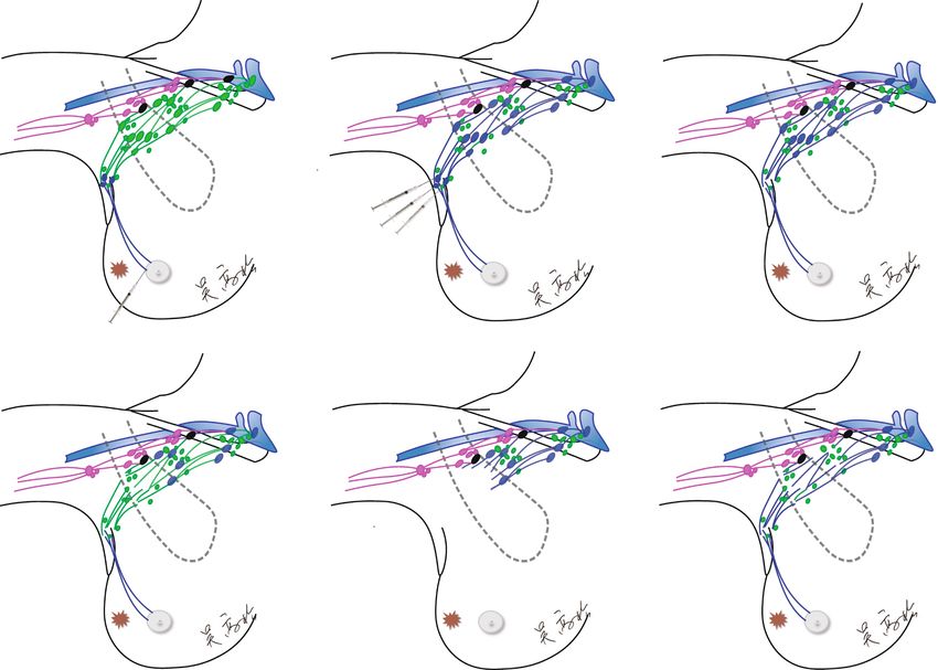
Yuan et al. BMC Cancer (2021) 21:293 Page 2 of 10
Background center, Zhongnan Hospital of Wuhan University.
The advent of sentinel lymph node biopsy (SLNB) has Women patients meeting the following criteria and
revolutionized the surgical management of axilla in undergoing ALND were included in the study: i) patients
breast cancer patients [1]. Complete axillary lymph node with clinically node-positive breast cancer, defined as
dissection (ALND) has been gradually replaced for select positive on preoperative axillary palpation, ultrasound
patients, owing to the substantial morbidity such as de- examinations or CT scans with contrast; ii) patients who
bilitating breast cancer-related lymphedema (BCRL), underwent mastectomy with a positive SLN; iii) patients
shoulder dysfunction, paresthesia and discomfort [2]. who underwent breast-conserving surgery (BCS) for
Damage to the lymphatic drainage system in the axilla more than two positive SLNs; iv) patients who did not
by multidisciplinary treatment contributes to the occur- receive neoadjuvant chemotherapy (NCT); v) patients
rence of BCRL [3]. Notably, extensive axillary dissection with no previous history of breast cancer. Those who
is related to a high occurrence rate of BCRL. A total of had benign tumors and those who did not meet the
77.8% of the patients who underwent ALND had fewer above criteria were excluded from the study. The Med-
than three additional involved nodes in the ACOSOG ical Ethics Committee of Zhongnan Hospital of Wuhan
Z0011 trial [4]. Thus, the complete removal all axillary University approved the routine use of the SLNB pro-
lymph nodes with ALND for those patients might be cedure for all patients. The trial was registered at the
over-treatment. For patients who were not eligible for Chinese Clinical Trial Registry (ChiCTR1800014247).
the ACOSOG Z0011 study criteria, especially clinically All patients provided written informed consent regard-
node-positive breast cancer patients, a method for de- ing SLNB and ALND.
escalating the surgical area of ALND would significantly
reduce the BCRL rate. Dual tracers for SLNB
The lymphatic system of the breast forms an extensive All patients underwent quadrantectomy or mastectomy
and complex network of periductal and perilobular ves- immediately followed by SLNB. Radioactive tracers were
sels that drain principally to the axillary nodes [5]. Breast unavailable in China. Instead, dual tracers, 1 ml (1 mg)
tumors commonly invade local structures and spread in indocyanine green (ICG) (H20045514; Weicai Pharma-
a progressive and sequential manner to regional nodes, ceutical Corporation, Liaoning, China) and 0.5 ml (5 mg)
and the lymphatic vessels provide anatomical continuity methylene blue (MB) (H32024827; Jichuan Pharmaceut-
for this process by acting as a link between the primary ical Corporation, Taixin, China) were administered for
tumor and the regional nodes. Hence, the sentinel lymph SLNB [9].
node (SLN) hypothesis presupposes an orderly spread of
cancer cells from the primary tumor to the first draining Surgical techniques
node, which has prevented clinically node-negative Before removal, all the identified SLNs were routinely
breast cancer patients from undergoing extensive axillary and meticulously injected with 0.1–0.2 ml MB using a 1-
dissection [6]. cc syringe with a 32-gauge needle, which was called
On the theoretical basis of SLNB, for node-positive ‘staged tracing’ (Supplement 1. MP4). The careful rein-
breast cancer patients, metastasis from breast tumors jection of MB was a key procedure to not artifactually
does not involve the axillary lymph nodes from the alter the lymphatic drainage patterns. MB could then
breast (bALNs) as a unit but instead progresses from the flow from the SLNs along several ascending lymphatic
primary tumor to the first draining nodes, then second channels towards the subclavian lymph nodes. Then, the
and third echelon nodes [7, 8]. Therefore, our institution blue-stained lymphatic channels were mapped by bluntly
implemented the approach of ALND based on the dissecting along the lymphatic drainage channels from
lymphatic drainage from the breast in order to limit sur- the breast to the axilla. After identifying the efferent and
gical resection in the axilla and reduce the morbidity of echelon nodes, the SLNs were ready to be harvested and
ALND. The primary aim was to accurately determine sent for immediate frozen sectioning (FS). Once the
the extent of every breast lymphatics level (BLL). The SLNs were confirmed positive in patients who under-
secondary aim was to determine the skip metastasis rate went mastectomy, ALND was performed with complete
to demonstrate the de-escalation feasibility of ALND resection of at least Berg’s levels I and II; the resection
based on lymphatic drainage from the breast. of Berg’s level III was performed only in patients with
gross disease in Berg’s level II and/or III. When patients
Methods with BCS had more than two positive SLNs, ALND was
Study cohort subsequently performed.
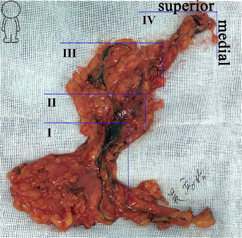
The prospective observational study enrolled consecutive After complete ALND, the removed specimen was dis-
breast cancer patients from November 2017 to March sected carefully ex vivo (Fig. 1). The lymph nodes adja-
2018 in a single tertiary referral academic medical cent to the blue-stained bALNs were defined as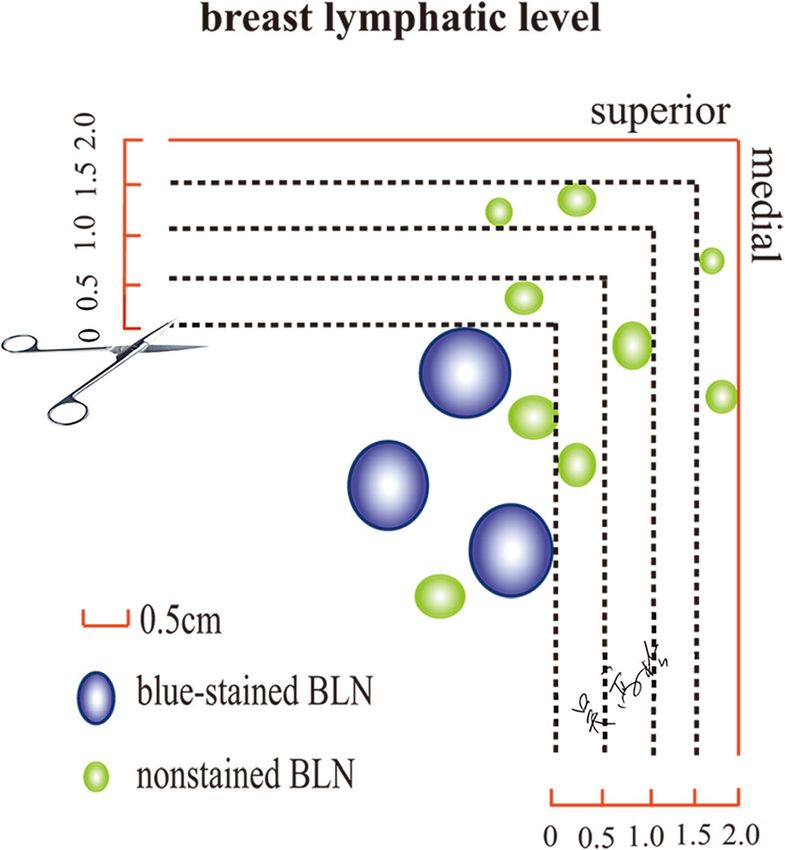
Yuan et al. BMC Cancer        (2021) 21:293                                                                               Page 3 of 10

                                                                         there was a predictable passage of efferent lymphatics
                                                                         towards nodes at BLL II and, in turn, nodes at BLL III
                                                                         and IV (Fig. 1). With efferent lymphatics, each BLL
                                                                         could be distinguished. The skip metastasis was defined
                                                                         as macrometastatic nodes that were found at a further
                                                                         site while the lymph nodes nearer to the primary breast
                                                                         tumor were negative, as it was assumed that nodal in-
                                                                         volvement occurred in a progressive manner.

                                                                         Pathological examination
                                                                         The removed SLNs were sent for immediate FS during
                                                                         the operation. After the operation, the nonstained
                                                                         bALNs were sent for pathological examination separ-
                                                                         ately by resecting serial tissue every 0.5 cm away from
                                                                         the horizontal line and vertical line along the marginal
                                                                         blue-stained bALN (Fig. 2) [10]. All the retrieved bALNs
                                                                         were processed for routine hematoxylin and eosin stain-
                                                                         ing for histology and immunohistochemically. The diam-
                                                                         eter, estrogen receptor status, progesterone receptor
 Fig. 1 Breast lymphatics level in this study (ex vivo, right axilla)
                                                                         status and human epidermal growth factor receptor-2
                                                                         status of the primary tumor were also assessed as part of
nonstained bALNs (Fig. 2). A horizontal line along the                   a routine pathology examination.
superior blue-stained bALN and a vertical line along the
medial blue-stained bALN formed a lower outer quad-                      Comparing ALND based on BLL with standard ALND
rant (LOQ) zone in the axilla, which was defined as the                  Patients with clinical T1–3 and node-positive axilla were
BLL (Fig. 2). The blue-stained lymphatic channels of the                 eligible and randomized to ALND based on BLL and
breast could converge towards a group of three to five                   standard ALND. Descriptive statistics were used to de-
lymph nodes, namely, SLNs, at BLL I. From these nodes,                   scribe the results of the pilot prospective study. The pri-
                                                                         mary objective of the pilot phase of the randomized
                                                                         controlled trial was to demonstrate feasibility of the pro-
                                                                         cedure of ALND based on BLL trial design, and to deter-
                                                                         mine if axillary recurrence rates for patients randomized
                                                                         to ALND based on BLL are equivalent to axillary recur-
                                                                         rence rates for patients randomized to standard ALND.

                                                                         Statistical analysis
                                                                         The demographic characteristics, tumor sizes, and num-
                                                                         ber of lymph nodes in each BLL were collected. Con-
                                                                         tinuous variables between groups were compared using
                                                                         a nonparametric test. Chi-squared tests or Fisher’s exact
                                                                         tests were run to compare the positive rate between the
                                                                         two groups. Two-sided p values< 0.05 were considered
                                                                         statistically significant. All statistical analyses were per-
                                                                         formed using SPSS17.0 for Windows (SPSS, Inc., Chi-
                                                                         cago, IL).

                                                                         Results
                                                                         Patients
                                                                         A total of 190 patients underwent SLNB with dual
 Fig. 2 Pathological examination method (right axilla). The              tracers. Of these patients, 136 had node-positive axilla,
 nonstained bALNs are sent for pathological examination separately       and 54 patients had node-negative axilla. Two patients
 by resecting serial tissue every 0.5 cm away from the horizontal line   who failed to have any SLNs identified, 26 patients who
 and vertical line along the superior blue-stained bALN and medial
                                                                         underwent mastectomy with negative SLNs, and 6 pa-
 blue-stained bALN. bALN: axillary lymph node from the breast
                                                                         tients who underwent BCS and planned whole-breast
Yuan et al. BMC Cancer (2021) 21:293 Page 4 of 10
irradiation with fewer than 3 macrometastastic SLNs minor, or both. In addition, BLL III was located behind
were excluded. Of the 26 patients with negative SLN the pectoralis minor, medial and superior the pectoralis
identified by FS, two (7.7%) false negative cases with one minor, or both; only later BLL IV lymph nodes were
lymph node were found to have macrometastatic (2 mm) medial and superior to the muscle.
disease after the postoperative H&E examination. A total
of 156 patients were entered into the study, and 78.8% Pathology
of the patients underwent mastectomy. Systemic therapy Ninety-seven (62.2%) of the 156 patients harbored fewer
was recommended according to the clinical guidelines. than four macrometastatic lymph nodes. cN+ patients
The baseline characteristics of the SLN+ and cN+ were more prone to have gross metastasis than SLN+ pa-
groups are shown in Table 1. Compared with the SLN+ tients (p < 0.001). The distribution of the positive nodes in
group, the cN+ group displayed larger tumors (p < the BLL between the SLN+ and cN+ groups was com-
0.001). All of the patients underwent staged tracing to pared and can be seen in Table 4. The cancer cells of
reveal the subsequent efferent and echelon bALNs, and 85.8% (121/141) of the patients with node-positive axilla
MB could flow from the SLNs along several ascending metastasized from the primary tumor to BLL II.
lymphatic channels towards the subclavian lymph nodes.
Twenty (100%) SLN+ patients and 134 (98.5%) cN+ pa- Outcome of the pilot study
tients successfully had the breast lymphatic vessels and The surgical procedure of ALND based on BLL was per-
bALNs identified (Fig. 1). formed as follows: (1) Perform SLNB with 0.5 ml MB
Supraclavicular and infraclavicular radiotherapy was (Fig. 3a). (2) Routinely and meticulously inject all the
performed for patients with one or more positive lymph identified SLNs with 0.1–0.2 ml of MB before removal
nodes. Chest wall radiotherapy was performed for pa- (Fig. 3b). (3) After the echelon nodes were identified
tients who underwent BCS. 20 (100%) SLN+ patients with injection of MB, SLNs would be harvested and
and 121 (89.0%) cN+ patients underwent radiotherapy. pathologically examined by immediate FS (Fig. 3c). (4) If
The dose was 25 fractions of 2 Gy. SLNs were positive, the blue-stained bALNs in BLL 2nd
would be removed and sent for immediate FS (Fig. 3d).
Breast lymphatics level (5) If the blue-stained bALNs in BLL 2nd were con-
The median number of BLL that could be classified firmed negative, the tissues in BLL 2nd were resected ‘en
was four, ranging from three to six (Fig. 1). The bloc’ (Fig. 3e); (6) If the blue-stained bALNs in BLL 2nd
mean number of lymph nodes in each BLL is pre- were macrometastic, the blue-stained bALNs in lymph-
sented in Table 2. The skip metastasis rate of differ- atic level 3rd would be removed and sent for immediate
ent serial distances from the marginal blue-stained FS (Fig. 3f). Finally, the limited axillary dissection would
bALNs is described in Table 3. While the cancer cells be performed upwards from the lowest BLL that con-
were not found in the blue-stained bALNs, the add- tained the first confirmed negative blue-stained bALNs,
itional skip macrometastatic lymph nodes were lo- and the tissues in the matched LOQ zone would be
cated within 1.0 cm distance away from the blue- resected en bloc.
stained bALNs. No additional macrometastatic nodes Twenty-seven patients were enrolled in the pilot
were identified beyond 1.0 cm away from the marginal study from March 2018 to July 2018. Fourteen par-
bALNs (Table 3). Therefore, a horizontal line 1.0 cm ticipants were randomized to ALND based on BLL
away from the superior blue-stained bALN and a ver- (study group), 13 subjects were randomized to stand-
tical line 1.0 cm away from the medial blue-stained ard ALND (controlled group), and two patients with-
bALN formed a LOQ zone in the axilla, which was drew from the trial. Thirteen patients from study
defined as BLL II, III, and IV (Fig. 3b). The skip me- group, and 11 patients from controlled group com-
tastasis rate of SLN was 9.6%, which was higher than pleted the study interventions and are included in
the subsequent lymph nodes (Table 3). And no add- the analysis. The median age in study group was
itional macrometastatic nodes were identified beyond 48.0 (range 39–62 years) and the median age in con-
1.5 cm away from the marginal blue-stained SLNs. trolled group was 46.0 (range 31–61). The two
Thus, a horizontal line 1.5 cm away from the superior groups are well-matched in terms of patient age,
blue-stained SLN and a vertical line 1.5 cm away from tumor size, biomarker profile and length of follow-
the medial blue-stained SLN formed a LOQ zone in up (Table 5). In the ALND based on BLL group, one
the axilla, which was defined as BLL I [10]. (1.3%) false negative case with two lymph nodes
The location of the BLL was dynamic and inconsistent. were found to have micrometastatic (1 mm) disease
The BLL I was lateral and inferior to the pectoralis after the postoperative H&E examination. After a
minor muscle; BLL II was either located lateral and in- median follow-up of 24 months (range 22–25
ferior to the pectoralis minor, behind the pectoralis months), no axillary recurrence and distantYuan et al. BMC Cancer (2021) 21:293 Page 5 of 10
Table 1 Characteristics of 156 breast cancer patients
SLN+ cN+ p value
N = 20 N = 136
Age (Mean) 49.45 51.1 0.627a
Tumor size, No. (%) < 0.001a
< 1 cm 4 (20.0) 6 (4.4)
1-2 cm 14 (70.0) 38 (27.9)
2-3 cm 2 (10.0) 59 (43.4)
> 3 cm 0 33 (24.3)
pN stage, No. (%) 0.03a
N0 0 15 (11.0)
N1 15 (75.0) 67 (49.3)
N2 5 (25.0) 45 (33.1)
N3 0 9 (6.6)
Surgery, No. (%) < 0.001b
Mastectomy (a SLN) 9 (45.0) 114 (83.8)
BCS (≥3 positive SLNs) 11 (55.0) 22 (16.2)
Histological type (%) 0.882a
ER/PR+, HER2- 12 (60.0) 79 (58.1)
ER/PR+, HER2+ 2 (10.0) 11 (8.1)
ER-, PR-, HER2+ 3 (15.0) 22 (16.2)
ER-, PR-, HER2- 3 (15.0) 24 (17.6)
Ki-67, No. (%) 0.699a
≥ 14% 11 (55.0) 81 (59.6)
< 14% 9 (45.0) 55 (40.4)
Lymphovascular invasion, No. (%) 11 (55.0) 83 (61.0) 0.607a
Chemotherapy, No. (%) 0.076a
Yes 16 (80.0) 292 (66.7)
No 4 (20.0) 74 (33.8)
Hormonal therapy, No. (%) 0.903a
Yes 14 (70.0) 97 (71.3)
No 6 (30.0) 39 (28.7)
Anti-HER2 therapy, No. (%) 1.000
Yes 5 (25.0) 33 (24.3)
No 15 (75.0) 103 (75.7)
Radiotherapy, No. (%) 0.248a
Yes 20 (100.0) 121 (89.0)
No 0 (0) 15 (11.0)
Identification rate of breast lymphatics level, No. (%) 20 (100) 134 (98.5) 1.000
Staged tracingc, No. (%) 17 (85.0) 91 (66.9) 0.102b
SLN+ positive sentinel lymph node, cN+ clinically node-positive axilla, BCS breast-conserving surgery
a
Nonparametric test; bChi-squire test
c
Staged tracing: injecting 0.1 ml blue dye into SLNs
metastasis event was observed in the ALND based Discussion
on BLL group and standard ALND group. One In this prospective observational study, ALND based on
(7.7%) patients in the ALND based on BLL group BLL attempted to resect potentially metastatic tissues
and two (18.2%) patients in the standard group oc- level by level to minimize the extent and morbidity of
curred arm lymphedema (Table 5). ALND. Staged tracing (injection of 0.1 ml MB into theYuan et al. BMC Cancer (2021) 21:293 Page 6 of 10
Table 2 Number of nodes obtained, blue-stained bALNs, and positive nodes for each breast lymphatics level
Breast lymphatics levela No. of nodes obtainedb No. of blue-stained bALNs No. positive nodes
Level I 5.0 (3–8) 3.5 (3–5) 3.0 (0–8)
Level II 6.5 (4–8) 5.5 (3–7) 1.0 (0–8)
Level III 5.5 (4–8) 5.0 (2–7) 0.8 (0–8)
Level IV 3.0 (2–5) 2.0 (1–3) 0.3 (0–2)
Total 19.5 (13–30) 16.5 (9–20) 5.1 (0–20)
a
Median breast lymphatics level was four, level V and level VI were not listed
b
No. of nodes obtained: blue-stained axillary lymph nodes from the breast (bALNs) plus non-stained bALNs, mean (mix- max)
SLNs) was utilized to reveal the breast lymphatic system [13] and Nos et al. [14]. have previously described a new
in the axilla basin. The median number of BLL was four, technique, axillary reverse mapping (ARM), to identify
ranging from three to six. A horizontal line 1.0 cm away and preserve arm lymph nodes, which reduced the num-
from the superior blue-stained bALN and a vertical line ber of arm lymphedema events [15]. A refined ARM
1.0 cm away from the medial blue-stained bALN formed technique was proposed in our institution to identify the
BLL II, III, IV. The skip metastasis rate was zero when arm lymphatic system and eliminate postoperative arm
an en bloc resection was performed upwards towards lymphedema [16, 17]. An intact pathway for lymphatic
the BLL that contained the first confirmed negative arm drainage is adjacent to the axillary vein and is usu-
blue-stained bALN. As described in a previous study, a ally located above the second intercostobrachial nerve.
horizontal line 1.5 cm away from the superior blue- Hence, a horizontal line in this study was designed as
stained bALN and a vertical line 1.5 cm away from the the upper landmark during ALND surgery based on BLL
medial blue-stained bALN formed BLL I, which was pro- to protect the arm lymphatic system. In addition, ac-
posed to be removed en bloc in breast cancer patients cording to the direction of the lymphatic drainage, the
with negative SLN to reduce the number of false-nega- medial and superior blue-stained bALNs were selected
tive events from SLNB [10]. In the present study, as the landmark.
through resecting the lymph nodes level by level for Over the past few years, axillary management has
breast cancer patients with node-positive axilla, the changed greatly [18]. With effective multidisciplinary
surgical approach of ALND based on BLL was valu- treatment, the theory of breast cancer surgery leans to-
able in reducing the BCRL rate without reducing can- wards “less is more” [19]. After the publication of the
cer control. ACOSOG-Z0011 and AMAROS trials, varieties of pat-
Depending on the various criteria of BCRL and the ex- terns of care for axillary surgery were present [20], par-
tent of axillary dissection, a pooled estimation of the ticularly for cT1-2N0M0 patients with positive SLNs,
arm lymphedema rate is 16.6% (95% CI 13.6–20.2) [11]. which aimed to decrease the treatment-related morbidity
The risk factors of BCRL can be affected by two aspects: without reducing cancer control. For breast cancer pa-
demographic and lifestyle [11], and breast cancer-related tients with cN+ axilla, NCT was often performed and
variables, including radiotherapy to the axilla, number of targeted axillary dissection was done to identify the pa-
nodes involved and removed, and taxane-based chemo- tients who might not require ALND [21]. The omission
therapy. In addition, a hypothesis was proposed that the of complete ALND in these studies was associated with
transection of lymphatic vessels that drain the arm dur- much lower rates of lymphedema. However, clinically
ing their course through the axilla during complete node-positive patients who undergo ALND and patients
ALND was associated with BCRL [12]. Thompson et al. who are not eligible based on the Z0011 criteria also
need de-escalate surgical areas for ALND. Approxi-
mately 25% of the patients who undergo SLNB have
Table 3 Skip metastasis rate of different serial distances from positive nodes, and these patients undergo ALND and
marginal blue-stained bALNs remain at risk for arm lymphedema [22].
Breast Skip metastasis rate (No.) As is well-known, metastasis from breast cancer does
lymphatics
0 cma 0.5 cm 1.0 cm 1.5 cm 2.0 cm not involve the breast regional lymph nodes as a unit
level
level I 9.6% (15) 7.7% (12) 3.8% (6) 0 0
but rather progresses from the primary tumor to the
first-line draining nodes and, in turn, sequentially to the
level II 6.5% (10) 3.9% (6) 0 0 0
second and third echelon nodes [23]. Based on the bio-
level III 4.5% (7) 1.3% (2) 0 0 0 logical and anatomical rationale of SLNB, the approach
level IV 3.9% (6) 0.6% (1) 0 0 0 of ALND based on BLL was proposed in our institution
a
Distance from visualized bALNs. bALN: breast lymph node to balance the demand of preventing axillary recurrenceYuan et al. BMC Cancer (2021) 21:293 Page 7 of 10
Fig. 3 Procedures of axillary lymph node dissection based on breast lymphatics level. a Identify the SLNs with MB. b Meticulously inject 0.1–0.2
ml MB to all the SLNs. c SLNs were removed and sent for pathological examination by frozen section. d If SLN contained macrometastasis, the
blue-stained bALNs in BLL II were resected and examined; e If the blue-stained bALNs in BLL II were confirmed negative, the tissues in BLL II
were resected ‘en bloc’. f If the blue-stained bALNs in BLL II confirmed positive, the blue-stained bALNs in BLL III were resected and examined.
SLN: sentinel lymph node; MB: methylene blue; bALNs: axillary lymph node from the breast. BLL: breast lymphatics level
and the wish of avoiding treatment-related morbidity, and vertical line along the marginal blue-stained bALN
particularly arm lymphedema. Classifying the bALNs ac- (Fig. 2). In cases of skip metastasis, additional involved
cording to lymphatic drainage is a feasible and dynamic nodes were found within the area 1.0 cm away from the
way to limit axillary surgical dissection. In the present marginal blue-stained bALNs (Table 3). Therefore, a
study, to dispel skip metastasis, nonstained bALNs were horizontal line 1.0 cm away from the superior blue-
sent for pathological examination separately by resecting stained bALN and a vertical line 1.0 cm away from the
serial tissue every 0.5 cm away from the horizontal line medial blue-stained bALN formed the BLL II, III, IV.
The skip metastasis rate was zero when en bloc resec-
Table 4 The distribution of the positive nodes in the breast
tion was performed upwards towards the BLL that con-
lymphatics level between the two groups
tained the first confirmed negative blue-stained bALN
Breast lymphatics level, No. (%) SLN+ cN+ Total
(n = 20) (n = 121) (n = 141) (Fig. 3f), which could limit the extent of axillary dissec-
level I 15 (75) 67 (55.4) 82 (58.1)
tion and reduce the number of BCRL events.
Potential limitations existed in the present study. Con-
level II 4 (20) 35 (28.9) 39 (27.7)
sidering that NCT could influence the structure of the
level III 1 (5) 14 (11.6) 15 (10.6) breast lymphatics and lead to an incomplete lymphatic
level IV 0 5 (4.1) 5 (3.5) pathway, patients who underwent NCT were excluded
SLN+ positive sentinel lymph node, cN+ clinically node-positive axilla from the study. The pilot phase of the randomizedYuan et al. BMC Cancer (2021) 21:293 Page 8 of 10
Table 5 Baseline characteristics of the two group participants in the pilot study
ALND based on BLL Standard ALND p value
N = 13 N = 11
Patients, No. (%) 0.478
SLN+ 4 (30.8) 2 (18.2)
cN+ 9 (69.2) 9 (81.8)
Age, Median (min, max) 48.0 (39, 62) 46.0 (31, 61) 0.239b
BMI, Mean (min, max) 24.1 (18.4, 28.4) 25.4 (19.1, 28.3) 0.364b
No. of nodes removed, mean (min, max) 8.0 (7, 17) 11.0 (10, 18) < 0.001b
Tumor size, n (%) 0.886a
T1b/a/mi 1 (7.7) 1 (9.1)
T1c 4 (19.2) 3 (27.3)
T2 7 (53.8) 5 (45.5)
T3 1 (7.7) 2 (18.2)
Pathological nodal status, No. (%) 0.531a
N0 0 0
N1 7 (53.8) 5 (45.5)
N2 6 (46.2) 5 (45.5)
N3 0 1 (9.1)
Tumor grade, No. (%) 0.850a
I 2 (15.4) 2 (18.2)
II a 4 (30.8) 3 (27.3)
II b 3 (23.1) 4 (36.4)
III 4 (30.8) 2 (18.2)
Tumor subtype, No. (%) 0.967a
ER/PR+, HER2- 6 (46.2) 5 (45.5)
ER/PR+, HER2+ 2 (15.4) 2 (18.2)
ER-, PR-, HER2+ 3 (23.1) 3 (27.3)
ER-, PR-, HER2- 2 (15.4) 1 (9.1)
Ki-67, No. (%) 0.973a
≥ 14% 7 (53.8) 6 (54.5)
< 14% 6 (46.2) 5 (45.5)
Lymphovascular invasion, No. (%) 8 (61.5) 7 (63.6) 1.000
Chemotherapy, No. (%) 0.834a
Yes 11 (84.6) 8 (72.7)
No 2 (15.4) 3 (27.3)
Hormonal therapy, No. (%) 1.000
Yes 8 (61.5) 7 (63.6)
No 5 (38.5) 4 (36.4)
Anti-HER2 therapy, No. (%) 1.000
Yes 5 (38.5) 5 (45.5)
No 8 (61.5) 6 (54.5)
Radiotherapy, No. (%) –
Yes 13 (100.0) 11 (100.0)
No 0 (0) 0 (0)
Arm lymphedema, No. (%) 1 (7.7) 2 (18.2) 0.877a
Locoregional recurrence, No. (%) 0 (0) 0 (0) –
cN+ clinically node-positive axilla, SLN+ positive sentinel lymph ndoe, ALND axillary lymph node dissection, BLL breast lymphatics level, BMI body mass
index, HER2 human epidermal growth factor receptor 2
a
Chi-square test, bnonparametric testYuan et al. BMC Cancer (2021) 21:293 Page 9 of 10
controlled trial comparing ALND based on BLL and Consent for publication
standard ALND revealed a satisfactory outcome. Fur- Not applicable.
ther randomized controlled trial was needed to con-
Competing interests
firm its effect. The authors declare that they have no competing interests.
Received: 28 August 2020 Accepted: 10 March 2021
Conclusion
With the ALND based on BLL approach, a more focused
and less radical axillary dissection to remove the disease References
can be performed. To determine the precise scope of the 1. Giuliano AE, Kirgan DM, Guenther JM, Morton DL. Lymphatic mapping and
sentinel lymphadenectomy for breast cancer. Ann Surg. 1994;220(3):391–8,
axillary dissection, a horizontal line 1.0 cm away from 398-401. https://doi.org/10.1097/00000658-199409000-00015.
the superior blue-stained bALN and a vertical line 1.0 2. Togawa K, Ma H, Sullivan-Halley J, Neuhouser ML, Imayama I, Baumgartner
cm away from the medial blue-stained bALN formed an KB, Smith AW, Alfano CM, McTiernan A, Ballard-Barbash R, Bernstein L. Risk
factors for self-reported arm lymphedema among female breast cancer
LOQ zone in the axilla, which was defined as BLL II, III, survivors: a prospective cohort study. Breast Cancer Res. 2014;16(4):414.
IV. This new classification of the breast lymphatics could https://doi.org/10.1186/s13058-014-0414-x.
minimize the axillary dissection for breast cancer pa- 3. Nguyen TT, Hoskin TL, Habermann EB, Cheville AL, Boughey JC. Breast
Cancer-related lymphedema risk is related to multidisciplinary treatment
tients with pathological node-positive axilla and the po- and not surgery alone: results from a large cohort study. Ann Surg Oncol.
tential to reduce BCRL events. 2017;24(10):2972–80. https://doi.org/10.1245/s10434-017-5960-x.
4. Giuliano AE, Ballman K, McCall L, Beitsch P, Whitworth PW, Blumencranz P,
Leitch AM, Saha S, Morrow M, Hunt KK. Locoregional recurrence after
Supplementary Information sentinel lymph node dissection with or without axillary dissection in
The online version contains supplementary material available at https://doi. patients with sentinel lymph node metastases. Ann Surg. 2016;264(3):413–
org/10.1186/s12885-021-08024-y. 20. https://doi.org/10.1097/SLA.0000000000001863.
5. Suami H, Pan W, Mann GB, Taylor GI. The lymphatic anatomy of the breast
Additional file 1: Supplement 1. The staged tracing procedure where and its implications for sentinel lymph node biopsy: a human cadaver
0.1 ml methylene blue is injected into the sentinel nodes study. Ann Surg Oncol. 2008;15(3):863–71. https://doi.org/10.1245/s10434-
007-9709-9.
6. Giuliano AE, Ballman KV, McCall L, Beitsch PD, Brennan MB, Kelemen PR,
Abbreviations Ollila DW, Hansen NM, Whitworth PW, Blumencranz PW, Leitch AM, Saha S,
SLNB: Sentinel lymph node biopsy; ALND: Axillary lymph node dissection; Hunt KK, Morrow M. Effect of axillary dissection vs no axillary dissection on
cN+: clinically node-positive axilla; BCRL: Breast cancer-related lymphedema; 10-year overall survival among women with invasive breast Cancer and
SLN: Sentinel lymph node; bALN: axillary lymph node from the breast; sentinel node metastasis: the ACOSOG Z0011 (Alliance) randomized clinical
BLL: Breast lymphatics level; NCT: Neoadjuvant chemotherapy; trial. JAMA. 2017;318(10):918–26. https://doi.org/10.1001/jama.2017.11470.
ICG: Indocyanine green; MB: Methylene blue; FS: Frozen sectioning; 7. Benson JR, Della RG. Management of the axilla in women with breast
LOQ: Lower outer quadrant; ARM: Axillary reverse mapping; cancer. Lancet Oncol. 2007;8(4):331–48. https://doi.org/10.1016/S1470-204
CARE: Conservative axillary regional excision 5(07)70103-1.
8. BERG JW. The significance of axillary node levels in the study of breast
Acknowledgements carcinoma. Cancer-Am Cancer Soc. 1955;8(4):776–8.
The authors thank the studied patients for their willingness to cooperate 9. Yamamoto S, Maeda N, Yoshimura K, Oka M. Intraoperative detection of
with our study. sentinel lymph nodes in breast cancer patients using ultrasonography-
guided direct indocyanine green dye-marking by real-time virtual
Authors’ contributions sonography constructed with three-dimensional computed tomography-
Conceptualization: WG, YQ; Investigation and Data curation: WG, YQ, HJ, HY, lymphography. Breast. 2013;22(5):933–7. https://doi.org/10.1016/j.breast.2
and ZL; Methodology, Formal analysis and Validation: WG, LY; Writing and 013.05.001.
editing: YQ, HY and ZL. Resources and Supervision: WG and HJ; All authors 10. Yuan Q, Wu G, Xiao S, He Y, Wang K, Zhang D. Surgical Management of the
have read and approved the manuscript. Axilla in breast Cancer patients with negative sentinel lymph node: a
method to reduce false-negative rate. World J Surg. 2019;43(4):1047–53.
https://doi.org/10.1007/s00268-018-4865-6.
Funding 11. DiSipio T, Rye S, Newman B, Hayes S. Incidence of unilateral arm
This study is supported by grants from Improving the Ability of Diagnosis lymphoedema after breast cancer: a systematic review and meta-analysis.
and Treatment of Difficult Diseases (Oncology) Registered Clinical Project of Lancet Oncol. 2013;14(6):500–15. https://doi.org/10.1016/S1470-2045(13
Wuhan University Zhongnan Hospital (ZLYNXM202014). None of the funding )70076-7.
bodies had any part in the design of the study, collection, analysis, and 12. Ochoa D, Korourian S, Boneti C, Adkins L, Badgwell B, Klimberg VS. Axillary
interpretation of data, or in writing the manuscript. reverse mapping: five-year experience. Surgery. 2014;156(5):1261–8. https://
doi.org/10.1016/j.surg.2014.05.011.
Availability of data and materials 13. Thompson M, Korourian S, Henry-Tillman R, Adkins L, Mumford S,
Due to the privacy of patients, the data related to patients cannot be Westbrook KC, Klimberg VS. Axillary reverse mapping (ARM): a new concept
available for public access but can be obtained from the corresponding to identify and enhance lymphatic preservation. Ann Surg Oncol. 2007;14(6):
author on reasonable request approved by the institutional review board of 1890–5. https://doi.org/10.1245/s10434-007-9412-x.
Wuhan University of Zhongnan Hospital. (wugaosong@whu.edu.cn). 14. Nos C, Lesieur B, Clough KB, Lecuru F. Blue dye injection in the arm in order
to conserve the lymphatic drainage of the arm in breast Cancer patients
Declarations requiring an axillary dissection. Ann Surg Oncol. 2007;14(9):2490–6. https://
doi.org/10.1245/s10434-007-9450-4.
Ethics approval and consent to participate 15. Tummel E, Ochoa D, Korourian S, Betzold R, Adkins L, McCarthy M, Hung S,
This research was comprised of human participants and was approved by Kalkwarf K, Gallagher K, Lee JY, Klimberg VS. Does axillary reverse mapping
Medical Ethics Committee of Wuhan University Zhongnan Hospital (ID: prevent lymphedema after lymphadenectomy? Ann Surg. 2017;265(5):987–
2017047). Written informed consent was obtained from all participants. 92. https://doi.org/10.1097/SLA.0000000000001778.Yuan et al. BMC Cancer (2021) 21:293 Page 10 of 10
16. Ochoa DA. Axillary lymphatic evaluation: a solution to a complex problem.
Ann Surg Oncol. 2019;26(11):3413–4. https://doi.org/10.1245/s10434-019-
07570-x.
17. Yuan Q, Wu G, Xiao S, Hou J, Ren Y, Wang H, et al. Identification and
preservation of arm lymphatic system in axillary dissection for breast Cancer
to reduce arm lymphedema events: a randomized clinical trial. Ann Surg
Oncol. 2019;26(11):3446–54. https://doi.org/10.1245/s10434-019-07569-4.
18. Beek MA, Verheuvel NC, Luiten EJ, Klompenhouwer EG, Rutten HJ, Roumen
RM, et al. Two decades of axillary management in breast cancer. Br J Surg.
2015;102(13):1658–64. https://doi.org/10.1002/bjs.9955.
19. Livingston EH, Li HC. Breast Cancer surgery: less is more. JAMA. 2017;
318(10):909–11. https://doi.org/10.1001/jama.2017.12890.
20. Poodt I, Spronk P, Vugts G, van Dalen T, Peeters M, Rots ML, et al. Trends on
Axillary Surgery in Nondistant Metastatic Breast Cancer Patients Treated
Between 2011 and 2015. Ann Surg. 2018;268(6):1084–90. https://doi.org/10.1
097/SLA.0000000000002440.
21. Caudle AS, Yang WT, Krishnamurthy S, Mittendorf EA, Black DM, Gilcrease
MZ, Bedrosian I, Hobbs BP, DeSnyder SM, Hwang RF, Adrada BE, Shaitelman
SF, Chavez-MacGregor M, Smith BD, Candelaria RP, Babiera GV, Dogan BE,
Santiago L, Hunt KK, Kuerer HM. Improved axillary evaluation following
Neoadjuvant therapy for patients with node-positive breast Cancer using
selective evaluation of clipped nodes: implementation of targeted axillary
dissection. J Clin Oncol. 2016;34(10):1072–8. https://doi.org/10.1200/JCO.201
5.64.0094.
22. Goyal A, Duley L, Fakis A. Axillary treatment for patients with early breast
cancer and lymph node metastasis: systematic review protocol. World J
Surg Oncol. 2013;11(1):6. https://doi.org/10.1186/1477-7819-11-6.
23. Martin RCG, Chagpar A, Scoggins CR, Edwards MJ, Hagendoorn L,
Stromberg AJ, et al. Clinicopathologic factors associated with false-negative
sentinel lymph-node biopsy in breast Cancer. Ann Surg. 2005;241(6):1005–
15. https://doi.org/10.1097/01.sla.0000165200.32722.02.
Publisher’s Note
Springer Nature remains neutral with regard to jurisdictional claims in
published maps and institutional affiliations.You can also read