COVID-19 Severity and Stroke: Correlation of Imaging and Laboratory Markers
←
→
Page content transcription
If your browser does not render page correctly, please read the page content below
ORIGINAL RESEARCH
                                                                                                                                              ADULT BRAIN

          COVID-19 Severity and Stroke: Correlation of Imaging and
                                               Laboratory Markers
       J.M. Katz,        R.B. Libman,         J.J. Wang,        C.G. Filippi,       P. Sanelli,     A. Zlochower,     M. Gribko,     S.V. Pacia, R.I. Kuzniecky,
                                                                                                                                        S. Najjar, and S. Azhar

      ABSTRACT
      BACKGROUND AND PURPOSE: Coronavirus disease 2019 (COVID-19) appears to be an independent risk factor for stroke. We
      hypothesize that patients who develop stroke while hospitalized for severe COVID-19 will have higher inflammatory markers and
      distinct stroke imaging patterns compared with patients positive for COVID-19 with out-of-hospital stroke onset and milder or no
      COVID-19 symptoms.

      MATERIALS AND METHODS: This is a retrospective case series of patients positive for COVID-19 on polymerase chain reaction test-
      ing with imaging-confirmed stroke treated within a large health care network in New York City and Long Island between March 14
      and April 26, 2020. Clinical and laboratory data collected retrospectively included complete blood counts and creatinine, alanine
      aminotransferase, lactate dehydrogenase, C-reactive protein, ferritin, and D-dimer levels. All CT and MR imaging studies were inde-
      pendently reviewed by 2 neuroradiologists who recorded stroke subtype and patterns of infarction and intracranial hemorrhage.

      RESULTS: Compared with patients with COVID-19 with outside-of-hospital stroke onset and milder or no COVID-19 symptoms
      (n ¼ 45, 52.3%), patients with stroke already hospitalized for severe COVID-19 (n ¼ 41, 47.7%) had significantly more frequent infarc-
      tions (95.1% versus 73.3%, P ¼ .006), with multivascular distributions (56.4% versus 33.3%, P ¼ .022) and associated hemorrhage (31.7%
      versus 4.4%, P ¼ .001). Patients with stroke admitted with more severe COVID-19 had significantly higher C-reactive protein and fer-
      ritin levels, elevated D-dimer levels, and more frequent lymphopenia and renal and hepatic injury (all, P , .003).

      CONCLUSIONS: Patients with stroke hospitalized with severe COVID-19 are characterized by higher inflammatory, coagulopathy,
      and tissue-damage biomarkers, supporting proposed pathogenic mechanisms of hyperinflammation activating a prothrombotic
      state. Cautious balancing of thrombosis and the risk of hemorrhagic transformation is warranted when considering anticoagulation.

      ABBREVIATIONS: COVID-19 ¼ coronavirus disease 2019; SARS-CoV-2 ¼ Severe Acute Respiratory Syndrome coronavirus 2

                                                                                            with the SARS-CoV-2 virus have mild or asymptomatic disease,3 a
F   irst reported as a respiratory illness in Wuhan, China, in
    December 2019,1 coronavirus disease 2019 (COVID-19), caused
by the novel coronavirus Severe Acute Respiratory Syndrome coro-
                                                                                            sizeable number of patients require hospitalization and frequently
                                                                                            develop multiorgan dysfunction secondary to a heightened immune
navirus 2 (SARS-CoV-2), has become a global pandemic. As of the                             response.4,5 Either due to this cytokine storm or direct viral or
end of August 2020, there were .33 million infections and .1 mil-                           immune-mediated endothelial injury, some patients with COVID-
lion deaths globally, including .7 million infections and .206,000                          19 develop prothrombotic and coagulopathic states, often simulta-
deaths in the United States alone.2 While many patients infected                            neously, and these phenomena may underlie the observed associa-
                                                                                            tion between COVID-19 and stroke.5-11
Received July 29, 2020; accepted after revision October 2.                                      In a previous study, we showed that COVID-19 is a strong in-
From the Departments of Neurology (J.M.K., R.B.L., M.G., S.V.P., R.I.K., S.N., S.A.), and   dependent risk factor for stroke in hospitalized patients.12 In this
Radiology (C.G.F., P.S., A.Z.), Donald and Barbara Zucker School of Medicine at
Hofstra/Northwell, Hempstead, New York; and Feinstein Institute for Medical                 study, we compare patients positive for COVID-19 with out-of-
Research at Northwell Health (J.J.W.), Manhasset, New York.                                 hospital stroke onset who had mild or no COVID-19 symptoms
Please address correspondence to Christopher G. Filippi, MD, Donald and Barbara
Zucker School of Medicine at Hofstra/Northwell, 500 Hofstra Blvd, Hempstead,
                                                                                            with patients diagnosed with stroke while already hospitalized for
NY 11549; e-mail: cfilippi@northwell.edu; @sairaallapeikko                                   severe COVID-19. We hypothesize that patients with stroke with
      Indicates open access to non-subscribers at www.ajnr.org                              severe COVID-19 will have significantly higher levels of inflamma-
     Indicates article with online supplemental data.                                       tory (C-reactive protein and ferritin) and coagulopathic markers
http://dx.doi.org/10.3174/ajnr.A6920                                                        (D-dimers) and distinctive stroke imaging patterns. Our aim is to
                                                                                                  AJNR Am J Neuroradiol 42:257–61   Feb 2021 www.ajnr.org   257
provide evidence that a hyperinflammatory and prothrombotic
state, such as seen in patients with severe SARS-CoV-2 infection,
underlies the mechanism linking COVID-19 and stroke.
MATERIALS AND METHODS
This is a retrospective study of patients with COVID-19 concur-
rently diagnosed with stroke, admitted between March 14 and
April 26, 2020, to 11 different Northwell Health hospitals in New
York City and Long Island. Northwell Health is the largest health
care network in New York, with multiple tertiary teaching hospitals
and community hospitals. The institutional review board approved
this Health Insurance Portability and Accountability Act–compliant
study as minimal risk and waived the requirement for informed
consent. Additional methods, as well as demographic, clinical, and
outcome details of this cohort, have been reported elsewhere.12
Study inclusion required the following: 1) polymerase chain reac-
tion–proved SARS-CoV-2 infection, 2) imaging-confirmed stroke,
3) documented stroke-symptom onset during a COVID-19 illness,
or 4) the onset of COVID-19 symptoms or SARS-CoV-2 polymer-
ase chain reaction positivity, within 14 days of stroke-symptom
onset. The last criterion was meant to capture patients who tested
positive for SARS-CoV-2 infection post–stroke hospitalization,
applying the standard definition used in clinical care in terms of the
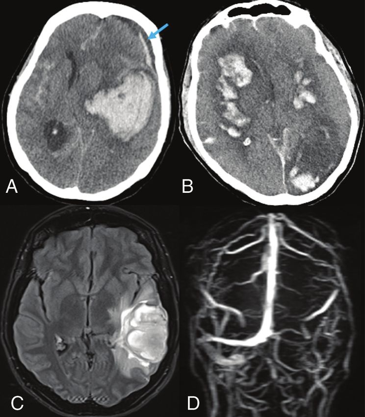
potential latency between infection and symptom onset. Using this FIG 1. Cerebral hemorrhage in patients with COVID-19. A, Noncontrast
criterion, we captured 6 likely patients with initially false-negative CT of the head shows a large left basal ganglia intraparenchymal hem-
orrhage, with diffuse subarachnoid and left frontal subdural (blue
results on polymerase chain reaction testing with a delay from
arrow) hemorrhages in a woman with asymptomatic COVID-19. B,
COVID-19 (n ¼ 4) or stroke (n ¼ two; 2- and 3-day delay) symp- Noncontrast CT of the head showing extensive bilateral multifocal cer-
tom onset to polymerase chain reaction positivity. Three additional ebral infarctions with hemorrhagic conversion in a comatose man with
patients who met this criterion possibly acquired COVID-19 while multiorgan failure. C, Brain MR imaging FLAIR and D, MR venogram
in the hospital, but this cannot be established with any certainty; show hemorrhagic venous infarction in the left temporal lobe (C) sec-
ondary to thrombosis of left transverse and sigmoid sinuses and inter-
therefore, these patients are included in our analysis. Clinical and
nal jugular vein (D) in a young woman presenting with seizures.
laboratory data were collected by retrospective chart review. Only
laboratory variables with at least 75% complete data were included
in our analysis. Two neuroradiologists with Certificates of Added subtype, infarction pattern, and the presence of hemorrhagic trans-
Qualification characterized the neuroimaging findings, blinded to formation and simultaneous infarction and hemorrhage.
clinical and laboratory data; in cases of disagreement, consensus
was reached. Statistical Analysis
In-hospital stroke onset was defined as a new-onset focal neu- A bivariate analysis of imaging and laboratory findings dichotom-
rologic deficit or altered mental status after hospital admission for ized by inpatient or outpatient stroke-onset location as a surrogate
COVID-19 and imaging confirmation of cerebral infarction or in- for COVID-19 severity was performed. Only laboratory variables
tracranial hemorrhage. During the pandemic peak, only patients with at least 75% complete data were included in our study.
with severe COVID-19 complications were hospitalized, and Statistical analyses were performed using the Wilcoxon rank sum
patients with less severe symptoms related to COVID-19 were test for continuous variables and the x 2 test for most categoric vari-
treated at home. Although hospitals in our system did not mandate
ables. The Fisher exact test was used when the cell number was ,5.
the use of specific hard criteria, in general, oxygen saturation
Statistical significance was considered for P values , .05. All statisti-
,90% or 92% or other signs of severe respiratory distress or severe
cal analyses were performed in SAS Version, 9.4 (SAS Institute).
systemic illness such as sepsis were used as guiding criteria for
COVID-19 admission during the crisis. Laboratory data included
complete blood count and coagulation profile at hospital admis- RESULTS
sion or in-hospital stroke onset, lowest lymphocyte count, signs Between March 14 and April 26, 2020, eighty-six patients (48
of noncerebral end-organ damage (elevated creatinine level men, 38 women) with mean age of 67.4 years (range, 25–94 years)
[.1.3 mg/dL], twice the normal alanine aminotransferase level met the inclusion criteria with imaging-confirmed infarction
[.90 U/L] and lactate dehydrogenase level [.484 U/L]), and the (83.7%) or pure intracranial hemorrhage (16.3%) (Fig 1A).
highest hospitalization inflammatory (C-reactive protein and ferri- Associated intracranial hemorrhage (Fig 1B) was found in 20.8%
tin) and coagulopathy markers (D-dimer). Diagnosis of coexistent of 72 patients with infarction, including 9 with simultaneous
deep vein thrombosis was recorded. Neuroimaging findings were hemorrhage and infarction and 6 with hemorrhagic transforma-
based on brain CT or MR imaging findings, including stroke tion, including 1 hemorrhagic venous infarction secondary to
258 Katz Feb 2021 www.ajnr.orgwith other patients with COVID-19 infection who develop stroke
as an outpatient with milder or no COVID-19 symptoms.
Although we are unable, in a retrospective study, to prove that our
findings are exclusively related to COVID-19, as opposed to other
intensive care unit–related variables, patients hospitalized with
severe COVID-19 had distinct stroke imaging patterns and even
more abnormal laboratory biomarkers, which support a potential
pathophysiologic link underpinning our previous finding that
COVID-19 is a strong independent risk factor for stroke in hospi-
talized patients.12 Patients with severe manifestations of COVID-19
had significantly more frequent ischemic strokes with multivascular
territory distributions, hemorrhagic transformation, and simultane-
ous infarction and intracranial hemorrhage. While many patients
FIG 2. Multivascular territory infarctions in COVID-19. MR imaging of
with COVID-19 and stroke had elevated serum inflammatory
the brain diffusion-weighted imaging demonstrates watershed-pat-
tern infarctions in a 52-year-old man with mild COVID-19 symptoms, markers (C-reactive protein and ferritin), lymphocytopenia, throm-
who awoke at home with left hemiparesis (A) and an 86-year-old bocytopenia, and coagulopathy (elevated D-dimer levels and inter-
woman admitted for hypoglycemia, dehydration, and COVID-19 national normalized ratio), those with severe consequences of
pneumonia (B) and found to have new-onset atrial fibrillation; 8 days COVID-19 had significantly higher serum markers of inflamma-
into her hospitalization, she developed lethargy, left hemiparesis, ex-
tion, with more frequent levels of ferritin .4 times normal and C-
pressive aphasia, and dysphagia.
reactive protein levels more than twice normal. These patients also
dural venous sinus thrombosis (Fig 1C, -D). Multivascular terri- had significantly higher markers of hypercoagulability with D-
tory infarction (33/72 patients; 45.8%) predominated, including dimer levels more frequently .4 times normal. Furthermore,
12 with a watershed pattern (Fig 2), followed by single vascular patients with severe COVID-19 and stroke had significant associa-
territory infarction (29/72 patients, 40.3%) and solitary small-ves- tions with greater frequencies of leukocytosis, lymphopenia, and
sel occlusion (10/72 patients, 13.9%). cytolysis (measured by lactate dehydrogenase), hepatic and renal
Forty-one patients (47.7%) were already hospitalized at stroke dysfunction, and deep vein thrombosis.
onset, whereas 45 patients (52.3%) developed out-of-hospital In a series of 214 patients hospitalized with COVID-19 from
Wuhan, China, acute cerebrovascular disease was first reported in
neurologic deficits, with ongoing mild or asymptomatic COVID-
6 patients (2.8%) and was associated with severe pulmonary infec-
19 infection. In-hospital stroke onset was associated with more
tion, older age, stroke risk factors, elevated inflammatory markers,
frequent mechanical ventilation (56.1% versus 33.3%, P ¼ .034),
and end-organ damage.8 Evidence of a concomitant prothrom-
admission tachypnea (respiratory rate, .20 breaths per minute;
botic state was proposed by others who describe a series of 6
61.0% versus 33.3%, P ¼ .016), and discharge to a rehabilitation
patients with COVID-19 and ischemic stroke who had infarctions
facility (51.2% versus 22.2%, P ¼ .011), supporting our hypothesis
suggesting large-artery occlusion.9 In that study, all cases had sig-
that these patients had more severe COVID-19 infection.
nificantly elevated D-dimer levels, 2 had venous thromboembolic
Supplemental Table 1 summarizes the bivariate analysis of
disease, and 2 developed large-artery occlusion despite therapeutic
imaging and laboratory findings dichotomized by patient location
anticoagulation. One patient had antiphospholipid antibodies.
at stroke onset (see Supplemental Table 2 for analysis of laboratory
Very high median D-dimer and C-reactive protein levels were also
values as continuous variables). In-hospital stroke onset was signifi-
reported in a larger retrospective study of 32 patients with
cantly associated with ischemic stroke (95.1% versus 73.3%,
COVID-19 with ischemic stroke in the New York metropolitan
P ¼ .006), particularly multivascular distribution (56.4% versus
area.10 In a study by others in the same region of 31 patients with
33.3%, P ¼ .022) and associated hemorrhagic transformation or si-
COVID-19 with ischemic stroke, higher D-dimer levels and eryth-
multaneous intracranial hemorrhage (31.7% versus 4.4%, P ¼ .001).
rocyte sedimentation rates were reported compared with concur-
These patients also had more frequent deep vein thrombosis
rent patients with COVID-19 without stroke and with a historical
(29.3% versus 2.2%, P ¼ .001). For those with laboratory values
sample of patients with influenza.13 In that study, when one
available, patients with in-hospital stroke onset had more frequent
adjusted for demographics, vascular risk factors, and critical care
leukocytosis (61.0% versus 26.7%, P ¼ .001) and lymphopenia
admission, COVID-19 still nearly quintupled the odds of ischemic
(90.2% versus 55.6%, P ¼ .001), abnormal creatinine levels (79.0%
stroke compared with influenza infection.
versus 43.8%, P ¼ .001), and severely elevated D-dimer (92.5% ver-
Our experience, like that of other centers,9-11,13,14 suggests
sus 54.8%, P ¼ .001), C-reactive protein (79.0% versus 43.8%,
that a hypercoagulable-prothrombotic state is a prevalent homeo-
P ¼ .002), ferritin (79.0% versus 31.1%, P , .001), lactate dehydro-
static complication among patients with COVID-19 and particu-
genase (73.0% versus 42.9%, P ¼ .014), and alanine aminotransfer-
larly in those with severe illness. Under these conditions, in
ase levels (78.1% versus 39.5%, P , .001).
which sepsis is common, ineffective host defense mechanisms to
limit crosstalk between coagulation and hyperinflammation can
DISCUSSION increase tissue injury, worsen organ dysfunction, and promote
Patients hospitalized for severe COVID-19 infection who develop thrombogenesis.15,16 Hyperinflammation can activate coagula-
stroke in the hospital demonstrate substantial differences compared tion pathways via several mechanisms. These include promoting
AJNR Am J Neuroradiol 42:257–61 Feb 2021 www.ajnr.org 259Supplemental Table 1: Bivariate analysis of imaging and laboratory findings in patients with COVID-19 dichotomized by location at stroke
onset
Variable Median (IQR) In-Hospital (No.) (%) Out-of-Hospital (No.) (%) P Value
Patients (No.) 86 41 45
Age (yr)
20–69 years of age 68 (60–76) 20 (48.8) 26 (57.8) .403
Sex, male 26 (63.4) 22 (48.9) .176
Imaging
Ischemic stroke 39 (95.1) 33 (73.3) .006
Infarction, no hemorrhage 26 (63.4) 31 (68.9) .001
Infarction with HT 5 (12.2) 1 (2.2)
Infarction and ICH 8 (19.5) 1 (2.2)
Pure ICH, no infarction 2 (4.9) 12 (26.8)
Multivascular territory infarction (n ¼ 72) 22 (56.4) 11 (33.3) .022
Deep vein thrombosis 12 (29.3) 1 (2.2) .001
Laboratory values
White blood cella
$10.5 K/uL 9.7 (7.2–14.7) 25 (61.0) 12 (26.7) .001
Lymphocytesb
,1.00 K/uL 0.6 (0.31–1.05) 37 (90.2) 25 (55.6) .001
Hemoglobina
,11.5 g/dL 12.3 (9.7–13.8) 19 (46.3) 16 (35.6) .309
Plateletsa
,150 K/uL 247 (152–335) 9 (22.0) 10 (22.2) .976
aPTTa,c
.36.3 seconds 30.9 (28.4–36.1) 13 (32.5) 7 (17.1) .108
INRa,c
.1.20 1.17 (1.07–1.31) 19 (48.7) 17 (39.5) .403
D-dimer,c 4 nl
.920 ng/mL 3060 (1064–24,211) 37 (92.5) 17 (54.8) .001
C-reactive protein,c 2 nl
.10.0 mg/L 17.5 (6.4–28.8) 30 (79.0) 14 (43.8) .002
Ferritin,c 4 nl
.1200 ng/mL 1382 (706–2424) 30 (79.0) 10 (31.1) ,.001
LDH,c 2 nl
.484 U/L 536 (357–815) 27 (73.0) 12 (42.9) .014
ALT,c 2 nl
.90.0 U/L 50 (29–136) 32 (78.1) 17 (39.5) ,.001
Creatininec
.1.30 mg/dL 1.30 (0.93–2.81) 28 (70.0) 15 (34.1) .001
Note:—HT indicates hemorrhagic transformation; ICH, intracranial hemorrhage; nl, upper limit of the normal value; aPTT, activated partial thromboplastin time; INR, inter-
national normalized ratio; LDH, lactate dehydrogenase; ALT, alanine aminotransferase; IQR, interquartile range.
a
White blood cell count, hemoglobin, platelets, activated aPTT, and INR are from admission or at the time of consultation for stroke. Remaining laboratory values
(except lymphocyte count) are the maximum level during the hospitalization.
b
Lowest lymphocyte count during admission.
c
Variable has missing values. The number and percentage (No.) (%) of patients with missing laboratory values are the following: aPTT (n ¼ 5, 5.8%), INR (n ¼ 4, 4.7%), D-
dimer (n ¼ 16, 18.6%), C-reactive protein (n ¼ 16, 18.6%), ferritin (n ¼ 16, 18.6%), LDH (n ¼ 21, 24.4%), ALT, (n ¼ 2, 2.3%), and creatinine (n ¼ 2, 2.3%). All other variables have no
missing values.
endothelial activation, increasing tissue factor expression on vas- infection of cerebrovascular endothelia expressing angiotensin-
cular endothelial and circulating inflammatory cells such as converting enzyme 2, causing endotheliitis and vascular injury,
monocytes, and downregulating the efficiency of the protein is postulated and may result in both ischemic and hemorrhagic
C–protein S thrombomodulin system to regulate the increased stroke, though this mechanism remains unproven. Finally, a
thrombogenesis associated with consumptive coagulation.14,17,18 potential downregulation of neurovascular endothelial angio-
Specifically, surges in proinflammatory cytokines and mediators, tensin-converting enzyme 2 expression in response to its bind-
related to an exaggerated innate immune response and extensive ing to SARS-CoV-2 may also be relevant to ischemic stroke
tissue damage associated with severe COVID-19, can downregu- pathogenesis, though this too is theoretic.
late the expression and function of thrombomodulin, an endoge- Our study has several strengths and limitations. As we previ-
nous protein with potent anticoagulant and anti-inflammatory ously reported,12 our series likely undercounted the actual
properties pivotal in preventing vascular occlusion.19,20 COVID-19-stroke burden during this timeframe. Many potential
Moreover, an autoimmune hypercoagulable state associated patients with stroke with normal initial brain imaging did not
with antiphospholipid antibodies, such as anti-cardiolipin and undergo repeat imaging because of clinical instability or concerns
anti– b 2-glycoprotein-1 antibodies, can contribute to the patho- for spreading infection while transporting patients with COVID-
physiology of a subset of strokes associated with COVID-19.21 19 off quarantined wards. For similar reasons, MR imaging use
As shown in the peripheral vasculature,22 direct SARS-CoV-2 was limited at numerous centers, especially earlier on in the local
260 Katz Feb 2021 www.ajnr.orgepidemic. Therefore, some patients with smaller and more acute 2. COVID-19 dashboard by the Center for Systems Science and
infarctions were not included in our study, and our results should Engineering at Johns Hopkins University. https://publichealthupdate.
be interpreted as only relevant to patients with COVID-19 who com/jhu/. Accessed October 1, 2020
3. Mizumoto K, Kagaya K, Zarebski A, et al. Estimating the asymptom-
have imaging-confirmed stroke.
atic proportion of coronavirus disease 2019 (COVID-19) cases on
A relative strength to our approach was the inclusion of board the Diamond Princess cruise ship, Yokohama, Japan, 2020.
patients with primary and secondary intracranial hemorrhage, Euro Surveill 2020;25:2000180 CrossRef Medline
allowing, for the first time, the demonstration of a frequent hem- 4. Richardson S, Hirsch JS, Narasimhan M, et al. Presenting characteris-
orrhagic component to infarctions seen in patients with severe tics, comorbidities and outcomes among 5700 patients hospitalized
COVID-19. Independent neuroradiologic review of the imaging- with COVID-19 in the New York City area. JAMA 2020;323:2052–59
CrossRef Medline
enabled confirmation of stroke diagnoses and characterization of
5. Mehta P, McAuley DF, Brown M, et al; HLH Across Speciality
imaging findings were blinded and uniform. As a retrospective Collaboration, UK. COVID-19: consider cytokine storm syndromes
study, some laboratory values were missing in a proportion of and immunosuppression. Lancet 2020;395:1033–34 CrossRef Medline
patients. To mitigate this issue, we analyzed only variables with at 6. Tang N, Li D, Wang X, et al. Abnormal coagulation parameters are
least 75% complete data. Consequently, some commonly cited associated with poor prognosis in patients with novel coronavirus
laboratory abnormalities associated with COVID-19 are not pneumonia. J Thromb Haemost 2020;18:844–47 CrossRef Medline
7. Cao W, Li T. COVID-19: towards understanding of pathogenesis.
included, such as fibrinogen, antiphospholipid antibodies, and
Cell Res 2020;30:367–69 CrossRef Medline
interleukin levels. In addition, given our small sample size, we 8. Mao L, Jin H, Wang M, et al. Neurologic manifestations of hospital-
were unable to perform an adjusted analysis. Nevertheless, the ized patients with coronavirus disease 2019 in Wuhan, China.
number of cases of COVID-19 stroke in our series permitted a JAMA Neurol 2020;77:683 CrossRef Medline
comprehensive analysis of neuroimaging and laboratory bio- 9. Beyrouti R, Adams ME, Benjamin L, et al. Characteristics of ischae-
markers, reinforcing potential pathophysiologic mechanisms mic stroke associated with COVID-19. J Neurol Neurosurg Psychiatry
2020;91:889–91 CrossRef Medline
supporting the association between COVID-19 and stroke.
10. Yaghi S, Ishida K, Torres J, et al. SARS2-CoV-2 and stroke in a New
York healthcare system. Stroke 2020;51:2002–11 CrossRef Medline
CONCLUSIONS 11. Arachchillage DR, Laffan M. Abnormal coagulation parameters are
Compared with those with mild or asymptomatic COVID-19 at associated with poor prognosis in patients with novel coronavirus
pneumonia. J Thromb Haemost 2020;18:1233–34 CrossRef Medline
stroke onset, patients who have strokes while hospitalized with
12. Katz JM, Libman RB, Wang JJ, et al. Cerebrovascular complications
severe manifestations of COVID-19 have significantly more inflam- of COVID-19. Stroke 2020;51:e227–31 CrossRef Medline
mation, multiorgan dysfunction, and coagulopathy, including deep 13. Merkler AE, Parikh NS, Mir S, et al. Risk of ischemic stroke in patients
vein thrombosis. These findings support proposed pathophysio- with coronavirus 2019 (COVID-19) vs patients with influenza. JAMA
logic mechanisms linking these diagnoses, though we cannot defini- Neurol 2020 Jul 2. [Epub ahead of print] CrossRef Medline
tively state that our findings are specific to SARS-CoV-2 infection 14. Lillicrap D. Disseminated intravascular coagulation in patients with
as opposed to the critical illness associated with severe COVID-19. 2019-nCoV pneumonia. J Thromb Haemost 2020;18:786–87 CrossRef
Medline
Infarctions with concurrent hemorrhage are also more frequent.
15. Foley JH, Conway EM. Cross talk pathways between coagulation
Given the associated hypercoagulability, clinical-treatment algo- and inflammation. Circ Res 2016;118:1392–1408 CrossRef Medline
rithms for patients hospitalized with COVID-19 have shifted to- 16. Levi M, van der Poll T. Coagulation and sepsis. Thromb Res
ward widespread use of full- or prophylactic-dose anticoagulation 2017;149:38–44 CrossRef Medline
to prevent thromboembolic complications, though the benefits of 17. Amiral J, Seghatchian J. Revisiting the activated protein C-protein
this approach remain unproven because it is based solely on obser- S-thrombomodulin ternary pathway: impact of new understand-
ing on its laboratory investigation. Transfus Apher Sci 2019;58:538–
vational data.23 In patients with COVID-19 with stroke, anticoagu-
44 CrossRef Medline
lation may be of benefit, but the approach may have to be more 18. Bode M, Mackman N. Regulation of tissue factor gene expression
nuanced, given the risks of associated cerebral hemorrhage, man- in monocytes and endothelial cells: thromboxane A2 as a new
dating a more cautious balancing of the risks of thrombosis and player. Vascul Pharmacol 2014;62:57–62 CrossRef Medline
hemorrhage. 19. Ikezoe T. Thrombomodulin/activated protein C system in septic
disseminated intravascular coagulation. J Intensive Care 2015;3:1
CrossRef Medline
Disclosures: Christopher G. Filippi—UNRELATED: Consultancy: Guerbet, Syntactx;
Grants/Grants Pending: Foundation of the American Society of Neuroradiology
20. Joseph L, Fink LM, Hauer-Jensen M. Cytokines in coagulation and
grant and National MS Society grant*; Payment for Manuscript Preparation: article thrombosis: a preclinical and clinical review. Blood Coagul Fibrinolysis
on topics in MRI journal (April 2020); Stock/Stock Options: minority stakeholder in 2002;13:10–16 CrossRef Medline
start-up Avicenna. Pina Sanelli—UNRELATED: Grants/Grants Pending: Siemens, 21. Zhang Y, Xiao M, Zhang S, et al. Coagulopathy and antiphospholi-
Harvey L. Neiman Sanell Health Policy Institute, Comments: research partnership.* pid antibodies in patients with COVID-19. N Engl J Med 2020;382:
Souhel Najjar—UNRELATED: Employment: Full-time and Chair of Neurology at e38 CrossRef Medline
Northwell Health. *Money paid to the institution.
22. Varga Z, Flammer AJ, Steiger P, et al. Endothelial cell infection and
endotheliitis in COVID-19. Lancet 2020;395:1417–18 CrossRef
REFERENCES Medline
1. Zhu N, Zhang D, Wang W, et al; China Novel Coronavirus 23. Nadkarni GN, Lala A, Bagiella E, et al. Anticoagulation, mortality,
Investigating and Research Team. A novel coronavirus from patients bleeding and pathology among patients hospitalized with COVID-
with pneumonia in China, 2019. N Engl J Med 2020;382:727–33 19: a single health system study. J Am Coll Cardiol 2020 Aug 24.
CrossRef Medline [Epub ahead of print] CrossRef Medline
AJNR Am J Neuroradiol 42:257–61 Feb 2021 www.ajnr.org 261You can also read