Parallel guidewire technique in acute ischemic stroke secondary to carotid artery dissection
←
→
Page content transcription
If your browser does not render page correctly, please read the page content below
Original Article

Parallel guidewire technique in acute ischemic stroke secondary
to carotid artery dissection
Xiongjun He#, Liang Zhang#, Kaifeng Li, Ming Hu, Hongxing Zhou, Jie Li, Yajie Liu

Department of Neurology, Shenzhen Hospital, Southern Medical University, Shenzhen, China
Contributions: (I) Conception and design: X He; (II) Administrative support: L Zhang; (III) Provision of study materials or patients: X He, Y Liu; (IV)
Collection and assembly of data: K Li, J Li; (V) Data analysis and interpretation: M Hu, H Zhou; (VI) Manuscript writing: All authors; (VII) Final
approval of manuscript: All authors.
#
These authors contributed equally to this work.
Correspondence to: Yajie Liu. Department of Neurology, Shenzhen Hospital of Southern Medical University, No.1333 Xinhu, Baoan District,
Shenzhen 518101, China. Email: docliu18@163.com.

                 Background: The aim of the study was to evaluate the influence of endovascular therapy (EVT) on the
                 acute ischemic stroke (AIS) secondary to carotid artery dissection (CAD).
                 Methods: This single-center retrospective study enrolled 17 patients admitted with AIS secondary to CAD
                 from January 2018 to November 2019 in the Department of Neurology, Shenzhen Hospital of Southern
                 Medical University where patients received EVT with guidewire or parallel guidewire. Outcomes including
                 postoperative complications were recorded, the prognostic factors of patients were explored, and the
                 effectiveness of single guidewire versus parallel guidewire on EVT was compared.
                 Results: Before treatment, the mean National Institute of Health stroke scale (NIHSS) was 14.4 in 10
                 cases (58.8%) of CAD complicated with intracranial artery embolism. All patients underwent EVT, and
                 the success rate of operation was 100%. After all interventions, modified thrombolysis in cerebral ischemia
                 (mTICI) 3 reperfusion was achieved in 14 patients (82.4%), and mTICI 2b reperfusion was achieved in
                 3 (17.6%). One patient had cerebral infarction and edema complicated with cerebral hernia, one patient
                 had cerebral hemorrhage, one patient had complicated subarachnoid hemorrhage, and five cases had
                 asymptomatic cerebral hemorrhage. Three months after treatment, 14 cases (82.3%) achieved a good clinical
                 outcome (modified Rankin scale, mRS: 0–2). Puncture-to-reperfusion time in the parallel guidewire group
                 was significantly shorter than that of the single guidewire group. However, the differences in NIHSS score,
                 postoperative mTICI, and mRS score between both groups did not reach significance.
                 Conclusions: CAD patients receiving EVT have a good prognosis, and application of a parallel guidewire
                 can reduce operation time.

                 Keywords: Carotid artery dissection (CAD); acute ischemic stroke (AIS); revascularization; parallel guidewire

                 Submitted Oct 14, 2020. Accepted for publication Dec 29, 2020.
                 doi: 10.21037/apm-20-2168
                 View this article at: http://dx.doi.org/10.21037/apm-20-2168

Introduction                                                                  but this disorder is the main cause of stroke in young
                                                                              people, accounting for 10–25% of all AIS cases (3).
Carotid artery dissection (CAD), a known causative                            Since five major clinical studies noted the efficacy of
factor of acute ischemic stroke (AIS), is characterized by                    endovascular therapy (EVT) in acute cerebral infarction
intramural hematoma (IMH) following carotid subintimal                        (ACI) in 2015, all countries have regarded EVT as the
tear (1). CAD is not commonly seen in general stroke                          first-level recommendation for ACI due to occlusion of
patients and only accounts for 2–2.5% of all AIS cases (2),                   the large vessel. However, there is still no report on the

© Annals of Palliative Medicine. All rights reserved.                 Ann Palliat Med 2021;10(1):266-277 | http://dx.doi.org/10.21037/apm-20-2168
Annals of Palliative Medicine, Vol 10, No 1 January 2021                                                                            267

operation and treatment guidelines for ACI with large               areas. For the judgment of the nature of carotid artery
vessel occlusion caused by CAD, especially concerning the           occlusion, all cases in this center are interpreted by two
dissection of lesions during the operation. In this single-         neuroimaging departments and neurointerventional
center retrospective study, we analyzed the prognosis of 17         doctors. The imaging diagnostic criteria for arterial
patients with AIS secondary to CAD from January 2018                dissection include the following: (I) fusiform or irregular
to November 2019 and compared the efficacy of single                tumor-like expansion of non-branched blood vessels; (II)
guide wire and parallel guide wire technique in EVT for             long, filamentous, and irregular stenosis, with one or more
CAD. We provide the following articles according to the             of the following manifestations: double lumen, Intramural
STROBE reporting checklist (available at http://dx.doi.             hematoma, endometrial valve, rapid morphological
org/10.21037/apm-20-2168).                                          changes, focal stenosis and dilation (bead-like changes); or
                                                                    (III) recanalization of non-branched vessels accompanied
                                                                    by fusiform or irregular aneurysm-like dilatation. These
Methods
                                                                    standards are mainly based on conventional angiography,
Clinical information                                                CTA or MRA imaging results.

Patients with incomplete information were excluded from
the study. A total of 17 patients (14 males and 3 females; age,     Treatments
29–58 years; mean age, 41.8 years) with CAD complicated             Patients detected within 6 hours after onset with National
with AIS were enrolled in this study from January 2018 to           Institute of Health stroke scale (NIHSS) scores >6 and
November 2019 in the Department of Neurology, Shenzhen              Alberta stroke program early CT (ASPECT) scores >6
Hospital of Southern Medical University. Of the 17 patients,        underwent EVT with bridging intravenous thrombolysis,
3 male patients had a history of smoking, 6 had a history of        in strict accordance with standards of EVT for AIS. Those
hypertension, 1 had a history of heart disease, 4 had a history     detected after more than 6 hours from onset with ASPECT
hyperlipidemia, 2 had a history of neck massage, and the rest       or pc-ASPECT >6, or NIHSS score >6, underwent
had no obvious risk factors.                                        multimodal imaging assessment. Patients with an obvious
                                                                    mismatch in the penumbral areas, according to the DAWN
Clinical symptoms                                                   or DIFFUSE3 trial standards, were given EVT. All
                                                                    operations received informed consent from the patients’
The 17 patients all had acute onset of ischemic stroke              family members, and all patients participated in the 13th
symptoms such as dysarthria, central facial and lingual             National Five-Year Plan Registered Study. All procedures
paralysis, hemianopia, hemiplegia, hemidysesthesia, and             performed in the studies involving human participants were
crossed paralysis. Eleven patients intravenously received           in accordance with the Helsinki Declaration (as revised in
thrombolysis with recombinant-tissue plasminogen activator          2013). This study was approved by the Clinical Research
(r-tPA) within 4.5 hours after onset, and with urokinase            Ethics Committee of Shenzhen Hospital of Southern
within 4.5–6 hours. The other patients were assigned with           Medical University. All operations received informed
treatment within 24 hours after onset.                              consent from the patients’ family members. Source data of
                                                                    this study are available from the corresponding author on
                                                                    reasonable request.
Clinical assessment

Patients who were diagnosed within 6 hours after onset
                                                                    Surgical procedure
underwent routine head computed tomography (CT). The
patients identified over 6 hours after onset were detected          Most of the patients undergoing surgery received local
through multimodal imaging: head CT angiography (CTA)               anesthesia and sedation, and only patients (29.4%) received
+ CT perfusion (CTP) or magnetic resonance imaging                  general anesthesia through the trachea cannula when
(MRI) diffusion-weighted imaging (DWI) + arterial                   sedation failed to control these patients.
spin labeling (ASL) + magnetic resonance angiography                   Aortography and intracranial angiography were initially
(MRA) to assess ischemic penumbra and great vessels and             performed with a 5-Fr pig tail catheter through the 8-Fr
to determine the presence of occlusion and mismatched               shuttle guiding sheath to evaluate vascular occlusion and

© Annals of Palliative Medicine. All rights reserved.        Ann Palliat Med 2021;10(1):266-277 | http://dx.doi.org/10.21037/apm-20-2168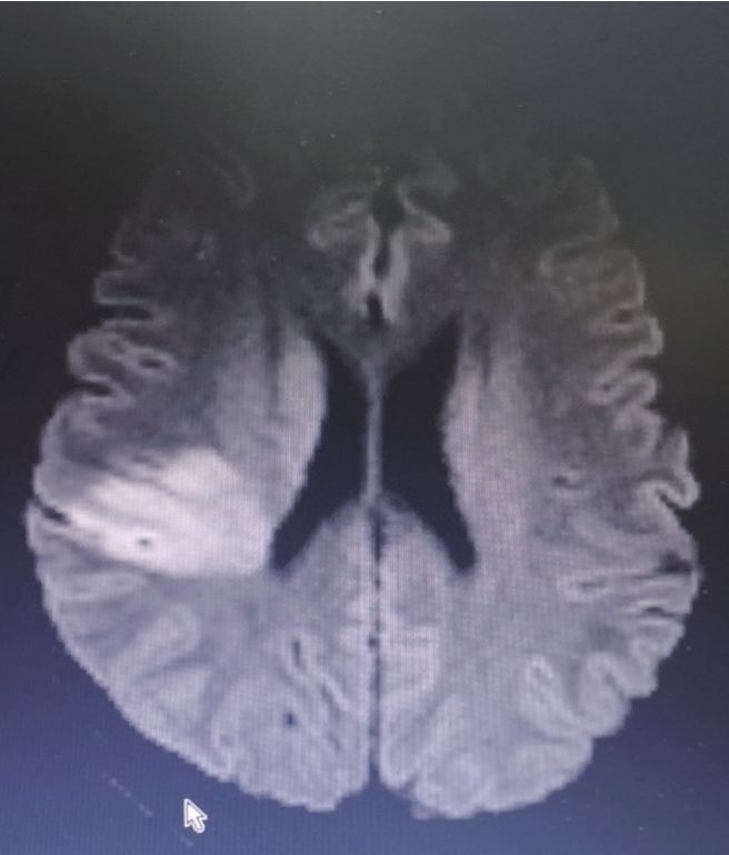
268                                                                                He et al. Parallel guidewire technique and stroke

general compensation. For patients with tortuous arcus             (Penumber) was passed through the proximal stent and
aortae, an 8-Fr NeuroMax (penumber) was inserted as                remained on the distal occlusion site, with ADAPT being
a guiding catheter under the guidance of a 5-Fr multi-             used to ensure the opening. If the intracranial blood vessel
function tube (Cordis) and the guide wire to locate vascular       thrombosis load was large, the Solumbra technique with
lesions and observe the morphology. Then, in one group,            stent was used to remove the embolus.
we inserted an SL-10 microcatheter (Stryer) or XT-27                  Technical results of recanalizing therapies were scored
microcatheter (Stryker) across the lesion using a 300 cm           by the modified thrombolysis in cerebral ischemia (mTICI)
single guide wire (Synchro or Transend 0.014, Stryker) until       scale. mTICI 2b and mTICI 3 are conventionally subsumed
the tip had passed sufficiently into the lesion. In another        under successful recanalization. Before stent implantation,
group, a parallel guide wire was used to pass through the          patients were administered with an intravenous injection
dissected lesion. We tried two times to first have a Synchro       of 6 mL of tirofiban and 5 mL/h of maintenance fluid.
or Transend guide wire pass through false lumen, but               Energy spectrum CT performed immediately after
this still failed, and the guide wire was left in the lumen        surgery to identify bleeding or contrast agent retention,
to block the pathway. The second guide wire was utilized           and exclude bleeding patients who were given continuous
to access true lumen from different angles for cross artery        pumping of tirofiban for 24 hours and then replaced with
dissection, and was left in the distal normal blood vessels.       dual antiplatelet therapy (DAPT, aspirin and Plavix), for
A microcatheter was inserted along the second guide wire           infarctions with a small amount of blood leakage Patients
across the dissection lesion and placed in the distal vascular     were treated with monoclonal Antiplatelet Therapy
cavity, which was followed by the withdrawal of the guide          (Polivix). If the bleeding is heavy, antiplatelet therapy is
wire. After the microcatheter was confirmed to be located          not given. After 3 months, the postoperative treatment of
in the true vascular cavity, the guide wire in the false lumen     DAPT was changed to monoclonal antiplatelet therapy.
was removed.
   For CAD complicated with distal thrombosis, different
                                                                   Clinical characteristics and outcome assessment
strategies for two distinct situations were used. (I) If the
thrombotic load of the proximal dissection was not large           The primary outcome was functional independence at
or did not affect the passage of the intermediate catheter         90 days. Functional independence was defined as an mRS
or aspiration catheter, the intracranial lesions was first         score of 0–2. The mRS score was assessed by a neurologist
treated, following insertion of the middle catheter or the         blinded to the treatment detail via telephone interview.
ACE aspiration catheter across the dissected lesion to             The secondary outcomes were successful recanalization at
the distal occlusion site, along the micro-guide wire and          the end of the procedure and puncture to recanalization
microcatheter. A direct aspiration first pass technique            time. Successful recanalization was defined mTICI ≥2b.
(ADAPT) was used for aspiration. If the intracranial               The safety outcome was symptomatic hemorrhage (sICH)
thrombus load was large, the Solumbra technique with               within 24 hours. sICH was evaluated on CT and regarded
a stent retriever was used to remove the embolus. After            as symptomatic if the patient’s neurologic symptoms
recanalizing the distal blood vessels, the protective umbrella     deteriorated with an increase of ≥4 on the NIHSS.
(Spider, Medtronic) was added to petrous bone segment
along the aspiration catheter. The intermediate catheter
                                                                   Statistical analysis
or the aspiration catheter was withdrawn into the guide
tube. We used the Wallstent stent (Boston Scientific) to            Baseline, clinical and procedural characteristics, outcomes
apply the CAD along the protective umbrella micro-guide            were compared between the single guide wire group and
wire. (II) If the cervical dissection affected the passage of      the parallel guide wire group. The Mann-Whitney U test,
the intermediate catheter or the suction catheter, or if a         χ2 test, and Fisher exact test were used for comparison. A
large thrombus load was present in the proximal treatment          value of P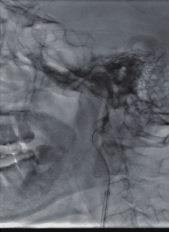
Annals of Palliative Medicine, Vol 10, No 1 January 2021                                                                             269

                         A                                       B

Figure 1 MRI imaging of the right temporal lobe (A) and the radiated coronary area (B) demonstrates cerebral infarction. MRI, magnetic
resonance imaging.

September 23, 2019 due to left limb weakness and slurred                  Intraoperative angiography confirmed that the initiation
speech. His symptoms appeared abruptly 8 hours earlier in             of the right internal carotid artery was occluded, and that
the workplace without obvious cause. He could not walk on             the left internal carotid artery was partially compensating
his own and had slurred speech, but no relief after rest. He          for this with anterior traffic to the right middle cerebral
was then rushed to Shenzhen Baoan Traditional Chinese                 artery area (Figure 2). The 0.014 in 300 mm Synchro micro-
Medicine Hospital. At that time, head CT indicated no                 guide wire was selected to pass through the CAD, but this
brain bleeding. Intravenous thrombolysis with 1×10⁶ IU of             failed (Figure 3A) and remained in the false cavity. Another
urokinase was administered to the patient at 16:30. After             0.014 300 mm Transend micro-guide wire successfully
thrombolysis at 17:00, slurred speech and weakness of the             crossed the dissection site (Figure 3B,C), and the XT
left limb were temporarily improved. About 20 minutes after           microcatheter was inserted along the Transend micro-
the patient completed the head MR scan + DWI + MRA,                   guide wire to the C2 section of the internal carotid artery,
the left limb fell limp again. The head MRI showed acute              followed by withdrawal of the micro-guide wire. Micro-
brain infarction in the right temporal lobe and a radiated            catheter angiography confirmed that it was located in the
coronary area (Figure 1). The condition worsened, and the             true lumen of the blood vessel (Figure 4A), and that no
patient was transferred to the emergency department of our            occlusion in the distal middle cerebral artery was present
hospital. He is a professional driver and has neck pain upon          (Figure 4B). The 0.014 300 mm Transend micro-guide
neck massage. In the emergency department, he had a blood             wire was placed in the C2 section, and the microcatheter
pressure of 140/75 mmHg, a clear mind, slurred speech, no             is removed. The Wallstent (7×50 mm) self-expanding
drooping of either eyelid, and a diameter in his pupils of 3.0        stent was placed in the cervical dissection and applied to
mm. His eyes had no nystagmus, his mouth was angled to                dissection. The blood vessel was formed (Figure 4C,D) and
the right, his uvula was centered, and his soft palate could          the mTICI score returned to grade 3.
be lifted. The muscle strength of the right limb was grade 5,             The next day after operation, head CT indicated no
the left upper limb was grade 1, and the left lower limb was          hemorrhage (Figure 5A,B). The patient was given tirofiban
grade 3. The pain and touch sensation on the left side were           for 24 hours, which was then was replaced with BsAb.
slightly weakened. The right side of his body could perform           Three months after surgery, the angiography suggested
coordinated movement. His neck was soft, and the Gram                 that the right carotid artery showed smooth blood
and Brinell signs were negative. The NIHSS score was 9,               circulation (Figure 5C,D), and the patient could speak
and the ASPECT score was 7.                                           clearly. The muscle strength had returned to level 5, and

© Annals of Palliative Medicine. All rights reserved.         Ann Palliat Med 2021;10(1):266-277 | http://dx.doi.org/10.21037/apm-20-2168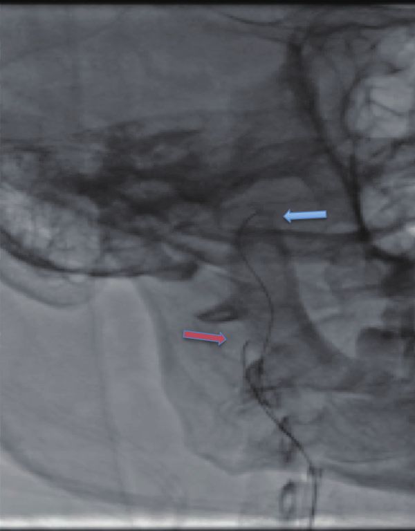
270                                                                                       He et al. Parallel guidewire technique and stroke

            A                                           B                                   C

Figure 2 MRA imaging indicates occlusion of the right internal carotid artery (A). Angiography shows the occlusion in the start of the
right internal carotid artery of a rat tail (B) and that the anterior communicating artery before the left internal carotid artery is open,
compensating for the right side anterior cerebral artery (C). MRA, magnetic resonance angiography.

                A                                       B                                     C

Figure 3 The micro-guide wire barely passes through the neck (A), the guide wire (blue arrow) is fixed in the false cavity (B), and another
guide wire (red arrow) is re-attached and passes through the distal true cavity (C).

the mRS score was 0.                                                       ASPECT score was 8.5 points. Eleven patients detected
                                                                           within 6 hours after onset were given thrombolysis. Of the
                                                                           17 patients, the success rate of the opening of an occluded
Results
                                                                           coronary vessel was 100%. The postoperative recanalization
In the postoperative effect observation and follow-up of                   was evaluated according to the mTICI classification, where
17 patients, hypertension appeared in 6 cases, smoking                     anterograde blood flow of mTICI 2b was considered partial
history in 3 cases, hyperlipidemia in 4 cases, and clear neck              recanalization, and mTICI 3 was considered complete
trauma or massage history in 2 cases. The preoperative                     recanalization. Of the 17 patients, 14 achieved mTICI
average NIHSS score of patients was 14.4 points, and the                   3 reperfusion, and 3 achieved mTICI 2b reperfusion.

© Annals of Palliative Medicine. All rights reserved.              Ann Palliat Med 2021;10(1):266-277 | http://dx.doi.org/10.21037/apm-20-2168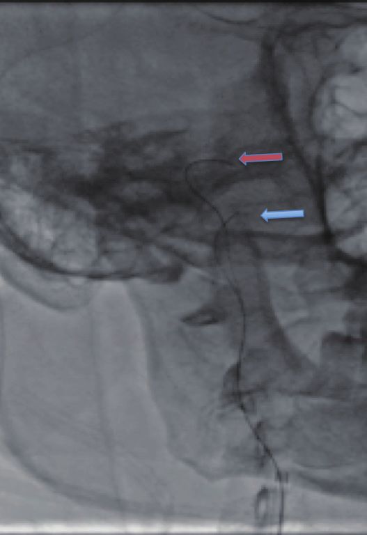
Annals of Palliative Medicine, Vol 10, No 1 January 2021                                                                                   271

                                 A                                       B

                                 C                                       D

Figure 4 Micro-catheter angiography imaging reveals that the micro-guide wire is located in the true cavity of the blood vessel (A, blue
arrow) and that the distal blood vessel is unobstructed (B, blue arrow). A Wallstent (7×50 mm) stent is implanted well and dissection repair is
normal (C, blue arrow), and the intracranial branches are visible (D).

Meanwhile, 5 patients developed asymptomatic brain                           was performed to these patients during treatment. Three
hemorrhage, and 1 had symptomatic cerebral hemorrhage,                       months later, the number of cases with an mRS score of 0–2
The hemorrhage transformation after cerebral infarction                      was 14 (82.3%).
is divided into HI type and PH type. HI (hemorrhagic                            Of the 17 patients (see Tables 1,2 for details), 7 patients
infarction), which means hemorrhagic cerebral infarction,                    (41.2%) presented with middle cerebral artery occlusion,
refers to small spotted hemorrhage with unclear borders;                     and 4 patients (23.5%) with internal carotid artery
PH (parenchymal hemorrhage), which means cerebral                            occlusion. Intraoperative digital subtraction angiography
parenchymal hemorrhage, refers to a mass hematoma                            (DSA) assessment of ASITN/SIR collateral circulation
with clear borders, with space-occupying effect. The two                     classification revealed the following: 11 grade 2 cases, 4
types can be divided into HI1 and HI2 PH1 and PH2                            grade 1 cases, and 2 grade 0 cases. The EVT strategy was
according to the degree of bleeding. The significance of                     performed as described above. A single guide wire was
the classification is that it is closely related to the prognosis.           used in 10 cases to pass the dissection lesion, and a parallel
The hematoma type, which is the PH type, has a worse                         guide wire was used in 7 cases. Stent angioplasty was first
prognosis, and the PH2 fatality rate reaches 50%. Suction                    performed in 9 patients where a self-expanding stent

© Annals of Palliative Medicine. All rights reserved.             Ann Palliat Med 2021;10(1):266-277 | http://dx.doi.org/10.21037/apm-20-2168
272 He et al. Parallel guidewire technique and stroke
A B
C D
Figure 5 Re-examination of head CT immediately after surgery reveals no intracranial hemorrhage (A), and the lesions are same as those
before the operation (B). At 3-month reexamination, the stent position is good without restenosis, and the dissection has healed (C,D).
was placed in the dissection to establish an opening. The were significantly relieved, and the vessel was not occluded
guide tube and the intermediate catheter were then passed as indicated by CT scans.
through the stent to the distal end to remove the thrombus. The comparison of CAD with a single guide wire to
Of these patients, a thrombus removal stent or aspiration that with a parallel guide wire (see Table 3) revealed no
catheter was used to open the distal occluded blood vessel in differences in risk factors, age, or ASPECT score. Of note,
5 cases, and in 4 cases the opening of the CAD allowed the the single guide wire group had a high preoperative NIHSS
distal thrombosis to canalize. The other 8 cases underwent score and thrombolysis rate. With time from admission
thrombectomy firstly via insertion of an ACE aspiration to puncture being insignificant between the groups, the
catheter into the middle cerebral artery occlusion and application of parallel guide wire shortened the time from
internal carotid artery occlusion, as the catheter was easy puncture to first recanalization, and from puncture to
to pass through the dissection. Of these 8 cases, 4 received complete recanalization. The difference between both
Wallstent self-expanding stent implantation at the later groups in postoperative mTICI blood flow and 3-month
stage, and the others were not further treated because their prognosis did not reach significance, as the rate of good
CAD did not affect the blood flow, their clinical symptoms prognosis was over 80% in both groups.
© Annals of Palliative Medicine. All rights reserved. Ann Palliat Med 2021;10(1):266-277 | http://dx.doi.org/10.21037/apm-20-2168Annals of Palliative Medicine, Vol 10, No 1 January 2021 273
Table 1 Clinical data of 17 patients with acute ischemic stroke (AIS) Table 2 Lesions and operation results
Number of patients Number of cases
Variable Surgical factors
(mean or ratio) (mean or ratio)
Age (year) 41.8 Tandem occlusion 11 (64.7%)
Gender (male) 14 (82.3%) Middle cerebral artery 7 (41.2%)
Hypertension 6 (35.3%) Intracranial internal carotid artery 4 (23.5%)
Smoking 3 (17.6%) Preferred strategy
Hyperlipidemia 4 (23.5%) Angioplasty and stenting 9 (52.9%)
History of trauma 2 (11.8%) Stent thrombectomy 2 (11.8%)
Preoperative NIHSS score 14.4 Suction 6 (35.3%)
ASPECT score 8.5 Remedy
ASITN/SIR collateral scale Angioplasty and stenting 4 (23.5%)
Grade 2 11 (64.7%) Suction 5 (29.4%)
Grade 1 4 (23.5%) Puncture-recanalization time (min) 142.47
Grade 0 2 (11.8%) mTICI grade
Thrombolysis 11 (64.7%) mTICI 3 14 (82.4%)
r-tPA 9 (52.9%) mTICI 2b 3 (17.6%)
Urokinase 2 (11.8%) IIb/IIIa use 12 (70.6%)
Anesthesia All intracranial hemorrhage 6 (35.3%)
Local anesthesia 12 (70.6%) Symptomatic intracranial hemorrhage 1 (5.9%)
General anesthesia 5 (29.4%) 3 months later (mRS 0–2 points) 14 (82.4%)
NIHSS score: National Institute of Health stroke scale. The Modified Rankin scale (mRA): 0–5 points, the higher the branch,
higher the score, the more serious the condition. mTICI: the worse the prognosis. 0: no symptoms; 1: no significant
modified treatment in cerebral infarction (mTICI) score (4); disability despite symptoms, able to carry out all usual duties
grade 0: no perfusion; grade 1: little blood passing through and activities; 2: slight disability, unable to carry out all previous
the occlusion segment, with little or no reperfusion; grade 2a: activities but not requiring assistance; 3: moderate disability,
antegrade reperfusion of less than half of the target artery requiring some help, but able to walk without assistance; 4:
previously in the ischemic territory; grade 2b: antegrade moderately severe disability, can walk with assistance but
reperfusion of more than half of the previously target artery unable to attend to own needs without assistance; 5: severe
ischemic territory; grade 3: complete antegrade reperfusion of disability, bedridden, incontinent, and requiring constant nursing
the downstream artery ischemic territory. The American Society care and attention. mTICI, modified thrombolysis in cerebral
of Intervention and Therapeutic Neuroradiology/Society of ischemia; MRA, magnetic resonance angiography.
Interventional Radiology (ASITN/SIR) collateral compensation
scale is a 5-point scale; grade 0: no collaterals visible to the
ischemic site; grade 1: slow collateral compensation to the
periphery of the ischemic site with persistence of some of the
defect; grade 2: rapid collaterals compensation to the periphery
of the ischemic site with persistence of some of the defect;
grade 3: collaterals compensation with slow but complete
angiographic blood flow of the ischemic bed by the late venous
phase; grade 4: complete and rapid collateral compensation
blood flow to the vascular bed in the entire ischemic territory.
© Annals of Palliative Medicine. All rights reserved. Ann Palliat Med 2021;10(1):266-277 | http://dx.doi.org/10.21037/apm-20-2168274 He et al. Parallel guidewire technique and stroke
Table 3 Comparison of the single guide wire and the parallel guide wire
Variable Single guide wire group (5) Parallel guide wire group (6) P value
Age 41.8 41.8 0.324
Gender (male) 9 (90%) 5 (71.4%) 0.537
Tandem lesions 7 (70%) 4 (57.1%) 0.644
Middle cerebral artery 5 (50%) 2 (28.6%)
Intracranial internal carotid artery 2 (20%) 2 (28.6%)
Collateral scale grade 2 5 (50%) 6 (85.8%) 0.304
ASPECT score 8.5 8.43 0.501
Preoperative NIHSS 16.8 10.8 0.281
Thrombolysis 8 (80%) 3 (42.9%) 0.058
From onset to puncture (min) 299 379 0.420
From admission to puncture (min) 53.5 37.6 0.339
From puncture to first recanalization (min) 112.5 58.7 0.136
From puncture to recanalization (min) 174.0 97.4 0.153
IIb/IIIa use 7 (70%) 5 (71.4%) 1.000
Postoperative cerebral hemorrhage 0.304
Asymptomatic 5 (50%) 1 (14.3%)
Symptomatic 1 (10%) 0
3 months after surgery mRS (0–2 points) 8 (80%) 6 (85.7%) 1.000
The ASPECT is a 10-point scale, where points are lost for each region affected. The lower the score, the more infarcted parts. ASPECT,
Alberta stroke program early CT; NIHSS, National Institute of Health stroke scale; mRS, modified Rankin scale.
After discharge, the telephone follow-up indicated Discussion
that patients’ symptoms had improved significantly. After
The annual incidence of CAD is approximately 10.4% (7).
successful opening of the CAD, patients did not have
Transcranial Doppler (TCD) ultrasound can detect
an ischemic attack in the ipsilateral carotid artery blood
cerebral micro-embolisms in 46% to 59% of patients with
supply area. All patients were rechecked with CTA or
cerebrovascular angiography 3 months later, and the results CAD, which suggests that patients with CAD are prone to
indicated that the original occluded carotid artery was cerebral embolisms in the arterial supply (6,8). In addition,
unobstructed, with the distal blood vessel having developed 20% of patients with CAD complicated by intracranial
well, with no collateral circulation. After surgery, 13 vascular occlusion also have severe internal carotid artery
patients with carotid artery stents were given antiplatelet stenosis or even complete occlusion (9). These patients have
drugs, lipid lowering therapy, and plaque stabilization. They a low recanalization rate upon intravenous thrombolysis
orally ingested statins (Lipitor 20–60 mg once a day), 75 mg with rt-PA (only 31%). Most patients with middle cerebral
of clopidogrel bisulfate, and aspirin in combination with artery occlusion have a poor prognosis (5,10). There
100 mg of BsAb or monoclonal antibody for 3 months. After were 11 patients in our study who received intravenous
3 months of the reexamination of blood vessels, treatment thrombolysis but whose blood vessels were not recanalized.
switched to monoclonal antibody; 4 patients without stent The subsequent bridging mechanical thrombectomy was
placement took anticoagulation therapy after discharge, and used to recanalize the blood vessels.
3 months later, reexamination of angiography indicated no One meta-analysis compared the efficacy of intravenous
stenosis of the blood vessel at the stent. thrombolysis and intravascular interventional therapy
© Annals of Palliative Medicine. All rights reserved. Ann Palliat Med 2021;10(1):266-277 | http://dx.doi.org/10.21037/apm-20-2168Annals of Palliative Medicine, Vol 10, No 1 January 2021 275
in patients with ACI secondary to CAD, revealing that middle cerebral artery stent embolization and internal
intravascular interventional therapy alone achieved better carotid artery stent implantation. Among these patients,
outcomes than intravenous thrombolysis alone, but there 62.50% (15/24) of patients achieved recanalization. (TICI
was no significant difference in cerebral hemorrhage, grade 2b–3), but only 29.17% (7/24) of patients had a good
mortality, or stroke recurrence (11). In another meta- clinical prognosis 3 months after operation (mRS score
analysis, 6 clinical data of ACI secondary to CAD ≤2 points). For tandem occlusion, some scholars suggest
were selected, comprising 193 patients who received that carotid artery disease should be routinely treated
interventional thrombectomy and 59 who received medical before the distal occlusion, so that the proximal and distal
treatment. After thrombectomy, 74% of patients were recanalization rates can exceed 84% and 33%, respectively
restored to grade 2b-3 blood flow. The interventional (15-19). The advantages of first restoring carotid artery
thrombectomy group (62.9%, 95% CI: 55.8–69.5%) blood flow are improving the safety of entering the distal
obtained better prognosis at 3 months (mRS 0–2) relative disease, the removal rate of distal thrombus, and the
to the drug treatment group (41.5%, 95% CI: 29.0–55.1%; simultaneous increase of arterial rt-PA activity due to the
P=0.006). The 90-day mortality upon interventional increase in anterograde blood flow, which in turn reduces
thrombectomy was similar to that obtained with drug the risk of distal re-occlusion and ischemia (16). Regardless
treatment (8.6% and 6.3%), and the difference in the of the surgical method, the main purpose is to open the
incidence of symptomatic intracranial hemorrhage (sICH) occluded blood vessels in a timely and effective manner
was not significant (5.9% vs. 4.2%, P=0.60) (12). In our and to quickly restore brain tissue perfusion. Given this,
study, the success rate of operation in the 17 patients was the EVT strategy involves passing the proximal dissection
100%. After all interventions, 1 patient had complicated lesion. In this study, we used a single guide wire to pass the
subarachnoid hemorrhage, and 5 had asymptomatic dissection lesion in 10 cases, and a parallel guide wire in
cerebral hemorrhage. Three months later, mRS scores were 7 cases. There were no differences in risk factors, age, or
improved (0–2) in 14 cases, which was higher than the rate ASPECT scores between both guide wire types. The single
of improvement of the Lin (11) and Dmytriw (12) study. guide wire group had a high preoperative NIHSS score and
This might be due to the fact that patients in our study had thrombolysis rate. As the time from admission to puncture
good collateral compensation. However, a 15-year follow-up was similar in both groups, it took less time to complete
study on ACI due to CAD in multiple centers revealed that the recanalization in the guide wire group. The difference
in 109 total patients, 24 received EVT (bridging treatment), between both groups in postoperative mTICI blood flow
38 received intravenous thrombolysis, and 47 received no and 3-month prognosis did not reach significance, as their
vascular recanalization treatment. Patients with EVT had a rate of good prognosis exceeded 80%.
higher rate of vascular recanalization within 24 hours, and Despite these findings, there remain some limitations
were more vulnerable to non-symptomatic bleeding when to our study which should be noted. First, the diagnosis
compared to patients given control treatment (P=0.026). of the cause of vascular occlusion mainly relies on the
There was no difference in sICH and mortality within comprehensive judgment of two neuro-interventional
7 days between the three groups. The rate of good clinical radiologists based on the patient's clinical data and
prognosis achieved at 3 months did not differ between the imaging data. There is a lack of a unified gold standard,
different subgroups (intravascular treatment: intravenous and there might have been a certain selection bias present.
thrombolysis P=0.407; EVT: no recanalization treatment Furthermore, this study is a single-center retrospective
P=0.580) (13). study with a small sample size. Some patients were excluded
CAD leads to acute occlusion, and is often complicated due to missing data, resulting in possible selection bias in
with intracranial artery embolism. At present, no research this study.
exists specifically focusing on the EVT of ACI caused
by CAD. Surgical methods for tandem lesions include
Conclusions
treating the proximal dissection lesion first, followed by
the opening of the distal occluded vessel, and vice versa. Our study indicates that CAD patients receiving EVT
A study by Stampfl et al. (14) enrolled 24 patients with may be reasonable, and application of a parallel guidewire
tandem occlusion of the internal carotid artery and middle will reduce operation time. A large prospective study is
cerebral artery, where patients were administrated with necessary for confirmation because of the small sample size
© Annals of Palliative Medicine. All rights reserved. Ann Palliat Med 2021;10(1):266-277 | http://dx.doi.org/10.21037/apm-20-2168276 He et al. Parallel guidewire technique and stroke
of this study. 1994;330:393-7.
2. Bassetti C, Carruzzo A, Sturzenegger M, et al. Recurrence
of cervical artery dissection. A prospective study of 81
Acknowledgments
patients. Stroke 1996;27:1804-7.
Funding: This study was supported by the Sanming 3. Mohr JP, Thompson JL, Lazar RM, et al. A comparison
Project of Medicine in Shenzhen (SZSM201812047) and of warfarin and aspirin for the prevention of recurrent
the Municipal Key Clinical Department in Shenzhen ischemic stroke. N Engl J Med 2001;345:1444-51.
(SZXK074). 4. Zaidat OO, Yoo AJ, Khatri P, et al. Recommendations
on angiographic revascularization grading standards for
acute ischemic stroke: a consensus statement. Stroke
Footnote
2013;44:2650-63.
Reporting Checklist: The authors have completed the 5. Rubiera M, Ribo M, Delgado-Mederos R, et al. Tandem
STROBE reporting checklist. Available at http://dx.doi. internal carotid artery/middle cerebral artery occlusion:
org/10.21037/apm-20-2168 an independent predictor of poor outcome after systemic
thrombolysis. Stroke 2006;37:2301-5.
Data Sharing Statement: Available at http://dx.doi. 6. Srinivasan J, Newell DW, Sturzenegger M, et al.
org/10.21037/apm-20-2168 Transcranial Doppler in the evaluation of internal carotid
artery dissection. Stroke 1996;27:1226-30.
Conflicts of Interest: All authors have completed the ICMJE 7. Beletsky V, Nadareishvili Z, Lynch J, et al. Cervical
uniform disclosure form (available at http://dx.doi. arterial dissection: time for a therapeutic trial? Stroke
org/10.21037/apm-20-2168). The authors have no conflicts 2003;34:2856-60.
of interest to declare. 8. Lucas C, Moulin T, Deplanque D, et al. Stroke patterns
of internal carotid artery dissection in 40 patients. Stroke
Ethical Statement: The authors are accountable for all 1998;29:2646-8.
aspects of the work in ensuring that questions related 9. Grau AJ, Weimar C, Buggle F, et al. Risk factors,
to the accuracy or integrity of any part of the work are outcome, and treatment in subtypes of ischemic stroke: the
appropriately investigated and resolved. All procedures German stroke data bank. Stroke 2001;32:2559-66.
performed in the studies involving human participants were 10. Tallarita T, Lanzino G, Rabinstein AA. Carotid
in accordance with the Helsinki Declaration (as revised in intervention in acute stroke. Perspect Vasc Surg Endovasc
2013). This study was approved by the Clinical Research Ther 2010;22:49-57.
Ethics Committee of the Shenzhen Hospital of Southern 11. Lin J, Liang Y, Lin J. Endovascular therapy versus
Medical University. All operations received informed intravenous thrombolysis in cervical artery dissection-
consent from the patients’ family members. related ischemic stroke: a meta-analysis. J Neurol
2020;267:1585-93.
Open Access Statement: This is an Open Access article 12. Dmytriw AA, Phan K, Maingard J, et al. Endovascular
distributed in accordance with the Creative Commons thrombectomy for tandem acute ischemic stroke associated
Attribution-NonCommercial-NoDerivs 4.0 International with cervical artery dissection: a systematic review and
License (CC BY-NC-ND 4.0), which permits the non- meta-analysis. Neuroradiology 2020;62:861-6.
commercial replication and distribution of the article with 13. Bernardo F, Nannoni S, Strambo D, et al. Efficacy and
the strict proviso that no changes or edits are made and the safety of endovascular treatment in acute ischemic stroke
original work is properly cited (including links to both the due to cervical artery dissection: A 15-year consecutive
formal publication through the relevant DOI and the license). case series. Int J Stroke 2019;14:381-9.
See: https://creativecommons.org/licenses/by-nc-nd/4.0/. 14. Stampfl S, Ringleb PA, Mohlenbruch M, et al. Emergency
cervical internal carotid artery stenting in combination
with intracranial thrombectomy in acute stroke. AJNR Am
References
J Neuroradiol 2014;35:741-6.
1. Schievink WI, Mokri B, O'Fallon WM. Recurrent 15. Jovin TG, Gupta R, Uchino K, et al. Emergent stenting of
spontaneous cervical-artery dissection. N Engl J Med extracranial internal carotid artery occlusion in acute stroke
© Annals of Palliative Medicine. All rights reserved. Ann Palliat Med 2021;10(1):266-277 | http://dx.doi.org/10.21037/apm-20-2168Annals of Palliative Medicine, Vol 10, No 1 January 2021 277
has a high revascularization rate. Stroke 2005;36:2426-30. in patients with severe stroke. Cardiovasc Intervent Radiol
16. Cohen JE, Gomori JM, Rajz G, et al. Extracranial 2007;30:34-41.
carotid artery stenting followed by intracranial stent- 19. Lavallee PC, Mazighi M, Saint-Maurice JP, et al. Stent-
based thrombectomy for acute tandem occlusive disease. J assisted endovascular thrombolysis versus intravenous
Neurointerv Surg 2015;7:412-7. thrombolysis in internal carotid artery dissection with
17. Nedeltchev K, Brekenfeld C, Remonda L, et al. Internal tandem internal carotid and middle cerebral artery
carotid artery stent implantation in 25 patients with acute occlusion. Stroke 2007;38:2270-4.
stroke: preliminary results. Radiology 2005;237:1029-37.
18. Dabitz R, Triebe S, Leppmeier U, et al. Percutaneous (English Language Editor: J. Gray)
recanalization of acute internal carotid artery occlusions
Cite this article as: He X, Zhang L, Li K, Hu M, Zhou H,
Li J, Liu Y. Parallel guidewire technique in acute ischemic
stroke secondary to carotid artery dissection. Ann Palliat Med
2021;10(1):266-277. doi: 10.21037/apm-20-2168
© Annals of Palliative Medicine. All rights reserved. Ann Palliat Med 2021;10(1):266-277 | http://dx.doi.org/10.21037/apm-20-2168You can also read