ANTIBODY-MEDIATED PROCOAGULANT PLATELETS IN SARS-COV-2-VACCINATION ASSOCIATED IMMUNE THROMBOTIC THROMBOCYTOPENIA
←
→
Page content transcription
If your browser does not render page correctly, please read the page content below
Antibody-mediated procoagulant platelets in
SARS-CoV-2-vaccination associated immune thrombotic
thrombocytopenia

by Karina Althaus, Peter Möller, Günalp Uzun, Anurag Singh, Annika Beck, Martin Bettag,
Hans Bösmüller, Martina Guthoff, Franziska Dorn, Gabor C. Petzold , Hans Henkes,
Nils Heyne, Hassan Jumaa, Kornelia Kreiser, Caroline Limpach, Beate Luz, Matthias Maschke,
Janis A. Müller, Jan Münch, Simon Nagel, Bernd Pötzsch, Jens Müller, Christoph Schlegel,
Andreas Viardot, Hansjörg Bäzner, Marc Wolf, Lisann Pelzl, Verena Warm, Winfried A.
Willinek, Jochen Steiner, Nicole Schneiderhan-Marra , Dominik Vollherbst, Ulrich J. Sachs,
Falko Fend, and Tamam Bakchoul

Haematologica 2021 [Epub ahead of print]

Citation: Karina Althaus, Peter Möller, Günalp Uzun, Anurag Singh, Annika Beck, Martin Bettag,
Hans Bösmüller, Martina Guthoff, Franziska Dorn, Gabor C. Petzold , Hans Henkes , Nils Heyne,
Hassan Jumaa, Kornelia Kreiser, Caroline Limpach, Beate Luz, Matthias Maschke, Janis A. Müller,
Jan Münch, Simon Nagel, Bernd Pötzsch, Jens Müller, Christoph Schlegel, Andreas Viardot,
Hansjörg Bäzner, Marc Wolf, Lisann Pelzl, Verena Warm, Winfried A. Willinek, Jochen Steiner,
Nicole Schneiderhan-Marra , Dominik Vollherbst, Ulrich J. Sachs, Falko Fend, and Tamam Bakchoul.
Antibody-mediated procoagulant platelets in SARS-CoV-2-vaccination associated immune thrombotic
thrombocytopenia.
Haematologica. 2021; 106:xxx
doi:10.3324/haematol.2021.279000

Publisher's Disclaimer.
E-publishing ahead of print is increasingly important for the rapid dissemination of science.
Haematologica is, therefore, E-publishing PDF files of an early version of manuscripts that
have completed a regular peer review and have been accepted for publication. E-publishing
of this PDF file has been approved by the authors. After having E-published Ahead of Print,
manuscripts will then undergo technical and English editing, typesetting, proof correction and
be presented for the authors' final approval; the final version of the manuscript will then
appear in print on a regular issue of the journal. All legal disclaimers that apply to the
journal also pertain to this production process.
Antibody-mediated procoagulant platelets in SARS-CoV-2-

vaccination associated immune thrombotic thrombocytopenia

Karina Althaus1,2*, Peter Möller3*, Günalp Uzun2, Anurag Singh1, Annika Beck3, Martin

Bettag4, Hans Bösmüller5, Martina Guthoff6, Franziska Dorn7, Gabor C. Petzold8, Hans

Henkes9, Nils Heyne6, Hassan Jumaa10, Kornelia Kreiser3, Caroline Limpach11, Beate Luz12,

Matthias Maschke13, Janis A. Müller14, Jan Münch14, Simon Nagel15, Bernd Pötzsch16, Jens

Müller16, Christoph Schlegel17, Andreas Viardot18, Hansjörg Bäzner19, Marc Wolf19, Lisann

Pelzl1, Verena Warm5, Winfried A. Willinek20, Jochen Steiner21, Nicole Schneiderhan-

Marra22, Dominik Vollherbst15, Ulrich J. Sachs23, Falko Fend5† and Tamam Bakchoul1,2†

1
    Institute for Clinical and Experimental Transfusion Medicine, Medical Faculty of Tuebingen,

University Hospital of Tuebingen
2
    Institute for Clinical Transfusion Medicine, University Hospital of Tuebingen
3
    Institute for Pathology, University Hospital of Ulm
4
    Department of Neurosurgery, Krankenhaus der Barmherzigen Brüder Trier, Trier, Germany
5
    Institute for Pathology and Neuropathology, University Hospital of Tuebingen
6
    Department of Internal Medicine IV, Section of Nephrology and Hypertension, University

Hospital of Tuebingen
7
    Department of Neuroradiology, University Hospital Bonn, Germany
8
    Division of Vascular Neurology, University Hospital Bonn, Germany
9
    Department of Neuroradiology, Klinikum Stuttgart, Stuttgart, Germany
10
     Institute for Immunology, University Hospital of Ulm
11
     Department of Neurology, Krankenhaus der Barmherzigen Brüder Trier, Trier, Germany
12
     Institute of Transfusion Medicine, Klinikum Stuttgart, Stuttgart, Germany
13
     Department of Neurology, Krankenhaus der Barmherzigen Brüder Trier, Trier, Germany
14
     Institute of Molecular Virology, Ulm University Medical Center, Ulm, Germany
15
 Department of Neurology, University Hospital Heidelberg, Germany
16
 Institute for Experimental Hematology and Transfusion Medicine, Bonn, Germany
17
 University Hospital of Ulm, Germany
18
 Internal Medicine III, University Hospital of Ulm, Germany
19
 Department of Neurology, Klinikum Stuttgart, Stuttgart, Germany
20
 Department of Radiology, Krankenhaus der Barmherzigen Brüder Trier, Trier, Germany
21
 Anaesthesiology and Intensive Care Medicine, University Hospital Tuebingen, Germany
22
 Natural and Medical Sciences Institute, University of Tuebingen, Reutlingen, Germany.
23
 Department of Thrombosis and Hemostasis and Institute of Immunology and Transfusion

Medicine, Giessen, Germany

* Indicates equal contribution, † Indicates shared senior authorship
Running title: Antibody-mediated procoagulant platelets in VITT

Corresponding authors:

Tamam Bakchoul, MD

Professor of Transfusion Medicine and Coagulation Disorders

and

Falko Fend, MD

Professor of Pathology

Medical Faculty of Tuebingen

Otfried-Müller Str. 4/1

72072 Tuebingen

Abstract: 212/250
Word Count: 2719/3,000
Number of Figures: 7
Number of Tables: 1
Number of Supplemental Figures: 5
Number of Supplemental Tables: 0
Reference count: 11

                                                                                         2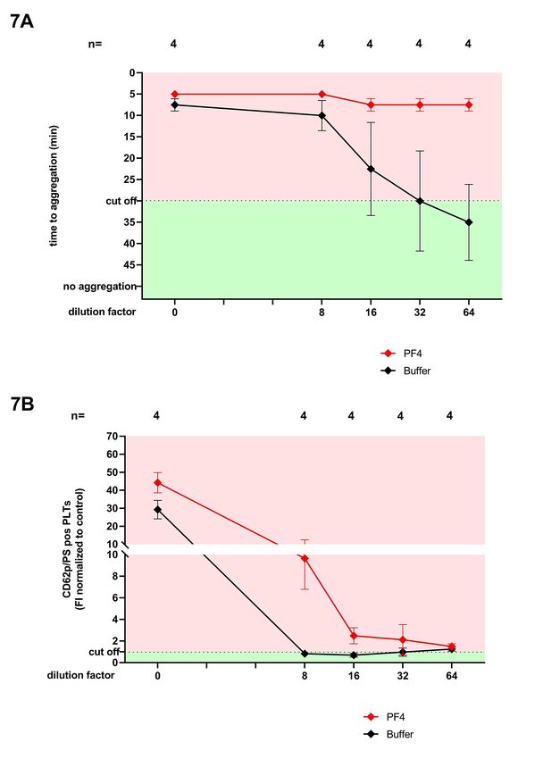
Acknowledgments

This work was supported by grants from the German Research Foundation and from the

Herzstiftung to T.B. (BA5158/4 and TSG-Study), by special funds from the state of Baden-

Württemberg for autopsy-based COVID-19 research and the DEFEAT PANDEMIcs network

funded by the BMBF to P.M. and FF. J.A.M is supported with a grant from German Research

Foundation. We thank our technicians Karoline Weich, Simone Weit, Franziska Lyshy, Marco

Mikus and Flavianna Rigoni for their excellent technical support. We also thank Harald Klüter

and Martin Holderried for vaccines.

Authorship contributions

K.A., P.M., U.J.S., F.F., and T.B. designed the study. J.S., M.G., M.P., M.B., A.B., H.B., F.D.,

A.V., H.H., N.H., K.K., C.L., B.L., J.M., M.M., S.N., C.S., B.P., U.J.S., M.W. and G.C.P. were

responsible for the treatment of the patients and collected and analyzed the clinical data.

T.B., G.U., K.A. and F.F. reviewed medical reports. K.A., G.U., J.A.M., A.S. and L.P.

performed the experiments. W.A.W., D.V., analyzed the radiological images. H.B., V.W., F.F.,

and P.M. analyzed the autopsy findings and performed tissue studies. H.J. produced Spike

protein. K.A., L.P., A.S., G.U., N.S., F.F. and T.B. analyzed the data, interpreted the results

and wrote the manuscript. All authors read and approved the manuscript.

Conflict of interest disclosures

The authors declare no competing financial interests.

Key points

   1- Vaccine-induced immune thrombotic thrombocytopenia (VITT) after ChAdOx1 nCoV-

       19 vaccination is associated with antibody-mediated procoagulant platelets

   2- Thrombosis of venous, arterial, and arteriolar vessels, including cerebral sinuses and

       glomeruli leads to fatal outcome in patients with VITT

                                                                                                   3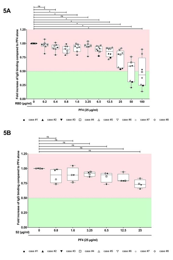
Abstract

The COVID-19 pandemic has resulted in significant morbidity and mortality worldwide. To

prevent severe infection, mass COVID-19 vaccination campaigns with several vaccine types

are currently underway. We report pathological and immunological findings in 8 patients who

developed vaccine-induced immune thrombotic thrombocytopenia (VITT) after administration

of SARS-CoV-2 vaccine ChAdOx1 nCoV-19. We analyzed patient material using enzyme

immune assays, flow cytometry and heparin-induced platelet aggregation assay and

performed autopsies on two fatal cases. Eight patients (5 female, 3 male) with a median age

of 41.5 years (range, 24 to 53) were referred to us with suspected thrombotic complications

6 to 20 days after ChAdOx1 nCoV-19 vaccination. All patients had thrombocytopenia at

admission. Patients had a median platelet count of 46.5 x10⁹/L (range, 8 to 92). Three had a

fatal outcome and 5 were successfully treated. Autopsies showed arterial and venous

thromboses in various organs and the occlusion of glomerular capillaries by hyaline thrombi.

Sera from VITT patients contain high titer antibodies against platelet factor 4 (PF4) (OD

2.59±0.64). PF4 antibodies in VITT patients induced significant increase in procoagulant

markers (P-selectin and phosphatidylserine externalization) compared to healthy volunteers

and healthy vaccinated volunteers. The generation of procoagulant platelets was PF4 and

heparin dependent. We demonstrate the contribution of antibody-mediated platelet activation

in the pathogenesis of VITT.

                                                                                              4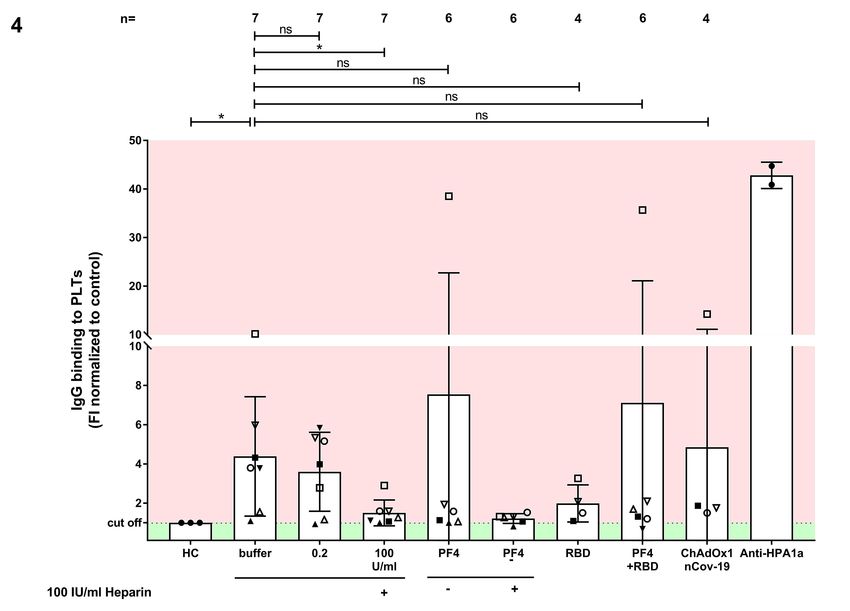
Introduction:

COVID-19 infection has resulted in considerable morbidity and mortality in the last 15

months.1 Within an exceptionally short time, several SARS-CoV-2 vaccines have been

licensed and used worldwide.2 Safety signals have been, however, noted. Center for disease

control and prevention (CDC) in the US reported in the beginning of March 26 cases of

venous thromboembolism, 20 cases of thrombosis and 41 ischemic strokes in individuals

vaccinated with mRNA vaccines in the US. More than 200 cases with thrombosis among 34

million persons vaccinated with ChAdOx1 nCoV-19 have been reported to European

database of suspected adverse reactions, EudraVigilance. After the investigation of reported

cases, European Medical Association (EMA) found a link between ChAdOx1 nCoV-19 and

unusual thrombotic events and concomitant thrombocytopenia. Although WHO and EMA

concluded that the benefit of vaccination with ChAdOx1 nCoV-19 outweighs the risks

associated with thrombosis and thrombocytopenia, several countries instituted restrictions

on the use of ChAdOx1 nCoV-19. The unusual clinical constellation of cerebral venous sinus

thrombosis (CVST) and thrombocytopenia is called vaccine-induced immune thrombotic

thrombocytopenia (VITT). We studied 8 cases with thrombocytopenia and primarily with

suspected CVST but also other thromboembolic complications to better understand the

pathophysiology of the VITT. In this study, we identified antibody-mediated procoagulant

platelets as a novel mechanism associated with VITT.

Methods:

Study cohort and evaluation of the clinical data

8 patients were referred to different university hospitals with neurological or hematological

symptoms after vaccination with ChAdOx1 nCoV-19 (AstraZeneca, London, UK) between

February 1st and April 6th 2021. Six patients admitted to university hospitals and two patients,

who admitted fırst to local hospitals, were later transferred to university hospital. Medical

records were used to collect treatments and outcome. Diagnosis of thromboembolic

                                                                                                1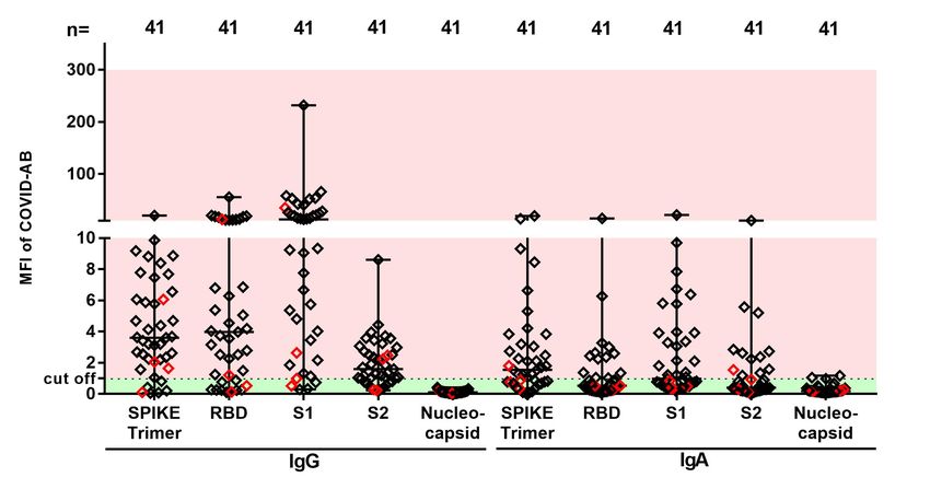
complications was made when indicated by clinical or laboratory findings and/or based on

computed tomography, ultrasound imaging or in case of death by autopsy.

       Blood samples were collected to exclude heparin-induced thrombocytopenia (HIT).

Blood samples from non-vaccinated healthy blood donors (n=24, 17 females, mean age

36.1±13.7 years) and from healthy vaccinated before and after the first vaccination with

ChAdOx1 nCoV-19 (n=41, 29 females, mean age 37.3 ± 10.9 years) served as health

controls.

       In addition, sera from 29 COVID-19 patients who had serial HIT EIA measurements

during hospitalization were also included in the study (7 females, mean age 65.3 ± 14.1

years). Clinical data from the ICU COVID-19 patients and a VITT-patient (case #7) were

reported in previous studies.3, 4

Bead-based multiplex assay for detection of COVID-19 antibodies

COVID-19 antibodies were measured with a multiplex assay (NMI, Reutlingen, Germany)

with the FLEXMAP 3D® system (Luminex Corporation, Austin, USA).5

Testing for anti-PF4/heparin antibodies

A commercially available IgG-Enzyme Immune Assay (EIA) was used in accordance to

manufacturer’s instructions (Hyphen Biomed, Neuville-sur-Oise, France). The ability of sera

to activate platelets was tested using the functional assay heparin induced platelet

aggregation assay (HIPA) as previously described.6 For more details, see supplemental

material.

Serological characterization of PF4-antibodies

Antibody binding to PF4 and Receptor Binding Domain of Spike Protein (Spike-RBD and S2

domaine) was analyzed using an In-House EIA.

Assessment of antibody-mediated procoagulant platelets

                                                                                              2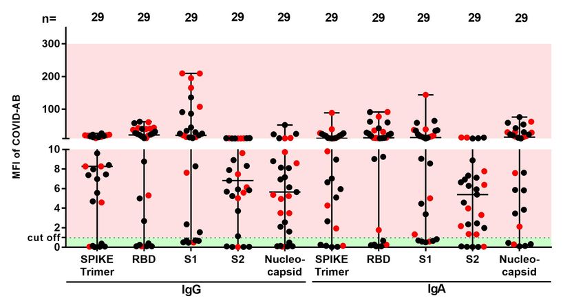
Patients’ sera were incubated with washed platelets (7.5x10⁶) for 1.5 h* under different

conditions at RT. Platelets were then stained with Annexin V-FITC and CD62-APC

(Immunotools, Friesoythe Germany) and directly analyzed by flow cytometry (FC). For more

details, see supplemental material.

Ethics Statement

The study was conducted in accordance with the declaration of Helsinki. The study protocol

was approved by the Institutional Review Board of the University of Tuebingen

(236/2021BO2, 224/2021BO2) and analysis of sera from ChAdOx1 nCoV-19 vaccinated

individuals were University of Ulm (99/21).

Statistical analyses

The statistical analysis was performed using GraphPad Prism, Version 7.0 (GraphPad, La

Jolla, USA). Since potential daily variations in FC measurements might result in bias in data

analysis, test results were normalized to two healthy donors tested in parallel at the same

time point (raw data are available in the supplemental data). Data in the text are presented

as median (range), mean ± standard deviation (SD) or n (%).

Data sharing statement

Data may be requested for academic collaboration from the corresponding author.

Results

Clinical and laboratory features of VITT

Eight patients (5 female, 3 male) with a median age of 41.5 years (range, 24 to 53) were

referred with suspected thrombotic complications after ChAdOx1 nCoV-19 vaccination.

Demographic data are summarized in Table 1. The patients were admitted to the hospital 6

to 20 days after ChAdOx1 nCoV-19 vaccination. All patients had thrombocytopenia at

admission with a median platelet count of 46.5 x10⁹/L (range, 8 to 92). D-dimer was

                                                                                                3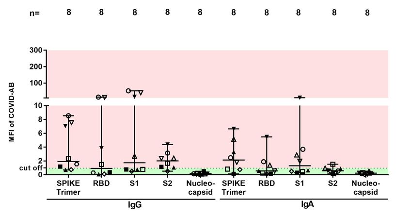
available in 5 patients, which was 9 µg/ml or higher. Thrombosis was detected in 6 patients
at admission and 2 developed thrombosis during hospitalization (Figure 1). Thrombotic
events included cerebral venous sinus thrombosis (5 patients), pulmonary embolism (4
patients), deep vein thrombosis (1 patient), and thrombosis in other organs (3 patients).
Three of 8 patients had more than one thrombotic event. Three patients presented initially
with bleeding signs with easy bruising and petechiae, which might be an early sign of VITT.
One patient underwent a successful thrombus removal by endovascular rheolysis. Three of
8 patients died (on day 6 [case #1], day 10 [case #2] and day 7 [case #3] of hospitalization).
All surviving patients received anticoagulation. Four patients received IVIG combined with
non-heparin anticoagulation.
Pathological findings
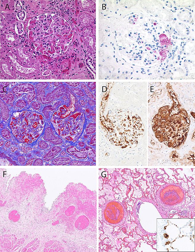
Autopsy was performed in 2 of 3 deceased patients. Autopsy of Case #2 showed complete
thrombotic obstruction of the straight, sagittal and transversal cerebral sinuses,
subarachnoidal haemorrhage, cerebral edema and bilateral pulmonary embolism in mid-
sized arteries and obstruction of glomerular arterioles and capillaries by hyaline
microthrombi containing fibrin and platelets (Figure 2 Panel A and B). Autopsy of Case #3
showed massive cerebral hemorrhage and cerebral edema, bilateral pulmonary
thromboembolism and obstruction of glomeruli by hyaline microthrombi (Figure 2 Panel C-
G). Histology of the bone marrow was normal in both cases without any hint of increased
thrombopoesis.
IgG binding profile of sera from VITT
High titer PF4/heparin antibodies were detected in all sera (8/8 100%) using the IgG
PF4/heparin EIA. Interestingly, binding of all sera was inhibited in the presence of high
concentration of heparin (mean optical density [OD] of IgG antibodies against PF4/heparin
complexes: 2.591±0.642 vs. 0.176±0.073, respectively, p < 0.0001, Figure 3A). No
correlation was found between the PF4/heparin antibodies and the detected COVID-19
antibodies in VITT patients and in vaccinated controls (Supplemental Figure 1 A-D [I-IV]).
4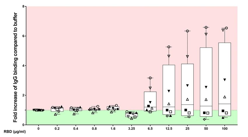
Among non-vaccinated controls only one subject (4%) had a PF4/heparin antibody in EIA

(Data not shown).

We next investigated the PF4-seroconversion after vaccination with ChAdOx1 nCoV-19, as

well as during severe SARS-CoV-2 infection (Figure 3 B). We found that 4 out of 41 (9.8%)

vaccinated healthy individuals and 4 out of 41 (9.8%) patients with severe COVID-19

seroconverted with IgG antibodies against PF4/heparin complexes within 14 days (Figure

3B). Next, we tested IgG binding to platelets by FC. An increase in IgG binding to test

platelets was observed (Fold increase in mean fluorescence intensity compared to healthy

controls [FI of MFI]: 4.39±1.15 vs. 1±1.10, p value 0.026, Figure 4, supplemental Figure 2A).

IgG binding to platelets was inhibited by heparin at high concentrations (FI of MFI IgG

binding: 1.51±0.66, p value 0.016), but not at low concentrations (FI of MFI of IgG binding:

3.60±2.01, p value 0.688). Only one serum showed increased binding to platelets in the

presence of PF4 and the vaccine ChAdOx1 nCoV-19 (case #4, Figure 4). Spike-RBD did not

induce a significant change in IgG binding in VITT patients (Figure 5A). Similar results were

observed when S2 protein is added (Figure 5B). IgG binding was also observed when sera

from ChAdOx1 nCoV-19 vaccinated volunteers with IgG PF4 antibodies were tested

(Supplemental Figure 2B). However, severe COVID-19 patients with IgG PF4 antibodies

showed no increase in IgG binding (Supplemental Figure 2C).

The impact of Spike-RBD on the binding of anti-PF4 antibodies

Compared to healthy controls, sera from VITT patients showed a strong binding to PF4 in

the in-house EIA (OD IgG antibodies against PF4: 1.03±0.04 vs. 0.110±0.002, respectively, p

value
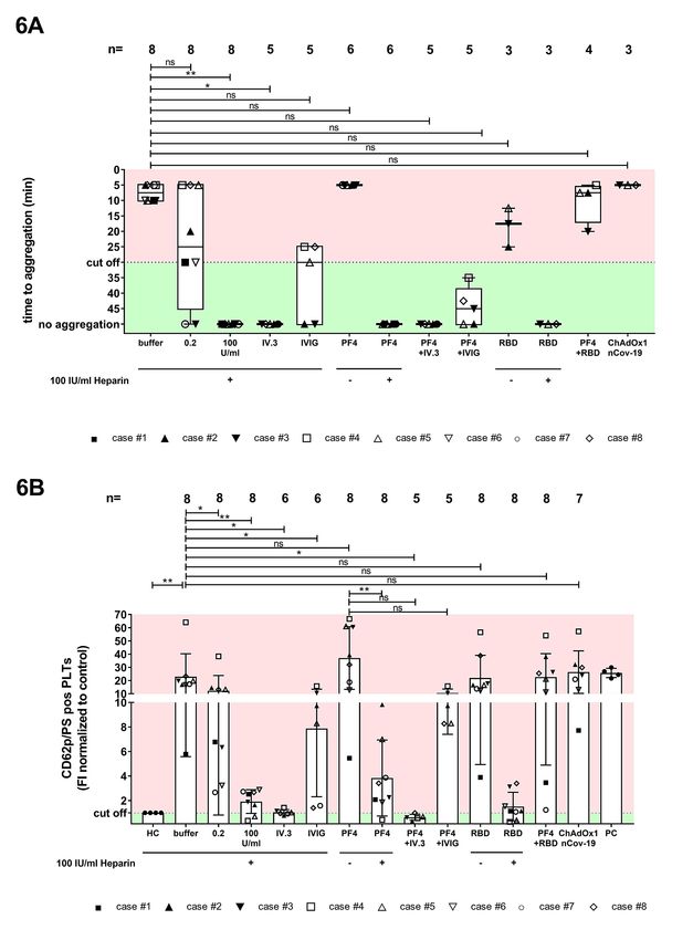
To investigate the ability of patients, sera to activate platelets, the HIPA assay was used with

several modifications. Sera were incubated with washed platelets in the presence of I) buffer,

II) 0.2 IU/mL LMWH, III) 100 IU/mL UFH, IV) an Fc gamma receptor IIa (FcγRIIA)-blocking

monoclonal antibody (mAb IV.3), VI) 30mg/mL IVIG, VII) 25 µg/mL PF4, VIII) 50 µg/mL

Spike-RBD, IX) PF4/Spike-RBD complexes, X) PF4+RBD or XI) ChAdOx1 nCoV-19 (XII).

Conditions with PF4 and RBD were also repeated in the presence of high concentration of

heparin (100 IU/mL UFH). We observed platelet activation in presence of buffer in 8/8 VITT

patients (Median time to platelet aggregation: 5, 5-10 minutes (min*), Figure 6A), but not in

sera from vaccinated individuals with anti PF4 antibodies, who did not develop any clinical

sign of thromboembolic complications, without side effects (Supplemental Figure 3A).

Moreover, only one of the sera from patients with severe COVID-19 who were tested positive

in PF4/heparin EIA showed platelet activation in the HIPA assay. (Supplemental Figure 3B).

Interestingly, the reaction was weaker in presence of low molecular weight heparin (Median

time to aggregate: 5 min, 5-10 min (no aggregation) vs. 30 min, 5->45min, Figure 6A). All

reactions were inhibited by high dose of heparin (p value 0.008, Figure 6A). In presence of

PF4, sera from VITT patients showed strong platelet activation (Median time to aggregate: 5

min, 5-5 min, Figure 6A). Most importantly, platelet activation was completely inhibited by the

mAb IV.3 that blocks the FcγRIIa and by high doses of IgG (>45 min, no aggregation, Figure

6A). No significant change was found when PF4/RBD-complexes were added. Antibody-

mediated platelet activation was inhibited when low molecular weight heparin (LMWH) was

added at low concentrations by testing three sera. When sera from VITT patients were

diluted, specific binding was observed to PF4 while no reaction in the buffer was found

(Figure 7A).

Sera of VITT patients induce PF4-dependent procoagulant phenotype

To explore the mechanism of coagulation dysregulation in VITT, sera were incubated with

washed platelets from healthy donors in the presence of buffer, heparin, mAb IV.3, IVIG,

PF4, PF4+IVIG, PF4+RBD, the Spike-RBD protein or the vaccine ChAdOx1 nCoV-19. FC

                                                                                                6
analyses revealed that sera from VITT patients induce remarkable changes in the
distribution of CD62p/PS positivity (FI CD62p/PS positive PLTs: 22.94±6.14 vs. 0.90±0.63,
respectively, p=0.009, Figure 6B, Supplemental Figure 4). In contrast, the platelet population
was almost non-affected after incubation with sera from vaccinated controls (Supplemental
Figure 5A). Interestingly, the generation of procoagulant platelets was reduced by 0.2 IU/mL
LMWH (FI CD62p/PS positive PLTs: 13.32±11.50, p=0.016, Figure 6B) and completely
inhibited by high concentration of unfractionated heparin (UFH) in VITT patients (FI
CD62p/PS positive PLTs: 1.92±0.96, p= 0.008, Figure 6B, supplemental Figure 4). These
reactions were inhibited by the FcγRIIA blocking with mAb IV.3 as well as by high
concentrations of IgG (FI CD62p/PS positive PLTs: 1.04±0.22, p= 0.031 and FI CD62p/PS
positive PLTs: 7.88±5.56, p= 0.031, respectively, Figure 6B). No significant increase of
procoagulant platelets was also observed in presence of PF4 (FI CD62p/PS positive PLTs:
37.07±23.73, p=0.078). and in the presence of Spike-RBD alone (FI CD62p/PS positive
PLTs: 22.02±17.09, p=0.195). While increased generation of procoagulant platelets was
observed after incubation of sera from severe COVID-19 patients, no significant change was
observed when sera from vaccinated individuals with anti-PF4 antibodies were tested
(Supplemental Figure 5A-B).
To identify the target antigen of the platelet activating antibodies, the HIPA and FACS testing
were repeated at different titrations of sera from VITT patients. Interestingly, diluted sera
(from 1:64) were able to activate platelets and induce procoagulant phenotype only in the
presence e of PF4 (Figure 7A and B, respectively).
Discussion
The increasing number of reports on rare thrombotic events after SARS-CoV-2 vaccination
draw public attention and led to concerns regarding the safety of this vaccine due to the
uncertainty of the origin of these undesired reactions.7-9 To understand the pathophysiology
of this phenomenon, the so-called vaccine-induced immune thrombotic thrombocytopenia
7(VITT), we analyzed sera from 8 patients. Our mostly young, generally fit cohort of patients,
presented acutely with atypical thrombosis, primarily, but not exclusively involving the
cerebral venous sinuses, an extremely rare manifestation of thrombosis in the general
population. All cases developed symptoms within 6-20 days after the ChAdOx1 nCoV-19
vaccination showing a temporal relationship between vaccination and symptoms. The main
findings in these cases were thrombocytopenia, high D-dimer, low fibrinogen, and high titer
IgG antibodies against PF4 that can induce procoagulant platelet phenotype.
After intensive laboratory investigations of the VITT cases, we were able to identify
the serological profile of the pathological antibodies. In a small cohort of vaccinated
volunteers, approximately 10% of the individuals developed IgG antibodies against
PF4/polyanion complexes within 14 days after the first vaccination; none of them was
exposed to heparin in the past 100 days. We observed that IgG binding to PF4 in these sera
as well as in VITT sera can be inhibited by heparin but also by increasing the concentration
of Spike-RBD. These data may suggest that these antibodies are specific for conformational
changes in PF4 that might be induced by negatively charged structures. Of note, no
significant IgG binding to platelets was observed in the presence of the vaccine ChAdOx1
nCoV-19. Accordingly, it is very unlikely that Vector (pCDNA4) may be responsible for the
high PF4-seroconversion rate in vaccinated individuals. Comparable data were reported by
Greinacher et al.7 and Schultz et al.9 in two very recent reports that appeared while our
manuscript was in preparation. In addition to their observations, we were also able to
demonstrate that sera from VITT patients directly induce procoagulant platelets, suggesting
a possible mechanism for thrombotic events seen in patients with VITT. This is further
corroborated by the pathological studies in two of our patients. Despite the distinct
immediate causes of death in these two fatal cases, namely fatal cerebral sinus thrombosis
and intracerebral hemorrhage, the two autopsy reports showed striking similarities. In
addition to arterial, arteriolar and venous thrombosis in various organs and pulmonary
thromboembolism, both cases showed a striking occlusion of multiple glomeruli and afferent
arterioles by hyaline thrombi composed of fibrin and platelets, but lacking erythrocytes. The
8kidney morphology bears resemblance to thrombotic microangiopathy, but we failed to
identify erythrocyte fragmentation, a key feature of thrombotic microangiopathy.10 Both
patients, however, had normal kidney function (highest creatinine level 0.5 mg/dL in Case #2
and 0.8 mg/dL in Case#3) until briefly before death, indicating rapid pre-terminal
development of glomerular microthrombosis. White thrombi have been associated with
antibody-mediated platelet activation.10, 11
Our data indicate that IgG antibodies against PF4 increase generation of
procoagulant platelets in VITT. However, we cannot exclude other co-factor(s) that could
also induce thromboembolic complications in vivo. We report on VITT after ChAdOx1 nCoV-
19, which is the only SARS-CoV-2 vaccine that includes a simian adenovirus. Disturbances
of platelets have been described in association with the intravenous administration of
adenovirus gene therapy vectors although it is unclear how that might relate to isolated
thrombocytopenia as an adverse event of the vaccine.8
Finally, the observed clinical and laboratory features of the VITT are exceptional and
rare. Therefore, the value of COVID-19 vaccination to provide critical protection should be
considered higher compared to significant health risk of COVID-19. With the better
recognition of this rare complication and the availability of efficient therapies, the risk-benefit
ratio of ChAdOx1 nCoV-19 might be reconsidered further.
Conclusion
Although the incidence of VITT after ChAdOx1 nCoV-19 vaccination is very low, the
mortality rate is high (37% in our case series). Since a global vaccination campaign is
underway and large numbers of people will be vaccinated, an increase in the number of
people with this side effect is to be expected, highlighting the importance of a better
understanding of the pathophysiology of VITT. In this study, we present immunological and
pathological findings in patients with VITT. Furthermore, we show the contribution of
antibody-mediated platelet activation in the pathogenesis of VITT.
9References:
1. Grasselli G, Greco M, Zanella A, et al. Risk Factors Associated With Mortality
Among Patients With COVID-19 in Intensive Care Units in Lombardy, Italy. JAMA
Intern Med. 2020;180(10):1345-1355.
2. Castells MC, Phillips EJ. Maintaining Safety with SARS-CoV-2 Vaccines. N
Engl J Med. 2021;384(7):643-649.
3. Althaus K, Marini I, Zlamal J, et al. Antibody-induced procoagulant platelets in
severe COVID-19 infection. Blood. 2021;137(8):1061-1071.
4. Wolf ME, Luz B, Niehaus L, Bhogal P, Bazner H, Henkes H.
Thrombocytopenia and Intracranial Venous Sinus Thrombosis after "COVID-19
Vaccine AstraZeneca" Exposure. J Clin Med. 2021;10(8):1599.
5. Becker M, Strengert M, Junker D, et al. Exploring beyond clinical routine
SARS-CoV-2 serology using MultiCoV-Ab to evaluate endemic coronavirus cross-
reactivity. Nat Commun. 2021;12(1):1152.
6. Greinacher A, Michels I, Kiefel V, Mueller-Eckhardt C. A rapid and sensitive
test for diagnosing heparin-associated thrombocytopenia. Thromb Haemost.
1991;66(6):734-736.
7. Greinacher A, Thiele T, Warkentin TE, Weisser K, Kyrle PA, Eichinger S.
Thrombotic Thrombocytopenia after ChAdOx1 nCov-19 Vaccination. N Engl J Med.
2021 Apr 9. [Epub ahead of print]
8. Othman M, Labelle A, Mazzetti I, Elbatarny HS, Lillicrap D. Adenovirus-
induced thrombocytopenia: the role of von Willebrand factor and P-selectin in
mediating accelerated platelet clearance. Blood. 2007;109(7):2832-2839.
9. Schultz NH, Sorvoll IH, Michelsen AE, et al. Thrombosis and
Thrombocytopenia after ChAdOx1 nCoV-19 Vaccination. N Engl J Med. 2021 Apr 9.
[Epub ahead of print]
10. Brocklebank V, Wood KM, Kavanagh D. Thrombotic Microangiopathy and the
Kidney. Clin J Am Soc Nephrol. 2018;13(2):300-317.
11. Pierangeli SS, Espinola RG, Liu X, Harris EN. Thrombogenic effects of
antiphospholipid antibodies are mediated by intercellular cell adhesion molecule-1,
vascular cell adhesion molecule-1, and P-selectin. Circ Res. 2001;88(2):245-250.
10Table 1: Demographic and clinical data of cases with vaccine induced immune thrombotic thrombocytopenia
Case age sex first thrombotic risk PLT, D-Dimer, Fibrinogen, INR aPTT,
# symptoms factors (150- (40s)
after 450 µg/ml) mg/dl)
vaccination Thrombosis x109/L)
(days) /Bleeding
1 47 f 7 CVST none 10 >35 128 1.30 23
2 48 f 6 CVST, PE n.a. 40 n.a. n.a. 1.16 22.9
3 24 m 10 bleeding, heterozygous FVL 22 n.a. 109 1.20 42
multiple mutation
thrombosis
4 53 m 9 DVT, PE none 8 >35 126 1.01 25
5 47 f 7 CVST none 56 9 263 1.25 35
6 32 m 20 PE none 71 n.a. n.a. n.a. n.a.
7 36 f 17 CVST none 92 13 n.a. 1.19 22
8 29 f 7 CVST contra-ception 53 32 274 1.00 23
CVST indicates Cerebral venous sinus thrombosis; DVT, deep vein thrombosis; FVL, Factor V Leiden; n.a., not available; PE, pulmonary
embolism; PLT, platelet.
11Figure Legends:
Figure 1: Imaging example of three illustrative cases
Imaging examples of Case #1 (A-C), Case #8 (D-F) and Case #4 (G, H). In case 1, non-
enhanced computed tomography imaging (A) showed a parenchymal and subdural
hemorrhage (arrows in A), causing a midline shift (arrowheads in A). Digital subtraction
angiography was performed (B) showing thrombosis of the right sigmoid and transverse
sinus, superior sagittal sinus (arrows in B), and straight sinus. Angiography after mechanical
recanalization (C) shows the recanalized cerebral sinuses (superior sagittal sinus marked
with arrows). In case #8, cerebral imaging 7 days after vaccination was unremarkable
(curved reconstruction of the left transverse and sigmoid sinus shown in the right upper
corner of D and F). She worsened, which led to a repeated cerebral imaging, showing a
large intraparenchymal hemorrhage in the left temporal lobe (arrow in E), causing midline
shift (arrowhead in E), caused by a thrombosis of the transverse and sigmoid sinus (arrows
in F), as well as of the adjacent tentorial veins. In case #4, a thrombus in the right pulmonary
artery was observed (arrows in E; coronal reconstruction shown in the right lower corner of
G). Further imaging also revealed thrombi in the femoral veins on both sides (arrows in H).
Figure 2: Histopathological findings in Case #2 and Case #3
Case #2. Occlusion of glomerular capillary loops by hyaline thrombi. Haematoxylin-Eosin
stain, original magnification 200x (Panel A). Deposition of platelets in glomerular vessels
documented by CD42b staining. (Immunoperoxidase, 200x) (Panel B).
Case #3. Occlusion of glomerular capillary loops by hyaline thrombi with fibrin deposits
highlighted in red (Masson’s trichrome stain, 200x) (Panel C). Immunostaining for CD61
(Panel D) and fibrin (Panel E) demonstrate the massive intravascular deposits of fibrin and
platelets (Immunoperoxidase, 200x). Thrombotic occlusions of submucosal vessels in the
urinary bladder with hemorrhage. H&E, 40x (Panel F)Thrombotic occlusion of medium-sized
12pulmonary vessels. H&E, 40x. Insert shows platelet deposits in pulmonary capillaries stained for CD61 (Immunoperoxidase, 400x) (Panel G). Figure 3: Binding profile of sera from VITT Results of the PF4/heparin enzyme linked immunosorbent assay (EIA) in patients with VITT with and without 100 IU/ml heparin (Panel A). All VITT patients showed an enhanced binding which was significantly inhibited at high dose of heparin (100 IU/ml). PF4-seroconversion after vaccination and severe SARS-CoV2 infection was followed up (Panel B). IgG PF4/Heparin antibody binding results in healthy volunteers before and after vaccination and COVID-19 patients in ICU showed four vaccinated volunteers displaying a positive EIA result after 7-14 days post-vaccination (red empty diamonds). Figure 4: IgG binding to platelets by flow cytometry in sera of VITT patients IgG binding to healthy washed PLTs after incubation with sera from VITT patients was measured (assessed by flow cytometry and expressed as fold increase normalized to controls). VITT patients showed significantly higher binding at the baseline in comparison to healthy controls, which was inhibited by high dose heparin. Abbreviation: ns: not significant, *p
Results of the platelet activation assay (HIPA) with modifications in the VITT-patients. Each

dot represents the median of four different donors. All VITT patients presented platelet

activation with buffer alone, which was significantly increased by PF4 heparin but inhibited

with high dose of heparin (Panel A). Procoagulant platelets (CD62P/Phosphatidylserine (PS)

positive) cells in different settings were analyzed via Annexin V-FITC and CD62p-APC

antibody staining. Where indicated, PLTs were treated with PF4, 0.2U/ml and 100U/ml

heparin, RBD and ChAdOx1 nCoV-19A (Panel B). Data are presented as mean ± standard

deviation (SD) of the measured fold increase compared to control, not significant, *p
Antibody-mediated procoagulant platelets in SARS-CoV-2-

vaccination associated immune thrombotic

thrombocytopenia

Supplemental material

Methods:

Testing for anti-PF4/heparin antibodies

A commercially available IgG-Enzym Immune assay (EIA) was used in accordance to

manufacturer’s instructions (Hyphen Biomed, Neuville-sur-Oise, France). Per manufacturer’s

recommendation, a sample was considered reactive if the optical density (OD) was greater

than 0.500. The ability of sera to activate platelets was tested using the functional assay

Heparin induced platelet aggregation assay (HIPA). In brief, serum was tested with washed

platelets from four different healthy donors in the absence (buffer alone) or in the presence

of heparin (0.2 IU/mL and 100 IU/mL). In addition, platelets were preincubated with PF4

(50µg/mL), Spike protein (50 µg/mL), a mixture of PF4 and Spike Receptor Binding Domain

(RBD) (50µg/mL and 0.82 µg/mL, respectively) or vaccine (1:750). To verify the charge

dependency, these conditions were also repeated in the presence of high concentration of

heparin (100 IU/mL UFH). Reactions were placed in microtiter wells containing spherical stir

bars and stirred at approximately 500 revolutions per minute (rpm). Wells were examined

optically at five-minutes interval for loss of turbidity. A serum was considered reactive

(positive) if a shift from turbidity to transparency occurred within 30 min in at least two

platelet suspensions. Observation time was 45 min. Each test included a diluted serum from

a patient with heparin induced thrombocytopenia (HIT) as a weak positive control, collagen

(5µg/mL) as strong positive control and a serum from a healthy donor as a negative control.

                                                1
Serological characterization of PF4-antibodies
Patient sera were tested for anti-PF4/RBD antibodies using an in-house PF4/RBD enzyme-
linked immunosorbent assay (EIA). In brief, PF4 (25 μg/ml) was incubated with or without
varying concentrations of RBD in carbonate coating buffer (0.05 M NaH2PO4, 0.1 % NaN3)
at 4 °C, overnight in a 96 well microtitre plate (Nunc MaxiSorp™, Langenselbold, Germany).
Then, plates were washed three times with PBS/Tween buffer (0.05 % Tween 20 in PBS)
before blocking with 3% BSA for 2h, at RT or overnight at 4 °C. Plates were again washed
three times and incubated (RT, 1 h) with patient sera (diluted 1:50 in PBS/Tween buffer). After
further washing three times, plates were incubated (RT, 1 h) with 1:1000 diluted peroxidase-
conjugated anti-human IgG (Jackson ImmunoResearch Laboratories Inc, USA). After five
times washing, plates were incubated (RT, 6 minutes [min], dark) with substrate
tetramethylbenzidine (TMB one, Kementec, Denmark). The reaction was stopped with H2SO4
and the absorbance was measured at 450 nm with 620 nm as a reference.
Assessment of antibody-mediated procoagulant platelets
To exclude unspecific effects like the activation of platelets via complement or non-specific
immune complexes, all sera were heat-inactivated (56°C for 30 min*), followed by a sharp
centrifugation step at 5,000g. The supernatant was collected. All experiments involving
patients’ sera were performed after incubation of 5 µL serum with 25 µL washed platelets
(7.5x10⁶) for 1.5 h* under rotating conditions at RT. When indicated, cell susspenions were
preincubated with PF4 (25 µg/ml), Spike protein (0-100 µg/mL) or vaccine (1:75, V:V).
Afterwards, samples were washed once (7 min*, 650g, RT, without brake) and gently
resuspended in 75 µL of phosphate-buffered saline (PBS, Biochrom, Berlin, Germany).
Platelets were then stained with Annexin V-FITC and CD62-APC (Immunotools, Friesoythe
Germany) and directly analyzed by flow cytometry (FC). As positive control, washed platelets
were incubated with ionomycin (5µM, 15 min at RT) and TRAP-6 (10 µM, 30 min at RT). Test
results were determined as fold increase of the percentage of double PS/CD62p positive
2events in platelets upon incubation with patients’ sera compared to cells incubated with
healthy donors tested in parallel.
Results
Response to SARS-CoV-2 vaccination
We compared three groups with antibody response to COVID-19 vaccination or infection
(Supplemental Figure 2 A-C). All VITT patients presented beginning immune response to the
vaccine in IgG and IgA (Supplemental Figure 2A). Nevertheless case #2 was negative with
all antigens. 6/8 patients presented antibodies against the Spike Trimer. For IgG only 4/8
patients presented antibodies against RBD and S1 domain. Most of the patients (6/8)
presented antibodies against the S2 domain. No antibodies were detected against the
nucleocapsid, so previous infection with SARS-CoV2 can be excluded. For IgA 5/8 patients
presented antibodies against the Spike Trimer, 3/8 presented antibodies against the RBD
and 4/8 against the S1 domain. Only case #4 presented IgA antibodies against the S2
domain and no antibodies were detected against the nucleocapsid structure. Immune
response in IgG was not different compared to the vaccinated volunteers (Spike, p=0.478;
RBD, p=0.215; S1, p=0.247; S2, p=0.639; Nucleocapsid, p=0.339). Compared to COVID-19
patients from ICU IgG antibodies against the Spike protein and S1 domain were not
significantly different (Spike, p=0.0753; S1, p=0.0659). Significant difference was found in
antibodies against RBD (p=0.139), S2 domain (p=0.0355) and IgG antibodies against the
nucleocapsid (p=0.0036).
3Supplemental figure legends: Supplemental Figure 1: Serum anti-COVID IgG and IgA antibody levels and correlation with HIT-EIA This figure shows the MFI of anti-COVID IgG and IgA antibodies values for Spike Trimer, RBD, S1, S2 and nucleocapsid quantified by using a bead-based Luminex assay in VITT patients (Panel A); vaccinated volunteers (Panel B) and COVID-19 patients (Panel C). Anti-PF4 antibody levels did not correlate with anti-COVID antibodies in VITT patients and vaccinated volunteers (Panel D). Each symbol shows an individual subject and numbers of tested subjects is indicated in the graphic. Supplemental Figure 2: IgG binding to healthy PLTs Panel A shows a representative flow cytometry histogram of AHG binding. The AHG- binding was measured in presence of buffer and high concentration of heparin (100 IU/ml heparin) after incubation of platelets from healthy donors (HC) with serum of VITT patients. The other panels show IgG binding to healthy PLTs (assessed by flow cytometry and expressed as fold increase normalized to controls) after incubation with sera from vaccinated volunteers (Panel B) and COVID-19 patients (Panel C). Where indicated, PLTs were treated with PF4, 0.2U/mL and 100U/mL Heparin, RBD and the vaccine ChAdOx1 nCoV-19. (Abbreviation: ns: not significant, and **p
SARS-CoV-2 S2 was not observed in VITT patients (Panel E). Each symbol

represents individual subject and the number subjects tested is reported in each

graphic.

Supplemental Figure 3: Heparin-induced platelet activation assay

(HIPA)

This figure shows the results of the platelet activation assay (HIPA) with

modifications in presence or absence of PF4, 0.2U/ml and 100U/ml Heparin, RBD

and vaccine ChAdOx1 nCoV-19. None of the subjects showed any platelet activation

under any conditions except one in presence of Spike RBD in vaccinated controls

(Panel A). Similar results were seen for COVID-19 patients except one patient who

showed enhanced activation in 2 out of 4 donors in the presence of 0.2U/ml heparin

(*p
Procoagulant platelets (CD62P/Phosphatidylserine (PS) positive cells in different

settings with sera from vaccinated volunteers (Panel A) and COVID-19 patients with

anti PF4 antibodies (Panel B). Data are presented as mean ± standard deviation

(SD) of the measured fold increase compared to control, not significant, and *p
Supp Figure 1A
■ case #1
▲ case #2
▼ case #3
case #4
△ case #5
▽ case #6
○ case #7
◇ case #8Supp Figure 1B
◇ vaccinated volunteers ◇ vaccinated volunteers EIA positiveSupp Figure 1C
● severe COVID-19 ● severe COVID-19 EIA positiveSupp Figure 1D
◇ vaccinated volunteers ◇ vaccinated volunteers EIA positive ◆ VITT EIA positiveSupp Figure 2A
case #8
◇ (buffer)
◇ case #8
(ChAdOx1nCov-19)
case #8
◇ (100 U/ml Heparin)
count
◇ case #8
(10 µg PF4)
● HCSupp Figure 2B
◇ vaccinated volunteers
EIA positiveSupp Figure 2C
● severe COVID-19 EIA positiveSupp Figure 2D
■ case #1
▲ case #2
▼ case #3
case #4
△ case #5
▽ case #6
○ case #7
◇ case #8Supp Figure 2E
▲ case #2
▼ case #3
case #4
○ case #7Supp Figure 3A
◇ vaccinated volunteers EIA positiveSupp Figure 3B
● severe COVID-19 EIA positiveSupp Figure 4
I buffer HC II buffer case #8 0.2 heparin case #8 IV 100 Heparin case #8
III
V IV.3 case #8 VI IVIG case #8 VII PF4 case #8 VIII IV.3+PF4 case #8Supp Figure 5A
◇ vaccinated volunteers
EIA positiveSupp Figure 5B
● severe COVID-19 EIA positiveYou can also read