Risk Factors and Anticoagulation Therapy in Patients With Isolated Distal Deep Vein Thrombosis in the Early Post-operative Period After Thoracic ...
←
→
Page content transcription
If your browser does not render page correctly, please read the page content below
ORIGINAL RESEARCH
                                                                                                                                              published: 28 April 2021
                                                                                                                                     doi: 10.3389/fsurg.2021.671165

                                             Risk Factors and Anticoagulation
                                             Therapy in Patients With Isolated
                                             Distal Deep Vein Thrombosis in the
                                             Early Post-operative Period After
                                             Thoracic Surgery
                                             Yuping Li 1 , Junrong Ding 1 , Lei Shen 1 , Jian Yang 1 , Haifeng Wang 1 , Yiming Zhou 1 ,
                                             Gening Jiang 1 , Yuming Zhu 1 and Yin Wang 2*
                                             1
                                              Department of General Thoracic Surgery, Shanghai Pulmonary Hospital, Tongji University School of Medicine, Shanghai,
                                             China, 2 Department of Ultrasound, Shanghai Pulmonary Hospital, Tongji University School of Medicine, Shanghai, China

                                             Background: Isolated distal deep vein thrombosis (IDDVT) accounts for ∼50% of all
                                             patients diagnosed with deep venous thrombosis (DVT), but the diagnosis and optimal
                         Edited by:          management of IDDVT remains unclear and controversial. The aim of this study was to
                       Marco Scarci,
                                             explore potential risk factors and predictors of IDDVT, and to evaluate different strategies
            San Gerardo Hospital, Italy
                                             of anticoagulation therapy.
                       Reviewed by:
                    Federico Raveglia,       Methods: A total of 310 consecutive patients after thoracic surgery, who underwent
                    ASST-Monza, Italy
                    Fabrizio Minervini,
                                             whole-leg ultrasonography as well as routine measurements of D-dimer levels before and
    University of Lucerne, Switzerland       after surgery were evaluated. The general clinical data, anticoagulant therapy, pre- and
                  *Correspondence:           postoperative D-dimer levels were collected. Differences between IDDVT, DVT and
                           Yin Wang
                                             non-DVT groups were calculated. Logistic regression analysis was used to analyze risk
                    lpbbl@aliyun.com
                                             factors of postoperative IDDVT.
                    Specialty section:       Results: Age and postoperative D-dimer levels were significantly higher in IDDVT group
          This article was submitted to
                       Thoracic Surgery,     than in non DVT group (p = 0.0053 and p < 0.001, respectively). Logistic regression
                a section of the journal     analysis showed that postoperative D-dimer level was a significant independent
                    Frontiers in Surgery
                                             predictor of IDDVT even when adjusted for age and operation method (p = 0.0003).
         Received: 23 February 2021
                                             There were no significant side effects associated with both full-dose and half-dose
            Accepted: 01 April 2021
            Published: 28 April 2021         anticoagulation regimens. Half-dose therapy was associated with a significant decrease
                              Citation:      in the requirement for anticoagulation medications after discharge (p = 0.0002).
Li Y, Ding J, Shen L, Yang J, Wang H,
  Zhou Y, Jiang G, Zhu Y and Wang Y
                                             Conclusion: Age and D-dimer levels after surgery are strong predictors of IDDVT
               (2021) Risk Factors and       following thoracic surgery. Half-dose therapeutic anticoagulation has the same efficiency
   Anticoagulation Therapy in Patients
                                             in preventing IDDVT progression, is not associated with any additional risks of adverse
         With Isolated Distal Deep Vein
                Thrombosis in the Early      effects compared to a full-dose regimen, and may be adopted for treating IDDVT patients
 Post-operative Period After Thoracic        after thoracic surgery.
       Surgery. Front. Surg. 8:671165.
      doi: 10.3389/fsurg.2021.671165         Keywords: anticoagulation therapy, IDDVT, D-dimer, thoracic surgery, risk factors

Frontiers in Surgery | www.frontiersin.org                                         1                                            April 2021 | Volume 8 | Article 671165
Li et al. Risk Factors and Anticoagulation Therapy
INTRODUCTION requirement was waived as this study was a retrospective
study of consecutive patients that underwent thoracic surgery
Deep venous thrombosis (DVT), a subset of venous for diagnosed thoracic tumors of different etiology from
thromboembolism (VTE), is a common complication of July to November 2018. Patients were subjected to complete
surgery with postoperative occurrence ranging from 0.7% to as compression ultrasonography (31) on the 1st day after surgery.
high as 48% (1–4). Almost 50% of DVT patients will develop
post-thrombotic syndrome symptoms, such as pain, swelling Patients
and venous ulcers in the most severe cases (5, 6). However, the Study period lasted from July 2018 to November 2018. All
key risk of untreated DVT is developing pulmonary embolism patients that underwent thoracic surgery during this time, a total
(PE) that occurs in up to one-third of cases and is the primary of 310 consecutive patients, were selected as potential candidates
contributor to DVT-related mortality (7). DVTs are classified for the study. A power analysis has been performed to ensure that
based on the anatomical site of involved venous segments. A the number of the included patients was sufficient. Forty-seven
proximal DVT may involve popliteal, femoral, and iliac veins, patients were excluded due to pre-existing DVT or PTE before
and the inferior vena cava (IVC) (8), while isolated distal deep surgery, or missing follow up data. Based on CUS examination
vein thrombosis (IDDVT), or calf DVT, that represents around of the compressibility of the whole leg’s veins, the remaining 263
30–50% of all lower-limb DVTs, does not extend to proximal patients were divided into non DVT and DVT groups. Of all
veins (9–11). Although there is evidence that IDDVT may patients with suspected DVT, 33 patients (84.6%), were further
spontaneously resolve (12), there are reports of it extending diagnosed with IDDVT based on the location of the detected
proximally and leading to PE with a combined risk of proximal thrombi. Plasma D-dimer levels of patients were assessed before
propagation and PE ranging between 7.8 and 11.4% (13–15). and after surgery, and clinical data were collected at follow up.
Proximal DVT and PE have been extensively studied, and
guidelines for their diagnosis and therapeutic anticoagulant Anticoagulation Therapy
management are well-established and optimized. According to All DVT patients received anticoagulation therapy after initial
the American College of Chest Physicians (ACCP) guidelines diagnosis. IDDVT patients were divided into two groups.
(16), DVT diagnosis is based on the Wells scoring system, which In Q12H group (n = 17) patients were treated with 6000
incorporates medical history and physical examination (17). international units subcutaneous injection of low molecular
Factors, such as advanced age, cancer, immobilization, recent weight heparin (LMWH) once every 12 h during hospitalization.
trauma or surgery, and hospitalization are all considered risk In QD group (n = 16) patients were treated with half-
factors for DVT progression into venous thromboembolism (18, dose therapeutic anticoagulation (4000–6000 IU once-daily
19). Risk stratification of DVT is further achieved by measuring administration of LMWH) during hospitalization.
serum levels of D-dimer, a degradation product of cross-linked After the discharge, patients were assessed by the physician,
fibrin. D-dimer levels are typically elevated in patients with acute and the risk of VTE was determined using the Caprini risk
thrombosis because of simultaneous activation of coagulation assessment score (32). If indicated by the physician, patients
and fibrinolysis (20). continued anticoagulation treatment at home using the following
High risk of IDDVT proximal extension is associated with regiments: Rivaroxaban (15 mg twice a day for the first 3 weeks,
thrombosis that is extensive or close to the proximal veins, followed by 20 mg once a day for a total of about 3 months);
unprovoked DVT, active cancer and inpatient status (21) Pradaxa (150 mg, twice a day for 3 months) or Aspirin (100 mg,
(Kearon). Although elevated D-dimer levels are associated with once a day for 3 months).
IDDVT, the usefulness of D-dimer in the diagnosis of IDDVT Follow up ultrasound was performed 1, 3, and 6 months
is limited with reported poor sensitivity (22). There have been after discharge.
only a few randomized clinical trials (RCTs) evaluating benefits
of anticoagulation therapy for IDDVT that yielded conflicting Statistical Analysis
results (14, 23–28). Statistical analyses were performed using R Software Version
One approach is to subject all the patients with suspected 3.5.3 (33). Binary and nominal discrete data was presented as
DVT to imaging of both the proximal and the calf veins, and to counts (% of total) and comparisons were made using chi-
treat diagnosed IDDVT patients with anticoagulant therapy (29). square tests. Continuous data was presented as mean (± standard
Another approach uses serial imaging of proximal veins only, deviation) for normally distributed variables and median (IQR)
thus leaving IDDVT untreated (30). The need to test and treat for non-parametric variables and comparisons were made using
IDDVT, therefore, remains a debated issue. two-tailed independent samples t-tests or Mann-Whitney U
In the recent study, we explore the potential risk factors tests, respectively. For variables with comparisons that were
of IDDVT following thoracic surgery and evaluate optimal significant, multiple logistic regression was used to compute odds
strategies of preventive anticoagulation therapy. ratios for predicting IDDVT with 95% confidence intervals to
adjust for demographic and clinical variables. Among these, Age
MATERIALS AND METHODS (1 case) and D-Dimer (2 cases) had missing data that were
imputed with regression imputation using formulas derived from
This investigation was approved by the Institutional Review the remaining demographic variables. For all tests, p < 0.05 were
Board of Shanghai Pulmonary Hospital. Informed consent considered statistically significant.
Frontiers in Surgery | www.frontiersin.org 2 April 2021 | Volume 8 | Article 671165Li et al. Risk Factors and Anticoagulation Therapy
RESULTS A diagnosis of lower limb DVT was established in 39 out of
263 patients (13.6%) based on the results of CUS. Comparison
Characteristics of Study Participants of baseline clinical characteristics of both DVT and non-DVT
Study design is shown in Figure 1. Of 331 patients considered for groups is shown in Table 2. DVT patients were significantly older
eligibility, 47 were excluded due to the previous history of VTE (mean age (± SD) of 66 (±8) vs. 58 (±2) years in non DVP group,
and insufficient follow up data. p = 0.001), had higher postoperative D-dimer levels (median
Baseline characteristics of the remaining 263 patients are (IQR) of 695 (1224) ng/mL as compared to 416 (353) ng/mL in
summarized in Table 1. The mean age of the patients was 58.8 non-DVT group, p = 0.012), and required longer hospitalization
years old, with BMI within the normal range. (9 (±8) days vs. 8 (±4) days in non-DVT group, p = 0.002).
Most of the patients (224, 85.2%) were diagnosed with Of 39 patients with established DVT diagnosis, 33 (84.6%)
malignant tumors, while 39 (14.8%) were diagnosed with were diagnosed with IDDVT. As summarized in Table 2, there
benign tumors. Out of 263 patients, 223 (84.7%) underwent was a significant correlation between the age of the patients
video-assisted thoracoscopic surgery (VATS), and 40 patients and IDDVT diagnosis (p = 0.0053). Similarly, IDDVT patients
(15.2%) underwent thoracotomy. exhibited elevated serum D-dimer levels post-surgery [median
FIGURE 1 | Study flow diagram. PTE, pulmonary thromboembolism; DVT, deep venous thrombosis; IDDVT, isolated distal deep vein thrombosis; Q12H, every 12 h;
QD, once-daily.
Frontiers in Surgery | www.frontiersin.org 3 April 2021 | Volume 8 | Article 671165Li et al. Risk Factors and Anticoagulation Therapy
(IQR=Q3-Q1) of 705 (1025) ng/mL as compared to 418 (348) after surgery was used as an independent variable and
ng/mL in the non-DVP group, p < 0.0001]. These results suggest subsequently adjusted for other significant and borderline-
that age and post-operative D-dimer levels were strongly and significant parameters, such as age, gender, and operative method
significantly associated with IDDVT. (VATS vs. thoracotomy). As shown in Table 4, D-dimer remained
Since elevated D-dimer levels are associated with various a strong predictor of IDDVT when adjusted for age [OR (95% CI)
pathological conditions, including cancer, we next evaluated 1.04 (1.01–1.09), p = 0.002], gender [OR (95% CI) 0.38 (0.16–
post-surgery D-dimer concentrations in patients, diagnosed 0.88), p = 0.021] or operative method alone [OR (95% CI) 8.41
with malignancies. As shown in Table 3, similarly to the (1.25–56.75), p = 0.028]. When all the variables were combined
general patient population, post-surgery D-Dimer was in a final model, gender was no longer a significant factor, with
significantly higher among patients with either DVT or OR (95% CI) of 0.44 (0.18–1.03), p > 0.05.
IDDVT outcomes (median 705 and 708, respectively) than those
without DVT (p < 0.0001).
Cut-Off Value of Post-operative D Dimer
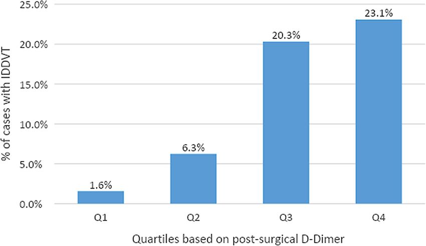
D-dimer as a Predictor of IDDVT We next analyzed the distribution of IDDVT rates according to
We next performed predictive analysis of the variables associated post-operative D-Dimer concentrations (Figure 2). Only 1.6%
with IDDVT incidence using logistic regression. D-dimer of patients with D-dimer values in the lowest quartile range
(0–306 ng/mL) had IDDVT diagnosis. IDDVT diagnosis rates
increased to 6.3% in the second (306–438 ng/mL) quartile. The
TABLE 1 | Baseline characteristics of patients.
most significant rates of IDDVT were associated with the third
Variables Data (N = 263) (438–717 ng/mL) and fourth (>717 ng/mL) quartiles of D-dimer
levels (20.3 and 23.1% of all cases, respectively).
Gender Male 125 (47.6) The predictive effect of D-Dimer on IDDVT was further
Female 138 (52.4) evaluated by calculating the odds ratio (unadjusted/adjusted for
Age* Years 58.8 (±10.1) age and operative method) for each quartile. As shown in Table 5,
BMI* kg/m2 23.5 (±2.7) cases in the lowest quartile of D-dimer have a very low risk of
Operative VATS 223 (84.7) IDDVT, both unadjusted and adjusted [OR (95% CI) 0.08 (0.01–
Method Thoracotomy 40 (15.2) 0.60), p = 0.0138, and OR (95% CI) 0.08 (0.01–0.62), p = 0.0157,
Cancer Malignant 224 (85.2) respectively]. The risk of IDDVT increases notably in the third
Histology Benign 39 (14.8) quartile [OR (95% CI) 2.20 (1.03–4.74), p = 0.0428 unadjusted,
and OR (95% CI) 2.32 (1.05–5.15), p = 0.0379 adjusted for age
Data are presented as No. (%) or *mean (± SD). and operation method]. Similar correlation between the risk of
TABLE 2 | Descriptive statistics with comparison of IDDVT and non-DVT cases.
Unit Non-DVT DVT IDDVT DVT IDDVT
(N = 224) (N = 39) (N = 33) vs. non-DVT vs. non-DVT
Gender† Male 109 (48.7) 16 (44.4) 11 (33.3) 0.378 0.0994
Female 115 (51.3) 23 (59.0) 22 (66.7)
Age‡ Years 58 (±11.8) 64 (±8.6) 64 (±9.1) 0.002* 0.0053*
BMI‡ kg/m2 23.3 (±3.1) 23.8(±2.54) 23.7 (±2.5) 0.206 0.5307
Operation time§ Min 120 (86–150) 127 (102–176) 127 (105–170) 0.482 0.4777
Blood loss in surgery§ mL 50 (50) 50 (50) 50 (50) 0.525 0.5687
D-Dimer before surgery§ ng/mL 133 (96–188) 170 (103–255) 170 (103–260) 0.113 0.2150
D-Dimer after surgery§ ng/mL 418 (298–646) 695 (479–1,597) 705 (472–1,497) 0.001*Li et al. Risk Factors and Anticoagulation Therapy
TABLE 3 | Comparison of post-surgical D-Dimer between DVT, IDDVT, and non-DVT cases among total cancer patients (N = 223).
Unit Non-DVT DVT IDDVT DVT vs. non-DVT IDDVT vs. non-DVT
(N = 188) (N = 35) (N = 30)
D-Dimer after surgery§ ng/mL 415 (299–651) 705 (501–1,780) 708 (479–1,646)Li et al. Risk Factors and Anticoagulation Therapy
FIGURE 2 | Rates of IDDVT by quartile of post-surgical D-Dimer. 1st quartile (0–306 ng/mL) had 1 case among 64 patients. 2nd quartile (306–438 ng/mL) had 4
cases among 64 patients. 3rd quartile (438–717 ng/mL) had 13 cases among 64 patients. 4th quartile (>717 ng/mL) had 15 cases among 65 patients.
those aged 45–54 years (40–43). The D-dimer levels as a TABLE 5 | Quartiles of post-surgical D-Dimer as predictors of IDDVT after thoracic
common screening test for DVT, has been shown to be highly surgery.
sensitive (>95%) in excluding acute VTE at the cutoff value Model OR (95% CI) p
of 500 ng/mL (44). However, elevated D-dimer levels are not
unique to venous thrombosis, and may rise with advanced D-Dimer 1st quartile† 0.08 (0.01–0.60) 0.0138*
age (>65), and in various pathologic conditions including D-Dimer 2nd quartile† 0.38 (0.13–1.12) 0.0784
malignancy (45, 46). Our data indicate that the postoperative D-Dimer 3rd quartile† 2.20 (1.03–4.74) 0.0428*
level of D-dimer in DVT and IDDVT patients with malignant D-Dimer 4th quartile† 2.90 (1.36–6.16) 0.0056*
tumors is still significantly higher (1.68- and 1.7-fold increase, D-Dimer 1st quartile‡ 0.08 (0.01–0.62) 0.0157*
respectively) than that before surgery, confirming the specific D-Dimer 2nd quartile‡ 0.35 (0.11–1.05) 0.0600
association of higher post-surgery D-dimer levels with DVT D-Dimer 3rd quartile‡ 2.32 (1.05–5.15) 0.0379*
in our study. D-Dimer 4th quartile‡ 2.93 (1.33–6.46) 0.0075*
The reports of the sensitivity and diagnostic accuracy of D-
dimer level measurements for IDDVT are scarce. In a large Models are calculated as odds ratios using logistic regression, with p < 0.05* considered
study of 393 outpatients with clinically suspected symptomatic statistically significant for each coefficient.
†
Unadjusted odds ratios are for each quartile of D-Dimer test results predicting IDDTV.
DVT (47–51), the D-dimer levels in patients with IDDVT ‡ Odds ratios, adjusted for age and operative method.
were, on average, almost twice as high for patients at least 60
years old compared with those younger than 60 years (median
D-dimer, 0.82 vs. 0.47). IDDVT patients in our study were
older (average of 64 years as compared to 58 in non-DVT its significance. Interestingly, among demographic variables in
group) and had over 1.6-fold higher postoperative D-dimer our study, gender had a borderline significant association with
serum concentrations. Since D-Dimer after surgery showed the IDDVT (IDDTV vs. non-DTV, p = 0.0994). Previous studies
strongest association with IDDVT incidence in our study, it was showed that male gender was related to a higher recurrence risk
used as an independent variable, and maintained significance of unprovoked DVT (52), but the overall role of gender in the
in all models, when adjusted for each one of the following causation of DVT is unclear. Borderline statistical significance
parameters: age, gender and operative method. However, when observed may be due, therefore, to the small sample size of
all 4 variables were combined in a final model, gender lost IDDVT patients in our study (n = 33).
Frontiers in Surgery | www.frontiersin.org 6 April 2021 | Volume 8 | Article 671165Li et al. Risk Factors and Anticoagulation Therapy
TABLE 6 | IDDVT statistics and anticoagulation therapy follow-up results.
Standard Q12H Half-dose QD Q12H vs. QD
(N = 17) (N = 16)
Multiple 3 (17.6) 1 (6.3)
Duration of admission§ days 10 (6–12) 8 (7–12) 0.7718
Duration of anticoagulation§ days 99 (8–103) 5 (4–13) 0.0040*
Total drainage§ mL 620 (450–900) 720 (450–993) 0.9283
Chest tube indwelling§ Days 4 (3–6) 5 (4–5) 0.6384
Average daily drainage‡ mL 177.9 (83.6) 162.9 (91.9) 0.6271
Medication after discharge† Medication 13 (76.5) 2 (12.5) 0.0002*
None 4 (23.5) 14 (87.5)
Progression of DVT† Progression 0 (0.0) 0 (0.0) N/A
None 17 (100.0) 16 (100.0)
Adverse events† Adverse events 0 (0.0) 1 (6.3) N/A
None 17 (100.0) 15 (93.8)
All variables compared between standard therapeutic anticoagulation with 6000 IU low-molecular-weight heparin (LMWH) twice per day (Q12H) and half-dose with 6000 IU LMWH
once per day (QD), with p < 0.05* considered statistically significant.
†
Binary or nominal discrete variables presented as N (%) and compared using a chi-square test.
‡ Normally distributed continuous variables were presented as mean (±SD) and compared using an independent samples t-test.
§ Non-parametric continuous variables were presented as median (IQR) and compared using a Mann-Whitney U test. Medication after discharge includes prescriptions for one of the
following anticoagulants: Rivaroxaban, Pradaxa, and Aspirin. Progression of DVT includes proximal progression, progression to pulmonary embolism, or recurrent VTE. Adverse events
include major bleeding, clinically relevant non-major bleeding, pulmonary embolism, or all-cause mortality (case listed was non-major bleeding).
D-dimer measurements appear to be less sensitive for the Postoperative bleeding is an important complication of
diagnosis of IDDVT than for the proximal DVT, with some pharmacoprophylaxis and may lead to serious complications
IDDVT patients having D-dimer levels below the usual cutoff and even death. Few studies have evaluated the risk of
value of 500 ng/mL (48, 49, 53). In our study the majority of postoperative bleeding with or without pharmacoprophylaxis
IDDVT incidences (over 40% combined) were associated with in an attempt to develop guidelines for proper prophylactic
the D-dimer levels in the ranges of 438–717 and over 717 ng/mL. strategies for VTE in general surgery (non-cancer-related).
Both these D-dimer ranges were significant predictors of IDDTV These guidlines take into account patient and surgery-specific
when adjusted for age and surgery method, with OR (95% CI) risks with consideration of the estimated bleeding risk. For
2.32 (1.05–5.15) and OR (CI 95%) 2.93 (1.33–6.46), respectively. instance, low-risk general abdominopelvic, gynecological and
Levels below 438 ng/mL were not predictive of IDDVT. These urological surgeries require either no prophylaxis or mechanical
results indicate that while D-dimer levels after surgery can quite prophylaxis only. In contrast, minimal prophylaxis requirement
reasonably predict the risk of IDDVT, the cutoff of elevated risk is for low-risk orthopedic, thoracic, neurologic, and vascular
not high, and may be just below 500 ng/mL. Further studies with surgeries includes mechanical prophylaxis (such as compression
larger cohorts of IDDVT patients are merited to define the exact stockings, active compression devices, and venous foot pumps)
cutoff of D-dimer levels. with/without pharmacological anticoagulation therapy (57).
Magnitude of surgical intervention have been shown to Recent randomized trials failed to reach a consensus on the
correlate with VTE recurrence (54). best approach for treating IDDVT due to variations in study
Patients who underwent a high-risk procedure had a 34% design, duration of treatment, and follow up (14, 23, 26–28,
incidence of DVT, while low-risk procedures were associated 58). The latest American College of Chest Physicians guidelines
with only 11% incidence. Interestingly, our study indicates that suggest anticoagulation therapy for IDDVT patients who are
Video-Assisted Thoracoscopic Surgery (VATS) (compared to symptomatic or high-risk for extension (based on criteria such
thoracotomy) can increase the risk of IDDVT [OR (95% CI) 8.41 as serum D-dimer levels, extensive disease, or proximity to
(1.25–56.75)]. Several large studies that addressed incidences of proximal veins, unprovoked DVT, malignancy, history of VTE,
VTE in patients following VATS, reported a relative decrease and inpatient status). All other cases are to be monitored by serial
in risk for vein thrombosis associated with this method, as ultrasound examinations (21, 59). The efficacy of this approach
compared to thoracotomy (55, 56). However, according to in treating IDDVT is confirmed by a number of studies and
these studies, surgical approach cannot serve as an independent trials (13, 26–28, 60–62). Longer treatment duration appears
predictor of VTE (56). In our study, a small sample size results to be necessary in higher risk subgroups of IDDVT patients,
in a very large confidence interval, and thus greater uncertainty but whether a treatment duration of 3 months is sufficient
associated with the estimated OR. Further studies with higher to prevent recurrences in these subgroups is unclear (63, 64).
sample sizes are needed to fully evaluate this potential effect of In our study, there was no difference in any of the adverse
the type of surgery on the risk of IDDVT. effects associated with DVT progression, such as bleeding, PE
Frontiers in Surgery | www.frontiersin.org 7 April 2021 | Volume 8 | Article 671165Li et al. Risk Factors and Anticoagulation Therapy
or mortality in patients, receiving half-dose (QD) of full dose IDDVT patients after thoracic surgery. Further large randomized
(Q12H) of anticoagulation therapy. Both groups reported similar controlled studies with higher sample sizes are needed to evaluate
volumes of average daily drainage, and requirement for chest the precise cut-off levels of post-operative D-dimer, and to
tube indwelling. Unsurprisingly, the duration of anticoagulation develop a standard anticoagulation program for IDDVT patients
time differed significantly between Q12H and QD groups, with with close follow-up.
median 99 and 5 days, respectively. However, the treatment
regimen correlated with the rate of medication required after DATA AVAILABILITY STATEMENT
the discharge. About one third of postoperative VTE events
occur after discharge, and patients who are at increased risk The raw data supporting the conclusions of this article will be
may benefit from extended anticoagulation prophylaxis (35). made available by the authors, without undue reservation.
Current guidelines recommend 7–10 days of total postoperative
prophylaxis for cancer surgery patients. The majority (76.5%) of ETHICS STATEMENT
the patients in the Q12H group were treated with anticoagulation
treatment at home, as compared to only 12.5% of patients in the All procedures performed in studies involving human
QD group. participants were in accordance with the ethical standards
Our study has several limitations. The most significant factor, of the ethics committee of Shanghai Pulmonary Hospital and
potentially impacting the outcomes of the study, is a small cohort with the 1964 Helsinki declaration and its later amendments
of DVT and IDDVT patients. This paper is a retrospective or comparable ethical standards (K20-454). Informed consent
study, which was originally an objective observation, not requirement was waived as this study was a retrospective study
a clinical study of intervention measures. Additionally, the of consecutive patients that underwent thoracic surgery for
anticoagulation regimen of study participants was inconsistent diagnosed thoracic tumors of different etiology from July to
and not standardized, especially the duration of anticoagulation. November 2018.
In conclusion, in the recent study, we show that the
D-dimer levels after surgery and the age of the patient are AUTHOR CONTRIBUTIONS
strong predictors of IDDVT following thoracic surgery. Half-
dose therapeutic anticoagulation is not associated with any YW and YL: conception and design. JY, GJ, YZhu, HW, and
additional risks of adverse effects, and is as efficient as a YZho: provision of study materials or patients. YL, JD, and
full-dose regimen in preventing IDDVT progression. Half- LS: collection and assembly of data. YL: data analysis and
dose therapy and ultrasound surveillance instead of prolonged interpretation. All authors: manuscript writing and final approval
full dose therapeutic anticoagulation might be an option for of manuscript.
REFERENCES inadequacy of the term “Proximal DVT.” Eur J Vasc Endovasc Surg. (2016)
51:415–20. doi: 10.1016/j.ejvs.2015.11.001
1. White RH. The epidemiology of venous thromboembolism. Circulation. 9. Elias A, Mallard L, Elias M, Alquier C, Guidolin F, Gauthier B, et al. A
(2003) 107:I4–8. doi: 10.1161/01.CIR.0000078468.11849.66 single complete ultrasound investigation of the venous network for the
2. Ambrosetti M, Salerno M, Zambelli M, Mastropasqua F, Tramarin R, diagnostic management of patients with a clinically suspected first episode
Pedretti RFE. Deep vein thrombosis among patients entering cardiac of deep venous thrombosis of the lower limbs. Thromb Haemost. (2003)
rehabilitation after coronary artery bypass surgery. Chest. (2004) 125:191– 89:221–7. doi: 10.1055/s-0037-1613435
6. doi: 10.1378/chest.125.1.191 10. Stevens SM, Elliott CG, Chan KJ, Egger MJ, Ahmed KM. Withholding
3. Goldhaber SZ, Schellong S, Kakkar A, Eriksson H, Feuring M, Kreuzer anticoagulation after a negative result on duplex ultrasonography for
J, et al. Treatment of acute pulmonary embolism with dabigatran suspected symptomatic deep venous thrombosis. Ann Intern Med. (2004)
versus warfarin. A pooled analysis of data from RE-COVER and RE- 140:985–91. doi: 10.7326/0003-4819-140-12-200406150-00007
COVER II. Thromb Haemost. (2016) 116:714–21. doi: 10.1160/TH16-04- 11. Schmidt B, Michler R, Klein M, Faulmann G, Weber C, Schellong S.
0271 Ultrasound screening for distal vein thrombosis is not beneficial after major
4. Reis SE, Polak JF, Hirsch DR, Cohn LH, Creager MA, Donovan orthopedic surgery. A randomized controlled trial. Thromb Haemost. (2003)
BC, et al. Frequency of deep venous thrombosis in asymptomatic 90:949–54. doi: 10.1160/TH03-03-0154
patients with coronary artery bypass grafts. Am Heart J. (1991) 122:478– 12. Garry J, Duke A, Labropoulos N. Systematic review of the complications
82. doi: 10.1016/0002-8703(91)91004-7 following isolated calf deep vein thrombosis. Br J Surg. (2016) 103:789–
5. Galanaud J-P, Sevestre M-A, Genty C, Kahn SR, Pernod G, Rolland C, et al. 96. doi: 10.1002/bjs.10152
Incidence and predictors of venous thromboembolism recurrence after a 13. De Martino RR, Wallaert JB, Rossi AP, Zbehlik AJ, Suckow B, Walsh DB. A
first isolated distal deep vein thrombosis. J Thromb Haemost. (2014) 12:436– meta-analysis of anticoagulation for calf deep venous thrombosis. J Vasc Surg.
43. doi: 10.1111/jth.12512 (2012) 56:228-237.e1; discussion 236–237. doi: 10.1016/j.jvs.2011.09.087
6. Kahn SR, Shrier I, Julian JA, Ducruet T, Arsenault L, Miron M-J, 14. Horner D, Hogg K, Body R, Nash MJ, Baglin T, Mackway-Jones K.
et al. Determinants and time course of the postthrombotic syndrome The anticoagulation of calf thrombosis (ACT) project: results from
after acute deep venous thrombosis. Ann Intern Med. (2008) 149:698– the randomized controlled external pilot trial. Chest. (2014) 146:1468–
707. doi: 10.7326/0003-4819-149-10-200811180-00004 77. doi: 10.1378/chest.14-0235
7. Kearon C. Natural history of venous thromboembolism. Circulation. (2003) 15. Palareti G, Cosmi B, Lessiani G, Rodorigo G, Guazzaloca G, Brusi C, et al.
107:I22–30. doi: 10.1161/01.CIR.0000078464.82671.78 Evolution of untreated calf deep-vein thrombosis in high risk symptomatic
8. De Maeseneer MGR, Bochanen N, van Rooijen G, Neglén P. Analysis of 1,338 outpatients: the blind, prospective CALTHRO study. Thromb Haemost. (2010)
patients with acute lower limb deep venous thrombosis (DVT) supports the 104:1063–70. doi: 10.1160/TH10-06-0351
Frontiers in Surgery | www.frontiersin.org 8 April 2021 | Volume 8 | Article 671165Li et al. Risk Factors and Anticoagulation Therapy
16. Kearon C, Akl EA, Ornelas J, Blaivas A, Jimenez D, Bounameaux H, et al. patients with resectable lung cancer. J Thorac Cardiovasc Surg. (2016)
Antithrombotic therapy for VTE disease: chest guideline and expert panel 151:37–44.e1. doi: 10.1016/j.jtcvs.2015.08.039
report. Chest. (2016) 149:315–52. doi: 10.1016/j.chest.2015.11.026 36. Trinh VQ, Karakiewicz PI, Sammon J, Sun M, Sukumar S, Gervais
17. Wells PS. Integrated strategies for the diagnosis of venous M-K, et al. Venous thromboembolism after major cancer surgery:
thromboembolism. J Thromb Haemost. (2007) 5(Suppl 1):41– temporal trends and patterns of care. JAMA Surg. (2014) 149:43–
50. doi: 10.1111/j.1538-7836.2007.02493.x 9. doi: 10.1001/jamasurg.2013.3172
18. Goldhaber SZ, Bounameaux H. Pulmonary embolism and deep vein 37. Blom JW, Doggen CJM, Osanto S, Rosendaal FR. Malignancies,
thrombosis. Lancet. (2012) 379:1835–46. doi: 10.1016/S0140-6736(11)61904-1 prothrombotic mutations, and the risk of venous thrombosis. JAMA.
19. Goldhaber SZ. Venous thromboembolism: epidemiology and (2005) 293:715–22. doi: 10.1001/jama.293.6.715
magnitude of the problem. Best Pract Res Clin Haematol. (2012) 38. Agnelli G, Verso M. Management of venous thromboembolism
25:235–42. doi: 10.1016/j.beha.2012.06.007 in patients with cancer. J Thromb Haemost. (2011) 9(Suppl
20. Wang K-L, Chu P-H, Lee C-H, Pai P-Y, Lin P-Y, Shyu K-G, et al. Management 1):316–24. doi: 10.1111/j.1538-7836.2011.04346.x
of venous thromboembolisms: part I. The consensus for deep vein thrombosis. 39. Kwaan HC, Parmar S, Wang J. Pathogenesis of increased risk of thrombosis in
Acta Cardiol Sin. (2016) 32:1–22. doi: 10.6515/acs20151228a cancer. Semin Thromb Hemost. (2003) 29:283–90. doi: 10.1055/s-2003-40966
21. Kearon C. Diagnosis of suspected venous thromboembolism. 40. Tsai AW, Cushman M, Rosamond WD, Heckbert SR, Polak JF, Folsom AR.
Hematol Am Soc Hematol Educ Program. (2016) 2016:397– Cardiovascular risk factors and venous thromboembolism incidence: the
403. doi: 10.1182/asheducation-2016.1.397 longitudinal investigation of thromboembolism etiology. Arch Intern Med.
22. Sartori M, Cosmi B, Legnani C, Favaretto E, Valdré L, Guazzaloca (2002) 162:1182–9. doi: 10.1001/archinte.162.10.1182
G, et al. The wells rule and D-dimer for the diagnosis of 41. Silverstein MD, Heit JA, Mohr DN, Petterson TM, O’Fallon WM, Melton
isolated distal deep vein thrombosis. J Thromb Haemost. (2012) LJ. Trends in the incidence of deep vein thrombosis and pulmonary
10:2264–9. doi: 10.1111/j.1538-7836.2012.04895.x embolism: a 25-year population-based study. Arch Intern Med. (1998)
23. Lagerstedt C, Olsson CG, Fagher B, Oqvist B, Albrechtsson 158:585–93. doi: 10.1001/archinte.158.6.585
U. Oral anticoagulants in calf-vein thrombosis. Lancet. (1985) 42. Khorana AA, Francis CW, Culakova E, Lyman GH. Risk factors for
2:1311–12. doi: 10.1016/S0140-6736(85)91600-9 chemotherapy-associated venous thromboembolism in a prospective
24. Lagerstedt CI, Olsson CG, Fagher BO, Oqvist BW, Albrechtsson U. Need observational study. Cancer. (2005) 104:2822–9. doi: 10.1002/cncr.21496
for long-term anticoagulant treatment in symptomatic calf-vein thrombosis. 43. Al Diab AI. Cancer-related venous thromboembolism: insight into
Lancet. (1985) 2:515–8. doi: 10.1016/S0140-6736(85)90459-3 underestimated risk factors. Hematol Oncol Stem Cell Ther. (2010)
25. Nielsen HK, Husted SE, Krusell LR, Fasting H, Charles P, Hansen HH, et al. 3:191–5. doi: 10.5144/1658-3876.2010.191
Anticoagulant therapy in deep venous thrombosis. A randomized controlled 44. Righini M, Perrier A, De Moerloose P, Bounameaux H. D-Dimer for venous
study. Thromb Res. (1994) 73:215–26. doi: 10.1016/0049-3848(94)90100-7 thromboembolism diagnosis:20 years later. J Thromb Haemost. (2008) 6:1059–
26. Righini M, Galanaud J-P, Guenneguez H, Brisot D, Diard A, Faisse P, et al. 71. doi: 10.1111/j.1538-7836.2008.02981.x
Anticoagulant therapy for symptomatic calf deep vein thrombosis (CACTUS): 45. Prisco D, Grifoni E. The role of D-dimer testing in patients with
a randomised, double-blind, placebo-controlled trial. Lancet Haematol. (2016) suspected venous thromboembolism. Semin Thromb Hemost. (2009) 35:50–
3:e556–62. doi: 10.1016/S2352-3026(16)30131-4 9. doi: 10.1055/s-0029-1214148
27. Pinede L, Ninet J, Duhaut P, Chabaud S, Demolombe-Rague S, 46. Lippi G, Franchini M, Targher G, Favaloro EJ. Help me, Doctor! My D-dimer
Durieu I, et al. Comparison of 3 and 6 months of oral anticoagulant is raised. Ann Med. (2008) 40:594–605. doi: 10.1080/07853890802161015
therapy after a first episode of proximal deep vein thrombosis 47. Jennersjö CM, Fagerberg IH, Karlander SG, Lindahl TL. Normal D-
or pulmonary embolism and comparison of 6 and 12 weeks of dimer concentration is a common finding in symptomatic outpatients
therapy after isolated calf deep vein thrombosis. Circulation. (2001) with distal deep vein thrombosis. Blood Coagul Fibrinolysis. (2005) 16:517–
103:2453–60. doi: 10.1161/01.CIR.103.20.2453 23. doi: 10.1097/01.mbc.0000187649.29204.f3
28. Schwarz T, Buschmann L, Beyer J, Halbritter K, Rastan A, Schellong S. 48. Lindahl TL, Lundahl TH, Ranby M, Fransson SG. Clinical evaluation of a
Therapy of isolated calf muscle vein thrombosis: a randomized, controlled diagnostic strategy for deep venous thrombosis with exclusion by low plasma
study. J Vasc Surg. (2010) 52:1246–50. doi: 10.1016/j.jvs.2010.05.094 levels of fibrin degradation product D-dimer. Scand J Clin Lab Invest. (1998)
29. Johnson SA, Stevens SM, Woller SC, Lake E, Donadini M, Cheng J, et al. Risk 58:307–16. doi: 10.1080/00365519850186481
of deep vein thrombosis following a single negative whole-leg compression 49. Lindahl TL, Lundahl TH, Fransson SG. Evaluation of an automated micro-
ultrasound: a systematic review and meta-analysis. JAMA. (2010) 303:438– latex D-dimer assay (Tina-quant on Hitachi 911 analyser) in symptomatic
45. doi: 10.1001/jama.2010.43 outpatients with suspected DVT. Thromb Haemost. (1999) 82:1772–
30. Wells PS, Hirsh J, Anderson DR, Lensing AW, Foster G, Kearon 3. doi: 10.1055/s-0037-1614916
C, et al. A simple clinical model for the diagnosis of deep-vein 50. Goodacre S, Sampson F, Thomas S, van Beek E, Sutton A. Systematic review
thrombosis combined with impedance plethysmography: potential for an and meta-analysis of the diagnostic accuracy of ultrasonography for deep vein
improvement in the diagnostic process. J Intern Med. (1998) 243:15– thrombosis. BMC Med Imaging. (2005) 5:6. doi: 10.1186/1471-2342-5-6
23. doi: 10.1046/j.1365-2796.1998.00249.x 51. Carrier M, Righini M, Djurabi RK, Huisman MV, Perrier A, Wells PS, et al.
31. Schellong SM, Schwarz T, Halbritter K, Beyer J, Siegert G, Oettler W, et al. VIDAS D-dimer in combination with clinical pre-test probability to rule out
Complete compression ultrasonography of the leg veins as a single test for pulmonary embolism. A systematic review of management outcome studies.
the diagnosis of deep vein thrombosis. Thromb Haemost. (2003) 89:228– Thromb Haemost. (2009) 101:886–92. doi: 10.1160/TH-08-10-0689
34. doi: 10.1055/s-0037-1613436 52. Eichinger S, Heinze G, Jandeck LM, Kyrle PA. Risk assessment of
32. Caprini JA, Arcelus JI, Hasty JH, Tamhane AC, Fabrega F. Clinical assessment recurrence in patients with unprovoked deep vein thrombosis or pulmonary
of venous thromboembolic risk in surgical patients. Semin Thromb Hemost. embolism: the Vienna prediction model. Circulation. (2010) 121:1630–
(1991) 17(Suppl 3):304–12. doi: 10.1055/s-2007-1002628 6. doi: 10.1161/CIRCULATIONAHA.109.925214
33. R Development Core Team. A Language and Environment for Statistical 53. Goodacre S, Sampson FC, Sutton AJ, Mason S, Morris F. Variation in the
Computing: Reference Index. Vienna: R Foundation for Statistical Computing diagnostic performance of D-dimer for suspected deep vein thrombosis. QJM.
(2010). Available online at: http://www.polsci.wvu.edu/duval/PS603/Notes/R/ (2005) 98:513–27. doi: 10.1093/qjmed/hci085
fullrefman.pdf (accessed at: February 2, 2021). 54. Liem TK, Huynh TM, Moseley SE, Minjarez RC, Landry GJ, Mitchell EL,
34. Phillippe HM. Overview of venous thromboembolism. Am J Manag Care. et al. Symptomatic perioperative venous thromboembolism is a frequent
(2017) 23:S376–82. doi: 10.2165/1158583-S0-000000000-00000 complication in patients with a history of deep vein thrombosis. J Vasc Surg.
35. Hachey KJ, Hewes PD, Porter LP, Ridyard DG, Rosenkranz P, (2010) 52:651–7. doi: 10.1016/j.jvs.2010.04.029
McAneny D, et al. Caprini venous thromboembolism risk assessment 55. Christensen TD, Vad H, Pedersen S, Licht PB, Nybo M, Hornbech
permits selection for postdischarge prophylactic anticoagulation in K, et al. Video-assisted thoracoscopic surgery (VATS) lobectomy for
Frontiers in Surgery | www.frontiersin.org 9 April 2021 | Volume 8 | Article 671165Li et al. Risk Factors and Anticoagulation Therapy
lung cancer does not induce a procoagulant state. Thromb J. (2017) 62. Sartori M, Migliaccio L, Favaretto E, Palareti G, Cosmi B. Two years
15:29. doi: 10.1186/s12959-017-0152-2 outcome of isolated distal deep vein thrombosis. Thromb Res. (2014) 134:36–
56. Thomas DC, Arnold BN, Hoag JR, Salazar MC, Detterbeck FC, 40. doi: 10.1016/j.thromres.2014.03.033
Boffa DJ, et al. Timing and risk factors associated with venous 63. Donadini MP, Dentali F, Pegoraro S, Pomero F, Brignone C, Guasti
thromboembolism after lung cancer resection. Ann Thorac Surg. (2018) L, et al. Long-term recurrence of venous thromboembolism after short-
105:1469–75. doi: 10.1016/j.athoracsur.2018.01.072 term treatment of symptomatic isolated distal deep vein thrombosis: a
57. Bartlett MA, Mauck KF, Stephenson CR, Ganesh R, Daniels PR. Perioperative cohort study. Vasc Med. (2017) 22:518–24. doi: 10.1177/1358863X1772
venous thromboembolism prophylaxis. Mayo Clin Proc. (2020) 95:2775– 0531
98. doi: 10.1016/j.mayocp.2020.06.015 64. Porfidia A, Carnicelli A, Bonadia N, Pola R, Landolfi R.
58. Galanaud J-P, Trujillo-Santos J, Bikdeli B, Di Micco P, Bortoluzzi C, Controversies in venous thromboembolism: the unique case of
Bertoletti L, et al. Management of isolated distal deep-vein thrombosis with isolated distal deep vein thrombosis. Intern Emerg Med. (2016)
direct oral anticoagulants in the RIETE registry. J Thromb Thrombolysis. 11:775–9. doi: 10.1007/s11739-016-1453-3
(2020). doi: 10.1007/s11239-020-02347-6. [Epub ahead of print].
59. Masuda EM, Kistner RL. The case for managing calf vein thrombi with Conflict of Interest: The authors declare that the research was conducted in the
duplex surveillance and selective anticoagulation. Dis Mon. (2010) 56:601– absence of any commercial or financial relationships that could be construed as a
13. doi: 10.1016/j.disamonth.2010.06.011 potential conflict of interest.
60. Parisi R, Visonà A, Camporese G, Verlato F, Lessiani G, Antignani PL, et al.
Isolated distal deep vein thrombosis: efficacy and safety of a protocol of Copyright © 2021 Li, Ding, Shen, Yang, Wang, Zhou, Jiang, Zhu and Wang. This
treatment. Treatment of Isolated Calf Thrombosis (TICT) study. Int Angiol. is an open-access article distributed under the terms of the Creative Commons
(2009) 28:68–72. Attribution License (CC BY). The use, distribution or reproduction in other forums
61. Ferrara F, Meli F, Amato C, Cospite V, Raimondi F, Novo G, et al. is permitted, provided the original author(s) and the copyright owner(s) are credited
Optimal duration of treatment in surgical patients with calf venous and that the original publication in this journal is cited, in accordance with accepted
thrombosis involving one or more veins. Angiology. (2006) 57:418– academic practice. No use, distribution or reproduction is permitted which does not
23. doi: 10.1177/0003319706290745 comply with these terms.
Frontiers in Surgery | www.frontiersin.org 10 April 2021 | Volume 8 | Article 671165You can also read