Book Chapter Acute Kidney Injury Risk in Orthopaedic Trauma Patients Pre and Post Surgery Using a Biomarker Algorithm and Clinical Risk Score
←
→
Page content transcription
If your browser does not render page correctly, please read the page content below
Prime Archives in Algorithms

Book Chapter

Acute Kidney Injury Risk in Orthopaedic
Trauma Patients Pre and Post Surgery
Using a Biomarker Algorithm and
Clinical Risk Score
Mary Jo Kurth1, William T McBride2, Gavin McLean3, Joanne
Watt1, Anna Domanska1, John V Lamont1, Daniel Maguire1,
Peter Fitzgerald1 and Mark W Ruddock1*
1
  Randox Laboratories Ltd, UK
2
  Department of Cardiac Anaesthesia, Royal Victoria Hospital,
Belfast Health and Social Care Trust, UK
3
  Trauma and Orthopaedics, Craigavon Area Hospital, UK

*Corresponding Author: Mark W Ruddock, Randox
Laboratories Ltd, 55 Diamond Road, Crumlin, County Antrim
BT29 4QY, Northern Ireland, UK

Published December 30, 2020

This Book Chapter is a republication of an article published by
Mark W Ruddock, et al. at Scientific Reports in November 2020.
(Kurth, M.J., McBride, W.T., McLean, G. et al. Acute kidney
injury risk in orthopaedic trauma patients pre and post surgery
using a biomarker algorithm and clinical risk score. Sci Rep 10,
20005 (2020). https://doi.org/10.1038/s41598-020-76929-y).

How to cite this book chapter: Mary Jo Kurth, William T
McBride, Gavin McLean, Joanne Watt, Anna Domanska, John V
Lamont, Daniel Maguire, Peter Fitzgerald, Mark W Ruddock.
Acute Kidney Injury Risk in Orthopaedic Trauma Patients Pre
and Post Surgery Using a Biomarker Algorithm and Clinical
Risk Score. In: Mark W Ruddock, editor. Prime Archives in
Algorithms. Hyderabad, India: Vide Leaf. 2020.

1                                            www.videleaf.com
Prime Archives in Algorithms

© The Author(s) 2020. This article is distributed under the terms
of the Creative Commons Attribution 4.0 International License
(http://creativecommons.org/licenses/by/4.0/), which permits
unrestricted use, distribution, and reproduction in any medium,
provided the original work is properly cited.

Acknowledgements: GMcL was funded by Randox
Laboratories Ltd, 55 Diamond Road, Crumlin BT29 4QY for 2
years to complete his MD. We thank Mr David Kealey
(Consultant Orthopaedic Surgeon, Department of Orthopaedic
Surgery, Royal Victoria Hospital, Belfast) and Professor Ian
Young (Professor of Medicine, Queen's University Belfast) for
their support throughout the project.

Contributions: W.M., M.J.K., G.M., M.R. and J.L. made
substantial contributions to conception and design, analysis and
interpretation of data, revising the manuscript for IP and given
final approval of the version to be published. Furthermore, G.M.
was responsible for data acquisition. A.D., D.M and J.W. have
made substantial contributions to analysis and interpretation of
data and manuscript revision. P.F. provided conceptual support
and contributed to manuscript revision. All authors have
reviewed the manuscript.

Competing Interests: YES, M.J.K., A.D., D.M., J.W., J.V.L.,
and M.W.R. are employees of Randox Laboratories Ltd but hold
no shares in the Company. P.F. is the Managing Director and
owner of Randox, a privately-owned Company. G.M. was
funded by Randox Laboratories Ltd, 55 Diamond Road, Crumlin
BT29 4QY for 2 years to complete his MD. W.M. has no
competing interests. A patent has been submitted by Randox to
protect the biomarkers identified from this work.

Data Availability: The datasets used and/or analysed during the
current study are available from the corresponding author on
reasonable request.

2                                             www.videleaf.com
Prime Archives in Algorithms

Abstract
Acute kidney injury (AKI) after major trauma is associated with
increased mortality. The aim of this study was to assess if
measurement of blood biomarkers in combination with clinical
characteristics could be used to develop a tool to assist clinicians
in identifying which orthopaedic trauma patients are at risk of
AKI. This is a prospective study of 237 orthopaedic trauma
patients who were consecutively scheduled for open reduction
and internal fixation of their fracture between May 2012 and
August 2013. Clinical characteristics were recorded, and 28
biomarkers were analysed in patient blood samples. Post
operatively a combination of H-FABP, sTNFR1 and MK had the
highest predictive ability to identify patients at risk of
developing AKI (AUROC 0.885). Three clinical characteristics;
age, dementia and hypertension were identified in the
orthopaedic trauma patients as potential risks for the
development of AKI. Combining biomarker data with clinical
characteristics allowed us to develop a proactive AKI clinical
tool, which grouped patients into four risk categories that were
associated with a clinical management regime that impacted
patient care, management, length of hospital stay, and efficient
use of hospital resources.

Introduction
Acute kidney injury (AKI) after major trauma is associated with
increased mortality. In a recent meta-analysis, from 17 articles
describing AKI outcomes in 24,246 trauma patients, the pooled
incidence of AKI was 20.4% [1]. The Kidney Disease:
Improving Global Outcomes (KDIGO) criteria have reported
that the incidence of AKI is 7–11% after orthopaedic surgery
[2,3]. Orthopaedic trauma patients in general already have longer
hospital stay, higher post-operative morbidity and mortality, and
reduced quality of life [4].

The most recent criteria developed for the diagnosis of AKI is
the      2012       KDIGO        [5],     which      combines
Risk/Injury/Failure/Loss/End-stage (RIFLE) and Acute Kidney
Injury Network (AKIN) and relies on changes in serum

3                                               www.videleaf.com
Prime Archives in Algorithms

creatinine (sCr) levels and urine output. Urine output may be
compromised as a result of surgery and if used as an indicator of
AKI could misclassify patients [6]. Creatinine measured in
serum or plasma is the most commonly used biomarker to
determine estimated glomerular filtration rate (eGFR). However,
an increase in creatinine levels after renal insult is often delayed
and a change in plasma creatinine levels may not be evident until
two to three days after the initial insult [7]. Work in animals and
humans has shown that although AKI due to ischemia can be
prevented and treated, preventative therapy must be started very
early after the renal injury [8].

Single biomarkers are unlikely to predict or diagnose AKI due to
the heterogeneity involved in the pathogenesis of AKI.
Identification of biomarkers for AKI, have focussed on cardiac
surgery populations [9-14]. Indeed, a recent publication
identified a biomarker combination (heart-type fatty acid binding
protein (H-FABP), midkine (MK) and soluble tumour necrosis
factor (sTNFR) 1 or 2) that predicted AKI both pre and post
cardiac surgery [11]. Clinical risk factors for AKI have been
reported to include age, male gender, pre-existing chronic kidney
disease (CKD), diabetes, heart failure and surgery [15-17].
Interestingly, clinical risk factors, pre and post cardiac surgery,
identified in a recent publication, when combined with
biomarkers could predict the risk of AKI [11]. However,
biomarker combinations together with clinical risk factors have
not been identified for orthopaedic trauma patients.

Therefore, we considered (1) could measurement of blood
biomarkers pre and post surgery be used to stratify risk of AKI in
orthopaedic trauma patients? and (2) could biomarker data
combined with clinical characteristics be used to develop a tool
to assist clinicians in identifying orthopaedic trauma patients at
risk of AKI and guide patient management?

Methods
Study Population

This prospective study of 237 patients was performed within the
Fracture Unit of the Royal Victoria Hospital, Belfast, UK

4                                               www.videleaf.com
Prime Archives in Algorithms

between May 2012 and August 2013. The study complied with
the Declaration of Helsinki, was approved by the Office for
Research Ethics Committee Northern Ireland, the Royal Victoria
Hospital Research Office Research Governance Committee and
written informed consent was obtained from all participating
patients. Orthopaedic trauma patients who were consecutively
scheduled for open reduction and internal fixation (ORIF) of
their fracture, were recruited into the study. Patients were
excluded if they were < 18 years of age, had preoperative or pre-
trauma dialysis-dependent renal failure or had a history of
significant renal disease prior to recruitment. Of the n = 237
patients recruited to the study, pre and post operative samples
were available for 201/237 (84.8%) patients. Patient samples
were not available for 36/237 (15.2%) and these patients were
excluded from the study (Figure 1).

Figure 1: Trial flow diagram. AKI acute kidney injury.

Clinical Data Collection

Clinical data was recorded for each patient from medical records
that included baseline demographic characteristics, comorbidities
and current medications.

Sampling and Laboratory Methods

Patient blood samples (10 ml) were collected preoperatively and
on day 1 post operatively. Patient blood samples were

5                                                        www.videleaf.com
Prime Archives in Algorithms

centrifuged, and serum and plasma were aliquoted within 30 min
of collection and stored at − 80 °C.

Patient blood samples were analysed in duplicate by Randox
Clinical Laboratory Services (RCLS) (Antrim, UK) using
cytokine arrays (Randox Laboratories Ltd, Crumlin, UK),
ELISAs or IT assays, as described previously [11]. Biomarker
values below the limit of detection (LOD) were recorded as 90%
of LOD. A total of 27 biomarkers (IL-2 (LOD 4.9 pg/ml), IL-4
(LOD 3.5 pg/ml), IL-6 (LOD 0.4 pg/ml), IL-8 (LOD 2.3 pg/ml),
VEGF (LOD 10.8 pg/ml), IFNγ (LOD 2.1 pg/ml), TNFα (LOD
3.7 pg/ml), IL-1α (LOD 0.9 pg/ml), MCP-1 (LOD 25.5 pg/ml),
EGF (LOD 2.5 pg/ml), IL-10 (LOD 1.1 pg/ml), IL-1β (LOD
1.3 pg/ml), IL-1Rα (LOD 16.83 pg/ml), PDGF-BB (LOD
16.16 pg/ml), IP-10 (LOD 7.81 pg/ml), IL12-p40 (LOD
7.81 pg/ml), sIL-2A (LOD 0.12 ng/ml), sIL-6R (LOD
0.62 ng/ml), sTNFR1 (LOD 0.09 ng/ml), sTNFR2 (LOD
0.2 ng/ml), MMP-9 (LOD 3.03 ng/ml), CRP (LOD 0.67 mg/l),
D-Dimer (LOD 2.1 ng/ml), NSE (LOD 0.26 ng/ml), NGAL
(LOD 17.8 ng/ml), MK (LOD 8.0 pg/ml) and H-FABP (LOD
2.94 ng/ml) were measured by RCLS. Serum creatinine (LOD
5 μm/L) was measured in the Kelvin Laboratory, Belfast Royal
Victoria Hospital, Belfast.

Outcome Definition

Patients did not have a baseline eGFR measurement prior to
trauma but were assumed to have a normal renal function with a
baseline eGFR of at least 60 ml/min/1.73m2 [18-20]. A value
of < 45 ml/min/1.73m2 was used to define a patient as AKI
positive on any of the recorded pre and post operative sampling
days, in accordance with the RIFLE classification [21]; any
patient with an eGFR result at any time (day 0, 1, 2, and
5) > 25% of 60 ml/min/1.73m2 (45 ml/min/1.73m2) were
determined to have AKI.

Statistical Analysis

Statistical analyses were performed using R [22]. Wilcoxon rank
sum test was used to identify differentially expressed

6                                           www.videleaf.com
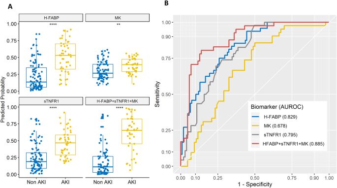
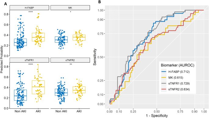
Prime Archives in Algorithms

biomarkers. Biomarkers with a p < 0.05 were considered
significant. The ability of the biomarkers to predict AKI was
further investigated using logistic regression (Lasso regression).
For each biomarker and biomarker combination, areas under the
receiver operator characteristic (AUROC), sensitivity,
specificity, positive predictive value (PPV) and negative
predictive value (NPV) were generated pre and post operatively
to identify models that differentiated between the two diagnostic
groups (non AKI vs. AKI).

Results
Clinical characteristics for patients involved in the study are
presented in Table 1. Of the 28 biomarkers that were
investigated, sTNFR1 and H-FABP had the highest AUROC pre
surgery to stratify risk of AKI in orthopaedic trauma patients
(Table 2) (sTNFR1 sensitivity 76.4%; specificity 59.7%;
AUROC 0.729 (CI 0.654–0.804); H-FABP sensitivity 62.1%;
specificity 71.2%; AUROC 0.712 (CI 0.637–0.786)
(Figure 2A,B)). Lasso regression identified a combination of 3
biomarkers post operatively to stratify risk of AKI, namely H-
FABP, sTNFR1 and MK (Table 2) (H-FABP, sTNFR1 and MK
combined sensitivity 80.5%; specificity 86.0%; AUROC 0.885
(CI 0.825-0.944) (Figure 3A,B)).

7                                              www.videleaf.com
Prime Archives in Algorithms

Table 1: Summary of clinical characteristics of the study patients.

                                Non AKI (n = 138)      AKI (n = 63)    p value
    Patient characteristics
    Age (years)                 78.7 ± 10.9            85.5 ± 6.1      0.000
    Gender (female)             109/138 (79.0%)        42/63 (55.7%)   0.089
    Comorbidities
    Hypertension                38/138 (27.5%)         27/63 (42.9%)   0.046
    Diabetes                    11/138 (8.0%)          4/63 (6.3%)     0.907
    Dementia                    15/138 (10.9%)         16/63 (25.4%)   0.015
    Pre surgery medications
    Hypertensive                46/116 (40.5%)         29/51 (56.9%)   0.074
    medications
    Intraoperative conditions
    Phenylephrine               19/115 (16.5%)         14/52 (26.9%)   0.176
    Packed red blood cells      6/115 (5.2%)           2/52 (3.8%)     1.000
    Fresh frozen plasma         0/115 (0.0%)           1/52 (1.9%)     0.683
    Platelet bags               4/115 (3.5%)           2/52 (3.8%)     1.000
    Operative method
    Hemiarthroplasty            30/138 (43.5%)         36/63 (57.1%)   0.100
    Intramedullary nailing      14/138 (10.1%)         1/63 (1.6%)     0.064
    Sliding hip screw           54/138 (39.1%)         26/63 (41.3%)   0.895
    Total hip replacement       10/138 (7.2%)          0/63 (0.0%)     0.065
    Post operative conditions
    Packed red blood cells      34/115 (29.6%)         16/52 (30.8%)   1.000
    Fresh frozen plasma         0/115 (0.0%)           1/52 (1.9%)     0.683
    Other
    Hospital stay (days)        9.8 ± 7.9              12.0 ± 8.3      0.020
    Operation time (minutes)    53.8 ± 19.1            52.4 ± 18.4     0.636
    Time between                2.1 ± 1.5              2.5 ± 2.0       0.138
    presentation and surgery
    (days)

Data presented as mean ± standard deviation or number/total (%). AKI acute
kidney injury.

8                                                        www.videleaf.com
Prime Archives in Algorithms
Table 2: Serum biomarkers for predicting AKI pre and post surgery.
Anytime
Biomarkers (n) AUROC CI Sensitivity (%) Specificity (%) PPV (%) NPV (%)
Pre operative MK (128) 0.615 0.513–0.718 57.1 67.4 46.2 76.3
sTNFR2 (174) 0.634 0.546–0.723 65.5 62.2 44.4 79.6
H-FABP (183) 0.712 0.637–0.786 62.1 71.2 50.0 80.2
sTNFR1 (174) 0.729 0.654–0.804 76.4 59.7 46.7 84.5
Post operative MK (128) 0.678 0.585–0.772 66.7 65.1 48.3 80.0
sTNFR2 (147) 0.734 0.648–0.821 67.4 71.3 51.7 82.8
sTNFR1 (147) 0.795 0.724–0.866 73.9 72.3 54.8 85.9
H-FABP (156) 0.829 0.764–0.893 75.0 74.1 56.3 87.0
H-FABP + sTNFR2 (147) 0.866 0.809–0.924 80.4 81.2 66.1 90.1
H-FABP + sTNFR2 + MK (127) 0.870 0.809–0.932 78.0 84.9 71.1 89.0
H-FABP + sTNFR1 (147) 0.881 0.825–0.937 78.3 87.1 73.5 89.8
H-FABP + sTNFR1 + MK (127) 0.885 0.825–0.944 80.5 86.0 73.3 90.2
AUROC, CI, sensitivity, specificity, PPV and NPV for serum biomarkers for predicting AKI pre and post surgery. 2. n number, AKI acute kidney
injury, AUROC area under the receiver operator characteristic, CI confidence interval, MK midkine, NPV negative predictive value, PPV positive
predictive value, sTNFR soluble tumour necrosis factor receptor, H-FABP heart-type fatty acid-binding protein.
9 www.videleaf.comPrime Archives in Algorithms Figure 2: (A) Pre surgery serum biomarker predicted probabilities for AKI development post operatively. H-FABP, MK, sTNFR1 and sTNFR2 predicted probabilities for preoperative serum levels. Wilcoxon rank sum statistical significance is indicated by *p ≤ 0.05, **p ≤ 0.01, ****p ≤ 0.0001. AKI acute kidney injury, H-FABP heart-type fatty acid-binding protein, MK midkine, sTNFR soluble tumour necrosis factor receptor. (B) Receiver operator characteristics for pre surgery serum biomarkers. H-FABP (AUROC 0.712), MK (AUROC 0.615), sTNFR1 (AUROC 0.729) and sTNFR2 (AUROC 0.634). AUROC area under the receiver operator characteristic, H- FABP heart-type fatty acid-binding protein, MK midkine, sTNFR soluble tumour necrosis factor receptor. Figure 3: (A) Post operative serum biomarker model predicted probabilities for non AKI and AKI patients. Predicted probabilities for post surgery serum levels for H-FABP, MK and sTNFR1 individually and combined. Wilcoxon rank sum 10 www.videleaf.com
Prime Archives in Algorithms

statistical     significance     is      indicated      by:     **p ≤= 0.01,
****p ≤ = 0.0001. AKI acute kidney injury, H-FABP heart-type fatty acid-
binding protein, MK midkine, sTNFR soluble tumour necrosis factor receptor.
(B) Receiver operator characteristics for post surgery serum biomarkers and
model. H-FABP (AUROC 0.829), MK (AUROC 0.678), sTNFR1 (AUROC
0.795) and model H-FABP + MK + sTNFR1 (AUROC 0.885). AUROC area
under the receiver operator characteristic, H-FABP heart-type fatty acid-
binding protein, MK midkine, sTNFR soluble tumour necrosis factor receptor.

To develop a tool to assist clinicians in identifying orthopaedic
trauma patients at risk of AKI and guide patient management,
the clinical characteristics between non AKI and AKI patients
were investigated. We used biomarker combinations to
determine a biomarker risk score (BRS) pre and post surgery
(based on AUROC determined by Lasso regression) that could
be used to identify patients at risk of AKI. Establishing a
biomarker set point (cut-off) pre and post surgery categorised
patients either positive or negative for AKI i.e. if a post surgery
patient had a BRS above the set point they would be predicted to
be positive for AKI (Table 3).

Table 3: Post surgery patient score calculation and BRS determination.

 BRS                                    Patient score*
 Negative                               < − 1.05
 Positive                               ≥ − 1.05

The patient score equation was derived from logistic regression. The cut-off
(closest top left) of − 1.05 was determined using the following equation:
closest top left = min((1 − sensitivities)2 + (1 − specificities)2).
If patient score < − 1.05 then BRS is negative, if patient score ≥ − 1.05 then
BRS is positive.
BRS biomarker risk score, H-FABP heart-type fatty acid-binding protein,
sTNFR soluble tumour necrosis factor receptor, MK midkine, min minimum.

*Patient Score = − 8.185 + 2.037*ln(H-FABP) + 2.373*ln(sTNFR1)
+ 0.056*ln(MK).

Three clinical characteristics were identified for patients at
potential risk of AKI pre and post operatively (Table 4). Each
clinical characteristic was given a score 0 or 1 (0 = no risk,
1 = risk). Each clinical characteristic was then added to give a
cumulative risk score (CRS). For example, pre surgery patients
who score ≥ 1 e.g. an ≥ 80-year-old patient with dementia and
hypertension would have a cumulative CRS of 3 and would

11                                                       www.videleaf.com
Prime Archives in Algorithms

therefore be categorized high risk for AKI. The cut-off for age
was based on significance, where patients > 80 years were at
greater risk of AKI, based on our patient cohort.

Table 4: Clinical risk factors.

     Clinical factor               Level              Clinical factor score
          Age                       < 80                        0
                                    ≥ 80                        1
       Dementia                     No                          0
                                    Yes                         1
     Hypertension                   No                          0
                                    Yes                         1

If total clinical factor score = 0 then CRS is low, if total clinical factor score ≥ 1
then CRS is high.
CRS clinical risk score.

To translate the results of the BRS and CRS into a proactive
clinical AKI tool, the BRS and CRS were combined. Combining
BRS with CRS either pre or post surgery identified 4 risk
categories for patient management (Table 5). Categories 1 and
2 = low risk; Categories 3 and 4 = high risk. Two worked
examples for a non AKI and AKI patient, are shown in the
Supplementary Notes S1–S3 and Supplementary Tables S1–S6.
The distribution of non AKI and AKI within the patient cohort,
for each risk category, is shown in Supplementary Note S4.

Table 5: Clinical management of patients using a combination of BRS and
CRS either pre or post surgery.

 Category       BRS               CRS         Clinical management
 1              Negative          Low         Routine management
 2              Negative          High        Assign to low risk management
 3              Positive          Low         Assign to higher risk management
 4              Positive          High        Assign to highest risk management

Combining BRS and CRS assigns a patient to a risk category.
BRS biomarker risk score, CRS clinical risk score.

Discussion
Hip fracture is the most common serious injury reported in the
elderly resulting in long hospital stays, high post-operative

12                                                            www.videleaf.com
Prime Archives in Algorithms morbidity and mortality, and reduced quality of life [4]. Furthermore, AKI after trauma, such as hip fracture is associated with a poor prognosis. Diagnosis of AKI using sCr and urine output can often result in misdiagnosis. The aim of this study was to further investigate if blood biomarkers and clinical risk factors could be used to identify AKI risk in orthopaedic trauma patients pre and post ORIF surgery in a similar fashion to those identified in patients undergoing cardiac surgery [11]. Interestingly, the same blood biomarkers, H-FABP, Midkine, sTNFR1 or sTNFR2, that predicted AKI in pre and post cardiac surgery patients also identified AKI risk in orthopaedic trauma patients undergoing ORIF surgery. The biomarkers combined with clinical characteristics (age, dementia and hypertension) identified from the study, delivered a proactive clinical AKI tool that could assist clinicians with patient management. A total of 28 blood biomarkers were investigated. However only two biomarkers, sTNFR1 or H-FABP, were identified as predictive of AKI pre surgery and a combination of three biomarkers, sTNFR1, H-FABP and MK, were predictive for AKI post surgery. Remarkably these biomarkers represent three main pathological processes of AKI. Mechanisms contributing to AKI include (1) perioperative episodes of under perfusion, followed by (2) ischemia reperfusion injury during restoration of normal blood pressure. Inflammatory mediators (3) contribute to and augment the renal injurious effects of this twofold process. Accordingly, an additional separate inflammatory insult arising from other perioperative factors such as coagulation disturbance (which is an important proinflammatory mechanism), can augment the renal injurious effect of hypotension and ischemia reperfusion. Biomarkers have been associated with identification of underlying processes of hypotension (VEFG and H-FABP), IRI (MK) and inflammation (sTNFR1 and 2), which as anti- inflammatory biomarkers are taken as surrogates for the underlying proinflammatory response which drives them [11]. Elevated sTNFR1 levels have been identified in many clinical conditions e.g. kidney disease [23], neuropathy, cardiovascular 13 www.videleaf.com
Prime Archives in Algorithms

disease and diabetes [24], and circulating levels of sTNFR1 have
been shown to be an independent predictor of CKD progression
in elderly patients [25]. Tumour necrosis factor alpha (TNFα)
and TNFR2 are almost undetectable in the kidneys of healthy
subjects unlike TNFR1 which is expressed within the trans-golgi
network of the glomerular endothelium [26]. An increase in the
level of sTNFRs in CKD patients has been implicated in
declining eGFR [27-29]. Moreover, TNFα acting through
TNFR1 has a damaging effect on renal endothelial cells [30],
possibly through iNOS, which would generate intratubular toxic
levels of NO, as demonstrated by increased urinary nitrate levels
in a porcine model of ischaemia reperfusion-mediated AKI [31].
The elevated anti-inflammatory sTNFR1 response in blood may
be driven by an underlying proinflammatory response which
includes TNFα [31]. Since monomeric TNFα is much smaller
than sTNFR1 and 2, it is more readily filtered by the glomerulus.
Accordingly, TNFα is able to cause glomerular injury once it
escapes from the moderating biological effect of sTNFR1 or 2.
This is consistent with orthopaedic trauma patients who develop
AKI having elevated levels of sTNFR1 when compared to non
AKI patients.

H-FABP was also predictive of AKI pre operatively, and in
combination with sTNFR1 and MK, post operatively. H-FABP,
associated with cardiac injury, is released into the bloodstream
30 min after an ischaemic event and peaks at 6 h before returning
to normal levels after 24 h [32]. H-FABP has been reported to
predict AKI pre and post cardiac surgery [7, 11, 33] however,
this is the first time that H-FABP has been demonstrated to
predict AKI in patients pre and post ORIF surgery.

H-FABP is predominantly expressed in the heart but also at
lower levels in skeletal muscle, kidney, stomach, brain and testis
[34, 35]. The levels of H-FABP in skeletal muscle have been
shown to be almost half that found in the heart. Moreover,
kidney H-FABP levels are almost two-thirds that found in
skeletal muscle [36]. While it is known that H-FABP levels
increase in the blood, this may arise from skeletal muscle or
renal sources, but it is more likely to be from the heart, which is
the largest reservoir of H-FABP in the body. In elderly patients,

14                                              www.videleaf.com
Prime Archives in Algorithms

acute coronary insufficiency is common and would be reflected
in elevated H-FABP. Any transient hypoperfusion, which such
an event would provoke, could result in a significantly
heightened risk of AKI. This is the most likely reason why H-
FABP was predictive in this orthopaedic trauma patient cohort.

In addition to sTNFR1 and H-FABP, MK was also identified in
the biomarker combination to predict AKI post operatively. The
pathophysiological roles of MK are diverse, ranging from AKI to
progression of CKD, accompanied by hypertension, renal
ischaemia and diabetic nephropathy [37,38]. After ischaemic
reperfusion MK is immediately induced in the proximal tubules,
leading to the upregulation of macrophage inflammatory protein-
2 for neutrophils and monocyte chemotactic protein-1 for
macrophages [38]. Eventually, infiltrated inflammatory cells
cause severe tubulointerstitial injury. Silencing renal MK
expression with anti-sense oligos prevents kidney damage and
increases osteogenic activity [39]. Midkine is also involved in
chondrogenesis and fracture healing [39]. Interestingly, MK-
deficient mice have been shown to display increased bone
formation rate and volume [39]. This is the first study, to our
knowledge which has identified MK as a biomarker for
stratifying patients at risk of AKI following orthopaedic trauma
and ORIF surgery.

Three important pathways involved in the pathogenesis of AKI
were identified, namely hypoperfusion (H-FABP), ischaemia
reperfusion injury (MK) and proinflammatory insult (sTNFR1)
(Figure 4).

Risk factors that have previously been reported for AKI include
age, pre-existing CKD, male gender, diabetes, heart failure and
surgery [15-17]. In this study, three clinical risk factors were
identified for patients who were at potential risk for the
development of AKI, pre and post operatively; age, dementia
and hypertension (Table 4). Using biomarker data and clinical
factors we developed a BRS and a CRS, respectively.
Combining BRS (Table 3) with CRS (Table 4) grouped patients
into four risk categories, each of which is associated with a
clinical management regime (Table 5). Deployment of this

15                                            www.videleaf.com
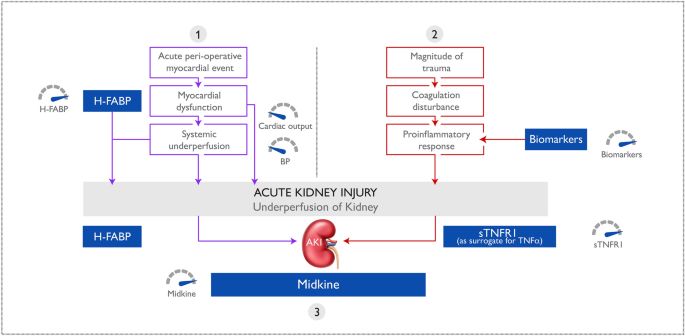
Prime Archives in Algorithms

proactive clinical AKI tool would allow clinicians to stratify
patients at risk of AKI enabling early intervention and improving
patient outcomes. Use of a cardiac proactive clinical AKI tool
has been described previously [11].

Figure 4: Pathogenesis of AKI. Three important pathways in the pathogenesis
of AKI are represented by biomarkers in the model: (1) hypoperfusion (H-
FABP), (2) proinflammation (sTNFR1 as a surrogate for the transient TNFα
response) and (3) ischaemia reperfusion injury (MK). Adapted from McBride
et al.11. AKI acute kidney injury, BP blood pressure, H-FABP heart-type fatty
acid-binding protein, MK midkine, sTNFR soluble tumour necrosis factor
receptor, TNFα tumour necrosis factor alpha.

In the study cohort 63/201 (31.3%) orthopaedic trauma patients
developed AKI post ORIF surgery. The incidence of AKI in this
patient cohort is higher than previously reported [40-43].
Patients who developed AKI were significantly older and more
likely to have hypertension and/or dementia. Advanced age is
frequently reported as a risk factor for AKI [1] however, to our
knowledge, this is the first report that identified dementia as a
potential risk factor in the development of AKI. Recently, an
association of heightened proinflammatory activity in patients
with dementia has been reported [44]. Our results are consistent
with these findings. Interestingly, patients surviving AKI have a
higher probability of developing dementia in the long-term
compared to patients who did not develop AKI [45].

The time between presentation and surgery was 2 days and was
not significant between non AKI and AKI patients. An optimal
operation time of between 24–48 h after orthopaedic trauma has

16                                                      www.videleaf.com
Prime Archives in Algorithms

been identified for lower extremity fracture fixation to reduce
complications. Operations performed outside of this timeframe
are associated with increased morbidity and mortality [46].

AKI patients stayed an additional two days in hospital compared
to non AKI patients (12.0 (3.7–20.3) days for AKI vs. 9.8 (1.9–
17.7) days for non AKI patients), consistent with previous
findings [47]. Patients that develop AKI following elective total
joint arthroplasty also have increased hospital stay [48]. The
management of patients with AKI is a significant burden to the
healthcare service [49]. Earlier diagnosis and management of
patients at risk of AKI will potentially reduce the financial
burden on healthcare systems in addition to improving patient
outcomes and welfare.

Surprisingly previous work has failed to identify hypotension as
a serious risk factor in AKI [50]. Since intraoperative blood
pressure modulation is a readily available strategy for
anaestheologists, inability to show a link between hypotension
and AKI at orthopaedic surgery could arguably generate a false
sense of complacency. However, the reason for lack of the
relationship between perioperative blood pressure and
subsequent AKI could be because most patients have non-
invasive blood pressure measurements where blood pressure
readings are obtained by an arm cuff measurement every 5 min
whereas more critically ill patients have continuous arterial
blood pressure measurements which detect and record all
hypotensive episodes. In Braüner’s study [50] they recorded
lowest blood pressure measurement intraoperatively. Their work
suggested that this was not a useful marker in terms of AKI
prediction. However, we argue that clinically significant
hypotensive episodes may have been missed in this study if they
happened in between measurements. This means that transient,
albeit clinically significant, hypotension could be missed in
between these times. In summary, the use of clinical data alone
(including perioperative hypotensive events) to predict
perioperative AKI is of limited usefulness in hip fracture
surgery. It has already been shown in cardiac surgery that
biomarkers of ischaemia reperfusion (MK) or hypotension
(VEGF or H-FABP) and inflammation augmented clinical

17                                             www.videleaf.com
Prime Archives in Algorithms

parameters [11]. This present work suggests that this principle is
also applicable to hip fracture surgery.

In a meta-analysis it was demonstrated that perioperative
hemodynamic optimization in surgery patients, reduces post-
operative acute renal injury [51]. Preoperative prediction would
allow for enhanced perioperative hemodynamic optimization i.e.
provision of Level 2 care provided post operatively, and invasive
hemodynamic monitoring intraoperatively rather than blood
pressure measurements every 5 min, as is routine for such cases
in many centres. It could also be taken as a contraindication to
non steroidal anti-inflammatory use post operation.

Biomarkers are not a substitute to the classical approach to using
low-cost information-but add to the information available to the
clinician. However, it must be noted that a clear clinical history
in these elderly patients can sometimes be unreliable. Hence the
need for the objective information that biomarkers provide.

Limitations of the study

Clinical characteristics were not reliably available for everyone
in this patient group including a guaranteed history of normal
renal function pre trauma; patients were assumed, based on
available clinical history, to have a normal renal function prior to
their trauma and a baseline eGFR of at least 60 ml/min/1.73m2.
Therefore, patients who had undiagnosed pre-trauma renal
dysfunction could have been included in the study. Nevertheless,
subsequent fluctuations in renal function were still detectable
using our proactive clinical AKI tool, demonstrating the clinical
utility of our proposed method in this patient cohort, where
obtaining clinical history is sometimes challenging and
unreliable.

Conclusion
In conclusion, serum H-FABP and sTNFR1 measured pre
operatively and serum H-FABP, MK and sTNFR1 measured
post operatively, identified orthopaedic trauma patients at risk of
developing AKI during ORIF surgery. Utilisation of the

18                                               www.videleaf.com
Prime Archives in Algorithms

proactive clinical AKI tool, which combines BRS with CRS,
would allow clinicians to stratify patients into one of four AKI
risk categories with related treatment regimens that could impact
patient care and management, length of hospital stay, and the
efficient use of hospital resources.

References

1. Haines RW, Fowler AJ, Kirwan CJ, Prowle JR. The
   incidence and associations of acute kidney injury in trauma
   patients admitted to critical care: a systematic review and
   meta-analysis. J. Trauma Acute Care Surg. 2019; 86: 141–
   147.
2. Bell, S. Risk of postoperative acute kidney injury in patients
   undergoing orthopaedic surgery–development and validation
   of a risk score and effect of acute kidney injury on survival:
   observational cohort study. BMJ. 2015; 351: h5639–h5639.
3. Grams ME. Acute kidney injury after major surgery: a
   retrospective analysis of veterans health administration data.
   Am. J. Kidney Dis. 2016; 67: 872–880.
4. Griffin XL, Parsons N, Achten J, Fernandez M, Costa ML.
   Recovery of health-related quality of life in a United
   Kingdom hip fracture population. Bone Joint J. 2015; 97:
   372–382.
5. Kellum JA, Lameire N. KDIGO AKI Guideline Work
   Group. Diagnosis, evaluation, and management of acute
   kidney injury: a KDIGO summary (Part 1). Crit. Care. 2013;
   17: 204.
6. Md Ralib A, Pickering JW, Shaw GM, Endre ZH. The urine
   output definition of acute kidney injury is too liberal. Crit.
   Care. 2013; 17: R112.
7. Parikh A. Does NGAL reduce costs? A cost analysis of
   urine NGAL (uNGAL) and serum creatinine (sCr) for acute
   kidney injury (AKI) diagnosis. PLoS ONE. 2017; 12:
   e0178091.
8. Bellomo R, Kellum JA, Ronco C. Defining acute renal
   failure: physiological principles. Intensive Care Med. 2004;
   30: 33–37.

19                                             www.videleaf.com
Prime Archives in Algorithms

9. Bennett M. Urine NGAL predicts severity of acute kidney
    injury after cardiac surgery: a prospective study. Clin. J. Am.
    Soc. Nephrol. 2008; 3: 665–673.
10. Liangos O. Comparative analysis of urinary biomarkers for
    early detection of acute kidney injury following
    cardiopulmonary bypass. Biomarkers. 2009; 14: 423–431.
11. McBride WT. Stratifying risk of acute kidney injury in pre
    and post cardiac surgery patients using a novel biomarker-
    based algorithm and clinical risk score. Sci. Rep. 2019; 9: 1–
    12.
12. McBride WT. Cytokine phenotype, genotype, and renal
    outcomes at cardiac surgery. Cytokine. 2013; 61: 275–284.
13. Mishra J. Neutrophil gelatinase-associated lipocalin (NGAL)
    as a biomarker for acute renal injury after cardiac surgery.
    Lancet. 2005; 365: 1231–1238.
14. Parikh CR. Postoperative biomarkers predict acute kidney
    injury and poor outcomes after adult cardiac surgery. J. Am.
    Soc. Nephrol. 2011; 22: 1748–1757.
15. Overview Acute kidney injury: prevention, detection and
    management NICE Guideline. 2019. Available Online at:
    https://www.nice.org.uk/guidance/ng148#:~:text=Acute%20
    kidney%20injury%3A%20prevention%2C%20detection%20
    and%20management,NICE%20guideline%20%5BNG148&t
    ext=It%20aims%20to%20improve%20assessment,people%2
    0with%20acute%20kidney%20injury
16. Kheterpal S. Predictors of postoperative acute renal failure
    after noncardiac surgery in patients with previously normal
    renal function. Anesthesiology. 2007; 107: 892–902.
17. Kheterpal S. Development and validation of an acute kidney
    injury risk index for patients undergoing general surgery.
    Anesthesiology. 2009; 110: 505–515.
18. Abdulla A. Proceedings from the symposium on kidney
    disease in older people: Royal Society of Medicine, London,
    January 19, 2017. Gerontol. Geriatr. Med. 2017; 3:
    233372141773685.
19. Myers GL. Recommendations for improving serum
    creatinine measurement: a report from the laboratory
    working group of the national kidney disease education
    program. Clin. Chem. 2006; 52: 5–18.

20                                              www.videleaf.com
Prime Archives in Algorithms
20. White SM, Rashid N, Chakladar A. An analysis of renal
dysfunction in 1511 patients with fractured neck of femur:
the implications for peri-operative analgesia. Anaesthesia.
2009; 64: 1061–1065.
21. Lopes JA, Jorge S. The RIFLE and AKIN classifications for
acute kidney injury: a critical and comprehensive review.
Clin. Kidney J. 2013; 6: 8–14.
22. R Core Team. R: A Language and Environment for
Statistical Computing. 2018.
23. Barr ELM. High baseline levels of tumor necrosis factor
receptor 1 are associated with progression of kidney disease
in indigenous Australians with diabetes: the eGFR follow-up
study. Diabetes Care. 2018; 41: 739–747.
24. Carlsson AC. Association of soluble tumor necrosis factor
receptors 1 and 2 with nephropathy, cardiovascular events,
and total mortality in type 2 diabetes. Cardiovasc. Diabetol.
201; 15.
25. Carlsson AC. Soluble tumor necrosis factor receptor 1 is
associated with glomerular filtration rate progression and
incidence of chronic kidney disease in two community-based
cohorts of elderly individuals. Cardiorenal Med. 2015; 5:
278–288.
26. Bradley JR, Thiru S, Pober JS. Disparate localization of 55-
kd and 75-kd tumor necrosis factor receptors in human
endothelial cells. Am. J. Pathol. 1995; 146: 27–32.
27. Al-Lamki RS, Mayadas TN. TNF receptors: signaling
pathways and contribution to renal dysfunction. Kidney Int.
2015; 87: 281–296.
28. Gohda T. Circulating TNF receptors 1 and 2 predict stage 3
CKD in type 1 diabetes. J. Am. Soc. Nephrol. 2012; 23:
516–524.
29. Miyazawa I. Association between serum soluble TNFα
receptors and renal dysfunction in type 2 diabetic patients
without proteinuria. Diabetes Res. Clin. Pract. 2011; 92:
174-180.
30. Wu X, Guo R, Chen P, Wang Q, Cunningham PN. TNF
induces caspase-dependent inflammation in renal endothelial
cells through a Rho- and myosin light chain kinase-
dependent mechanism. Am. J. Physiol. Renal Physiol. 2009;
297: F316–F326.
21 www.videleaf.comPrime Archives in Algorithms
31. Baker RC. Methylprednisolone increases urinary nitrate
concentrations and reduces subclinical renal injury during
infrarenal aortic ischemia reperfusion. Ann. Surg. 2006; 244:
821–826.
32. Muehlschlegel JD. Heart-type fatty acid binding protein is an
independent predictor of death and ventricular dysfunction
after coronary artery bypass graft surgery. Anesth. Analg.
2010; 111: 1101–1109.
33. Oezkur M. Preoperative serum h-FABP concentration is
associated with postoperative incidence of acute kidney
injury in patients undergoing cardiac surgery. BMC
Cardiovasc. Disord. 2014; 14: 117.
34. Kamijo-Ikemori A, Sugaya T, Kimura K. Urinary fatty acid
binding protein in renal disease. Clin. Chim. Acta. 2006;
374: 1–7.
35. Pelsers MMAL, Hermens WT, Glatz JFC. Fatty acid-binding
proteins as plasma markers of tissue injury. Clin. Chim.
Acta. 2005; 352: 15–35.
36. Veerkamp JH. Detection, tissue distribution and
(sub)cellular localization of fatty acid-binding protein types.
Mol. Cell. Biochem. 1990; 98: 11–18.
37. Andreucci M, Faga T, Pisani A, Perticone M, Michael A.
The ischemic/nephrotoxic acute kidney injury and the use of
renal biomarkers in clinical practice. Eur. J. Intern. Med.
2017; 39: 1–8.
38. Sato W, Sato Y. Midkine in nephrogenesis, hypertension and
kidney diseases. Br. J. Pharmacol. 2014; 171: 879–887.
39. Haffner-Luntzer M. Midkine-deficiency delays
chondrogenesis during the early phase of fracture healing in
mice. PLoS ONE. 2014; 9: e116282.
40. Bennet SJ, Berry OMB, Goddard J, Keating JF. Acute renal
dysfunction following hip fracture. Injury. 2010; 41: 335–
338.
41. Ersoy A. Survival analysis of the factors affecting in
mortality in injured patients requiring dialysis due to acute
renal failure during the Marmara earthquake: survivors vs
non-survivors. Clin. Nephrol. 2003; 59: 334–340.
42. Kimmel LA, Wilson S, Janardan JD, Liew SM, Walker RG.
Incidence of acute kidney injury following total joint
22 www.videleaf.comPrime Archives in Algorithms
arthroplasty: a retrospective review by RIFLE criteria. Clin.
Kidney J. 2014; 7: 546–551.
43. Sykes L, Kalra PA, Green D. Comparison of impact on death
and critical care admission of acute kidney injury between
common medical and surgical diagnoses. PLoS ONE. 2019;
14: e0215105.
44. Kim JW. Longitudinal associations between serum cytokine
levels and dementia. Front. Psychiatry. 2018; 9: 1.
45. Tsai HH, Yen RF, Lin CL, Kao CH. Increased risk of
dementia in patients hospitalized with acute kidney injury: a
nationwide population-based cohort study. PLoS ONE.
2017; 12: e0171671.
46. Sangkomkamhang T, Thinkhamrop W, Thinkhamrop B,
Laohasiriwong W. Incidence and risk factors for
complications after definitive skeletal fixation of lower
extremity in multiple injury patients: a retrospective chart
review. F1000Research. 2018; 7: 612.
47. Porter CJ. Acute and chronic kidney disease in elderly
patients with hip fracture: Prevalence, risk factors and
outcome with development and validation of a risk
prediction model for acute kidney injury. BMC Nephrol.
2017; 18: 1–11.
48. Abar O, Toossi N, Johanson N. Cost and determinants of
acute kidney injury after elective primary total joint
arthroplasty. Arthroplast. Today. 2018; 4: 335–339.
49. College of Physicians R. Falls and fragility fracture audit
programme national hip fracture database (NHFD) annual
report 2017. 2017.
50. Braüner Christensen J. Predictors of acute kidney injury after
hip fracture in older adults. Geriatr. Orthop. Surg. Rehabil.
2020; 11: 215145932092008.
51. Brienza N, Giglio MT, Marucci M, Fiore T. Does
perioperative hemodynamic optimization protect renal
function in surgical patients? A meta-analytic study. Crit.
Care Med. 2009; 37: 2079–2090.
23 www.videleaf.comYou can also read