Eosinophilic esophagitis in Egyptian adult patients presenting with upper gastrointestinal symptoms
←
→
Page content transcription
If your browser does not render page correctly, please read the page content below
Open Journal of Gastroenterology, 2014, 4, 88-95                                                                            OJGas
http://dx.doi.org/10.4236/ojgas.2014.42015 Published Online February 2014 (http://www.scirp.org/journal/ojgas/)

Eosinophilic esophagitis in Egyptian adult patients
presenting with upper gastrointestinal symptoms
Shereen Shoukry Hunter1, Dina Omar Helmy2, Naglaa Aly Zayed1, Tamer Mohamed El-Tayeb3,
Magdy Amin El-Serafy1
1
  Endemic Medicine Department and Gastrointestinal Endoscopy and Liver Unit, Faculty of Medicine, Cairo University, Cairo, Egypt
2
  Pathology Department, Faculty of Medicine, Cairo University, Cairo, Egypt
3
  Endoscopy Unit, New Kasr El-Ainy Teaching Hospital, Cairo, Egypt
Email: shereenhunter@hotmail.com

Received 4 January 2014; revised 3 February 2014; accepted 9 February 2014

Copyright © 2014 Shereen Shoukry Hunter et al. This is an open access article distributed under the Creative Commons Attribution
License, which permits unrestricted use, distribution, and reproduction in any medium, provided the original work is properly cited.
In accordance of the Creative Commons Attribution License all Copyrights © 2014 are reserved for SCIRP and the owner of the
intellectual property Shereen Shoukry Hunter et al. All Copyright © 2014 are guarded by law and by SCIRP as a guardian.

ABSTRACT                                                            clusion: The prevalence of EoE is low in adult Egyp-
                                                                    tian patients presenting with upper gastrointestinal
Background and Study Aim: Eosinophilic esophagitis
                                                                    symptoms. Dysphagia is the main presenting symp-
(EoE) is a clinicopathological disease characterized by
                                                                    tom of EoE while heartburn is not characteristic of
esophageal dysfunction and marked esophageal eosi-
                                                                    the disease. Normal esophagus endoscopically does
nophilic infiltration. It shows a marked increase in in-
                                                                    not exclude EoE.
cidence and prevalence and has been associated with
gastroesophageal reflux disease (GERD). The aim of
this work was to detect the prevalence of EoE in Egy-
                                                                    KEYWORDS
ptian adult patients presenting with upper gastroin-                Eosinophilic Esophagitis (EoE); Adults;
testinal symptoms and to clarify its clinical pattern               Gastroesophageal Reflux Disease (GERD); Egypt
and the possibility of its overlap with GERD. Patients
and Methods: The study included 91 adult patients
presenting with various upper gastrointestinal symp-
                                                                    1. INTRODUCTION
toms. Upper gastrointestinal endoscopy was done and                 Eosinophilic esophagitis (EoE) is a chronic immune/anti-
esophageal biopsies were taken. The presence of >15                 gen-mediated esophageal disease characterized clinically
eosinophils per high power field together with a histo-             by symptoms related to esophageal dysfunction and his-
ry of intake of proton pump inhibitors for at least 3               tologically by eosinophil-predominant inflammation [1].
weeks without improvement was used as prerequisite                  It was thought to be a rare inflammatory condition in
diagnostic criteria for EoE. Results: Classification of             adults [2] with an estimated prevalence 0.2 - 3 in 10,000
the patients was based on both endoscopic and histo-                [3]. However, the epidemiology of EoE has been rap-
pathological findings. Accordingly, out of the 91 pa-               idly evolving over the past two decades, with a marked
tients, 70 had GERD (76.9%); 58 of them had erosive
                                                                    increase in incidence and prevalence [4] and it has be-
reflux disease (ERD) and 12 had endoscopically nor-
                                                                    come increasingly recognized as an important cause of
mal esophagus but with histopathological changes
                                                                    dysphagia and food impaction in adults [5]. This is likely
compatible with reflux esophagitis and were classified
                                                                    attributable to a combination of an increasing incidence
as non erosive reflux disease (NERD). Eighteen pa-
tients had normal endoscopic and histopathological                  and a growing awareness of the condition amongst gas-
esophagus (19.8%), and 3 patients had EoE (3.3%),                   troenterologists and pathologists [6]. EoE has recently
with an overlap between ERD and EoE in one patient.                 been associated with gastroesophageal reflux disease
The mean age of EoE patients was 41.6 ± 11.7 years.                 (GERD) [7-9]. It mimics GERD and may result in nar-
Two of them were males and one was a female. All of                 rowing and stricture of the esophagus [10-12]. This dis-
the 3 patients complained of dysphagia and none com-                ease is differentiated from reflux esophagitis on the basis
plained of heartburn. The endoscopists did not report               of the magnitude of mucosal eosinophilia and a lack of
any endoscopic findings characteristic of EoE. Con-                 response to acid suppression [13].

OPEN ACCESS
S. S. Hunter et al. / Open Journal of Gastroenterology 4 (2014) 88-95 89
2. AIM OF THE WORK (number of cases) and percentages. Comparison between
the study groups was done using Chi-square (χ2) test.
The aim of this study was to detect the prevalence of EoE
Exact test was used instead when the expected frequency
in Egyptian adult patients presenting with upper gastro-
was less than 5. P value less than 0.05 was considered
intestinal symptoms and to clarify its clinical pattern as
well as the possibility of its overlap with GERD. statistically significant. All statistical calculations were
done using computer programs SPSS (Statistical Package
3. PATIENTS AND METHODS for the Social Science; SPSS Inc., Chicago, IL, USA)
version 15 for Microsoft Windows.
The study included 91 adult patients presenting with va-
rious upper gastrointestinal symptoms as heartburn, dys- 5. RESULTS
phagia/odynophagia, eructation, epigastric pain and vo-
miting, and who were on acid suppression therapy [either The mean age of all patients was 43.06 ± 9.9 years (mi-
proton pump inhibitors (PPIs) or histamine H2 receptor nimum = 21, maximum = 65 years). They were 56 males
antagonists (H2RA)]. Patients on any form of steroids (61.5%) and 35 females (38.4%). Thirty patients (32.9%)
were excluded. Patients were recruited from the Endos- gave history of smoking, 65 (71%) gave history of use of
copy Units of Cairo University Hospital and New Kasr PPIs and 26 (28.5%) gave history of use of H2RA.
El-Ainy Teaching Hospital, Cairo, Egypt in the period The presenting symptoms of the patients were epigas-
from December 2009 to December 2011 and the study tric pain in 56 (61%), vomiting in 49 (53.8%), dysphagia/
was approved by the local ethical committee. odynophagia in 33 (36.2%), heartburn in 27 (29.7%) and
Patients were clinically assessed with special emphasis food impaction in 3 (3.8%) of patients.
on history of smoking, any allergic manifestations such Endoscopically, a total of 58 patients had ERD; 38
as bronchial asthma and allergic rhinitis, and relevant (41.8%) had grade A, 19 (20.9%) had grade B and only 1
drug history. patient (1.1%) had grade C. No cases with grade D were
Midazolam or propofol were used for sedation then encountered. Eleven of them (12% of patients) had also
upper gastrointestinal endoscopy was done. The endosco- hiatus hernia. Thirty eight patients (41.7%) had erythe-
pist noted the following: matous gastritis, 32 (35.1%) had endoscopically normal
• The presence of endoscopic findings of EoE which esophagus, 19 (20.8%) had erosive gastritis, 14 (15.3%)
include esophageal rings, strictures, narrow-caliber had duodenal ulcer or erosion, and 9 (9.8%) had gastric
esophagus, linear furrows, white plaques or exudates ulcer. One patient had only hiatus hernia and only 1 was
and pallor or decreased vasculature [14,15]. found to have Barrett’s esophagus. The endoscopists did
• The presence of erosive reflux disease (ERD) which not report any endoscopic findings characteristic of EoE.
was classified according to the Los Angeles Classi- Histopathological examination of esophageal biopsies
fication System [16]. revealed histopathological findings of reflux esophagitis
• Any other finding in the esophagus, stomach or duo- in 69 (75.8%) of patients, normal stratified squamous
denum was reported. epithelium in 18 (20%), EoE in 3 (3.3%) and Barrett’s
Biopsies were taken from mid-esophagus and, in cases esophagus in only 1 patient. One of the EoE cases had
of ERD, lower esophageal biopsies were done as well. histopathological findings of reflux esophagitis.
All esophageal mucosal biopsy specimens were fixed in Our classification of the patients was based on both
formalin, routinely processed, embedded in paraffin and histopathological and endoscopic findings as shown in
cut serially in 5-micron sections. Biopsies were examin- Table 1. Accordingly, out of the 91 patients, 70 had
ed by the same histopathologist after staining with hae- GERD (76.9%); 58 of them had ERD (82.9% of GERD
matoxylin and eosin. GERD was diagnosed after fulfill- patients; 63.7% of all patients) and 12 (17.1% of GERD
ment of its histopathological criteria such as epithelial patients; 13.2% of all patients) had endoscopically nor-
edema and basal cell hyperplasia, elongation of the papil- mal esophagus but with histopathological changes com-
lae, thinning of the squamous cell layer plus lymphocytic patible with reflux esophagitis and were classified as
or neutrophilic inflammation. Goblet cell metaplasia or non-erosive reflux disease (NERD) as they all also com-
Barrett’s esophagus was identified and recorded. plained of heart-burn. Eighteen patients had normal en-
On the high power field (HPF) the pathologist counted doscopic and histopathological esophagus (19.8%), and 3
the eosinophils in all biopsies. The presence of >15 eo-
patients had EoE (3.3%) (Figures 1 and 2), with an over-
sinophils/HPF together with a history of intake of PPIs
lap between ERD and EoE in one patient.
for at least 3 weeks without improvement were used as
The mean age of EoE patients was 41.6 ± 11.7 years
prerequisite diagnostic criteria for EoE.
(minimum = 33, maximum = 55 years). Two were males
and one was a female. All 3 patients complained of dys-
4. STATISTICAL ANALYSIS
phagia. None of them was a smoker and one (33.3%) had
Data were statistically described in terms of frequencies history of bronchial asthma. Two had a normally appear-
Copyright © 2014 SciRes. OPEN ACCESS90 S. S. Hunter et al. / Open Journal of Gastroenterology 4 (2014) 88-95
Table 1. Patterns of esophageal mucosal injury and the subsequent group classification.
Pattern of mucosal Positive endoscopy and Negative endoscopy and Negative endoscopy/positive
injury histopathology (N = 59) histopathology (N = 18) histopathology (N = 14)
ERD (N = 57)
NERD (N = 12)
Group classification ERD + EE (N = 1) Normal esophagus
EoE (N = 2)
Barrett’s esophagus (N = 1)
ERD: erosive reflux disease. NERD: non erosive reflux disease. EoE: eosinophilic esophagitis.
cal findings with the presenting symptoms are shown in
Table 2. Dysphagia occurred more significantly in EoE
patients (p value = 0.020) and heartburn occurred more
significantly in patients with NERD in relation to other
presenting symptoms. None of the symptoms showed sta-
tistically significant occurrence among different groups.
The patient with Barrett’s esophagus manifested clini-
cally by heartburn.
6. DISCUSSION
EoE is a clinicopathologic disease characterized clinica-
lly by symptoms related to esophageal dysfunction. Pa-
thologically there must be eosinophil-predominant in-
flammation in one or more biopsy specimens. The dis-
ease is isolated to the esophagus, and other causes of eso-
phageal eosinophilia should be excluded, specifically PPI-
Figure 1. Case histologically diagnosed as eosinophilic esopha- responsive esophageal eosinophilia. The disease should
gitis (EoE): a section in esophageal mucosa shows many intra-
epithelial eosinophils (23 eosinophils). The superficial position remit with treatments of dietary exclusion, topical corti-
of the eosinophilic infiltrate is also noted (hematoxylin and eo- costeroids, or both [1]. The epidemiology of EoE may be
sin stained section, original magnification ×200). changing; several case reports and case series suggest
that either the incidence is increasing or the disease is
now recognised more often [14,17].
The aim of this work was to detect the prevalence of
EoE in adult Egyptian patients presenting with various
upper gastrointestinal symptoms as well as to clarify its
clinical pattern as well as the possibility of its overlap
with GERD.
The lack of a clinicopathologic response to PPI treat-
ment in patients adherent to the treatment regimen with
compatible symptoms of EoE and isolated esophageal
eosinophilia is consistent with the diagnosis of EoE [18].
With few exceptions, 15 eosinophils/HPF (peak value) is
considered a minimum threshold for a diagnosis of EoE
[1]. In our study, the presence of >15 eosinophils/HPF
together with a history of intake of PPIs for at least 3
weeks without improvement were used as prerequisite
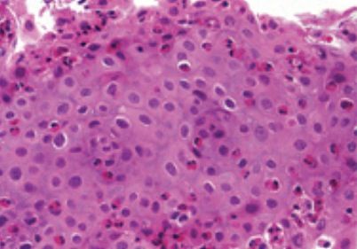
Figure 2. Esophageal mucosa from another case of eosinophil- diagnostic criteria for EoE.
ic esophagitis (EoE): intraepithelial eosinophils (58 eosinophils)
were detected. Of notice also is the intercellular edema giving We found 3 cases of EoE out of 91 adult patients pre-
the spongy or moth eaten appearance (hematoxylin and eosin senting with various upper gastrointestinal symptoms
stained section, original magnification ×200). (3.3%). The prevalence of EoE varies with the popula-
tion studied. For example, it has been estimated to be 0.4%
ing esophagus endoscopically, 1 had additional ERD and - 1.1% in the general population [19,20], in an out-patient
non had any detectable endoscopic findings in stomach population undergoing routine upper endoscopy the pre-
or duodenum. valence increased to 6.5% [21], and in those undergoing
Associations between endoscopic and histopathologi- an endoscopy for dysphagia, the prevalence was 10% -
Copyright © 2014 SciRes. OPEN ACCESSS. S. Hunter et al. / Open Journal of Gastroenterology 4 (2014) 88-95 91
Table 2. Association of histopathological and endoscopic find- of GERD patients are heartburn and regurgitation [31].
ings with presenting symptoms. In the current study, the main presenting symptom of
Findings EoE patients was dysphagia which was present in all 3
NERD ERD EoE patients with EoE (100%) (p value = 0.020). None of
Symptoms EoE patients complained of food impaction. In an earlier
(N = 12) (N = 58) (N = 3)
Dysphagia 6 20 3* study, dysphagia was documented in 26 of 31 EoE pa-
% 50% 34.4% 100% tients (89%) [2]. Also, food impaction was found in 32.0%
Food impaction 1 1 0 of EoE positive patients and dysphagia in 64.0% [21]
% 8.3% 1.7% 0% and the most common endoscopy indications in adults
Heartburn 12* 21 0
with EoE were dysphagia (70.1%) and GERD/heartburn
(27.1%) [32]. Heartburn was the presenting symptom in
% 100% 36.2% 0%
36.2% of ERD patients in our study, in 100% of NERD
Epigastric pain 9 36 2
patients (which was a statistically significant finding in
% 75% 62% 66.6%
this group), and in none of EoE patients. In the study by
Vomiting 11 32 1
Parfitt et al. [25], dysphagia was more common in EoE
% 91.6% 55.1% 33.3% patients (63%), while heartburn was more common in
Chi-square test. (*) denotes statistically significant occurrence of the rele- none EoE patients (53%) who were regarded to represent
vant symptom within the same studied group. NERD: non erosive reflux di-
sease. ERD: erosive reflux disease. EoE: eosinophilic esophagitis.
patients with GERD.
Endoscopic findings of EoE include esophageal rings,
15% [22-24]. strictures, narrow-caliber esophagus, linear furrows, whi-
Most EoE studies report a male predominance, inclu- te plaques or exudates, and pallor or decreased vascula-
ding >75% of reported adult and child cases [10]. In the ture [14,15]. Two of the 3 positive cases of EoE in our
current study, the mean age of EoE patients was 41.6 ± study (66%) showed normal endoscopic appearance of
11.7 years, and EoE was detected in males more than fe- the esophagus and the third had an overlap with ERD.
males (2 males and 1 female). Compared with EoE nega- However, the small number of patients found to have
tive patients, EoE positive patients in the study by Vee- EoE in our study may have precluded us from stating a
rappan et al, [21] were more likely to be male and youn- specific endoscopic finding for this disease. Besides, the
ger than 50 years. Among 41 subjects with histological endoscopic features of EoE may be subtle and over-
EoE, the ratio of males to females was 4:1 and the aver- looked at endoscopy [2,33]. One adult series of histolo-
age age at diagnosis was 45 years [25]. gically confirmed EoE reported 8.8% of patients without
Several lines of evidence support a role for allergic in- any apparent endoscopic features [14]. In a meta-analysis,
flammation in the pathogenesis of EoE. The most ob- the endoscopic examination was normal in 17% of cases
vious evidence for such involvement is the central role of [34]. However, esophageal mucosal furrows were present
the eosinophil which is often considered synonymous in 30 of 31 EoE patients (97%) [2] and the presence of
with allergic disease because of its accumulation in spu- classic findings of EoE on endoscopy (rings, furrows,
tum in asthma, in nasal secretions in allergic rhinitis and plaques, or strictures) was the strongest predictor of this
in the skin during flares of acute eczema [26]. Among disease process with a sensitivity of 72%, specificity of
adults with EoE, studies report personal or family histo- 89%, and negative predictive value of 98% [21]. On the
ries of allergies ranging from 50 to 90%, including up to other hand, Machenzie et al. [35] found that 13/31 (42%)
60% with asthma and up to 25% with food allergies [2, of EoE patients did not have the classic endoscopic find-
12,27]. Although clearly an atopic condition, the role of ings (rings +/− furrows) and would have been missed
specific allergic triggers in EoE remains unclear [28]. In without esophageal biopsies. Consequently, although a
our study, 33.3% of the EoE patients had history of bron- high degree of suspicion for EoE must be maintained for
chial asthma. Compared with EoE negative patients, EoE patients that have endoscopic features of this disease, the
positive patients were more likely to have asthma (32.0% presence or absence of endoscopic findings is insuffi-
vs 10.8%) [21] and 14 of 29 patients (48%) with docu- cient to make a diagnosis. Esophageal biopsies should be
mented EoE had a history of asthma, environmental al- obtained from all patients who present with symptoms of
lergy, or atopy [27]. EoE, regardless of the endoscopic appearance of the eso-
The symptom profile is similar to that of severe GERD, phagus [34]. Also, it is advised that esophageal biopsies
but unlike GERD, EoE is not resolved with acid reduc- routinely be taken in the clinical setting of unexplained
tion therapy, such as antacids, PPIs, and H2RA. Symp- dysphagia, refractory heartburn, or chest pain regardless
toms of EoE vary with age [5,29,30]. Common presen- of endoscopic findings as endoscopic mucosal biopsy re-
ting symptoms in adults include dysphagia, food impac- mains the most important diagnostic test for EoE and the
tion, heartburn and chest pain [5]. The typical symptoms diagnosis of EoE is ultimately established histologically
Copyright © 2014 SciRes. OPEN ACCESS92 S. S. Hunter et al. / Open Journal of Gastroenterology 4 (2014) 88-95
[5]. Esophageal biopsies demonstrate often marked epi- tients have typical reflux symptoms (i.e., heartburn and/
thelial basal hyperplasia and extensive infiltration of the or regurgitation) in the absence of endoscopically visible
epithelium by eosinophils. The changes occur not just in esophageal mucosal injuries, making NERD the more
the distal esophagus, as in GERD, but also in the mid and common form of GERD [47,48]. In the current study,
upper esophageal mucosa, a feature that is often useful in only 17.1% of GERD patients had NERD diagnosed on
the differentiation of EoE from reflux esophagitis. Eosi- the basis of the presence of heartburn and histopathologi-
nophils generally number in excess of 20 to 24/HPF [36, cal changes compatible with reflux esophagitis in a nor-
37]. There are limited data to support routine gastric or mal endoscopic esophagus. The changes were detected in
duodenal biopsies in adults in the absence of symptoms mid-esophageal biopsies. There has been little standardi-
or endoscopic abnormalities suggesting other gastrointes- zation of biopsy techniques or tissue processing in GERD
tinal disorders, although it is reasonable for these biopsi- and NERD patients. Biopsies have been obtained at the
es to be performed [1]. squamocolumnar junction, or at 1, 2, 3 and 5 cm above it.
In 1985, Lee [38] reported on 11 patients with obvious Furthermore, there is no consensus on the number of bi-
esophageal eosinophil infiltration, 10 of whom had re- opsy specimens obtained, or the location around the in-
flux esophagitis. Since then, the connection between ner circumference of the esophagus at which biopsies
GERD and EoE has been under debate [3,27,33,39]. should be taken. This issue is especially important since
Historically, the diagnosis of EoE was often overlooked the severity of exposure to refluxate decreases with in-
in adults with many patients alternatively diagnosed as creasing distance from squamocolumnar junction and the
having GERD or a Schatzki ring. In some instances, distribution of mucosal injury may be patchy [49]. How-
these patients had undergone repeated endoscopies and ever, in the attempt to better understand the mechanisms
involved in the perception of gastroesophageal reflux,
dilation prior to accurate diagnosis [40,41]. Another his-
some observations have pointed out the role of the acid
torical explanation for the delayed diagnosis of EoE is
extent into the middle-proximal esophagus [50-55]. In-
that eosinophilic infiltrate in the esophageal mucosa was
deed, in NERD patients, independently of the acid expo-
previously equated with GERD [42]. Accepting that that
sure time, reflux episodes reaching the proximal eso-
can be the case, the current strategy for making this dis-
phagus were perceived more than those confined to the
tinction is to rely on a quantitative threshold of eosino-
distal esophagus [51,55].
philic infiltration (currently ≥15/HPF in the area of great-
est eosinophilic infiltration); lower counts are presumed
7. CONCLUSION
related to GERD whilst higher counts are diagnostic of
EoE [5]. In conclusion, the prevalence of EoE is low in adult Egy-
GERD is extremely common, with an incidence of 10 ptian patients presenting with upper gastrointestinal symp-
to 20% in Western adults presenting with reflux symp- toms. Dyshagia is the main presenting symptom of EoE
toms and heartburn [43] and is the most common disease while heartburn is more common in GERD. Normal en-
in patients referred for upper endoscopy [44]. The pre- doscopic esophagus does not exclude EoE.
valence of GERD in our patients was 76.9%. In the 3
cases of EoE, one (33.3%) had also features of ERD. The REFERENCES
overlap between GERD and EoE continues to be enig-
matic because of the high prevalence of GERD in the [1] Liacouras, C.A., Furuta, G.T., Hirano, I., Atkins, D., At-
twood, S.E., Bonis, P.A., Burks, A.W., Chehade, M., Col-
adult population. In nine adult EoE studies reporting pH
lins, M.H., Dellon, E.S., Dohil, R., Falk, G.W., Gonsalves,
monitoring data, abnormal results were reported in 18% N., Gupta, S.K., Katzka, D.A., Lucendo, A.J., Markowitz,
of patients [5]. In a systematic review, pathological acid J.E., Noel, R.J., Odze, R.D., Putnam, P.E., Richter, J.E.,
reflux was found in only 10% of cases of EoE [14]. Romero, Y., Ruchelli, E., Sampson, H.A., Schoepfer, A.,
Among patients with GERD, 8.8% had EoE [45]. It is Shaheen, N.J., Sicherer, S.H., Spechler, S., Spergel, J.M.,
now suggested that EoE is more prevalent among GERD Straumann, A., Wershil, B.K., Rothenberg, M.E. and Ace-
patients who do not respond to treatment with PPIs [5, ves, S.S. (2011) Eosinophilic esophagitis: Updated con-
sensus recommendations for children and adults. Journal
46]. An initial trial of PPI therapy in patients with clini-
of Allergy and Clinical Immunology, 128, 3-20.
cal, endoscopic and pathologic findings of EoE is thus http://dx.doi.org/10.1016/j.jaci.2011.02.040
warranted. Lack of a response to PPI may reinforce a
[2] Croese, J., Fairley, S.K., Masson, J.W., Chong, A.K., Whi-
diagnosis of EoE, but a clinical response to PPI may not taker, D.A., Kanowski, P.A. and Walker, N.I. (2003) Cli-
rule out quiescent EoE. Esophageal pH measurements nical and endoscopic features of eosinophilic esophagitis
and histopathologic data on patients on PPI treatment are in adults. Gastrointest Endosc, 58, 516-522.
pivotal in cases with overlapping GERD and EoE in or- http://dx.doi.org/10.1067/S0016-5107(03)01870-4
der to evaluate the role of each disease [46]. [3] Straumann, A. and Beglinger, C. (2006) Eosinophilic eso-
It has been documented that up to 70% of reflux pa- phagitis: The endoscopist’s enigma. Gastrointest Endosc,
Copyright © 2014 SciRes. OPEN ACCESSS. S. Hunter et al. / Open Journal of Gastroenterology 4 (2014) 88-95 93
63, 13-15. http://dx.doi.org/10.1016/j.gie.2005.09.010 ease. Clinical Gastroenterology and Hepatology, 7, 1305-
[4] Dellon, E.S, Erichsen, R., Pedersen, L., Shaheen, N.J., 1313. http://dx.doi.org/10.1016/j.cgh.2009.08.030
Baron, J.A., Sørensen, H.T. and Vyberg, M. (2013) De- [16] Armstrong, D., Bennett, J.R., Blum, A.L., Dent, J., De
velopment and validation of a registry-based definition of Dombal, F.T., Galmiche, J.P., Lundell, L., Margulies, M.,
eosinophilic esophagitis in Denmark. World Journal of Richter, J.E., Spechler, S.J., Tytgat, G.N. and Wallin, L.
Gastroenterology, 19, 503-510. (1996) The endoscopic assesment of esophagitis: A pro-
http://dx.doi.org/10.3748/wjg.v19.i4.503 gress report on observer agreement. Gastroenterology,
111, 85-92.
[5] Furuta, G.T., Liacouras, C.A., Collins, M.H., Gupta, S.K.,
http://dx.doi.org/10.1053/gast.1996.v111.pm8698230
Justinich, C., Putnam, P.E., Bonis, P., Hassall, E., Strau-
mann, A., Rothenberg, M.E. and Members of the First [17] Straumann, A. and Simon, H.U. (2005) Eosinophilic eso-
International Gastrointestinal Eosinophil Research Sym- phagitis: Escalating epidemiology? Journal of Allergy
posium (FIGERS) Subcommittees (2007) Eosinophilic and Clinical Immunology, 115, 418-419.
esophagitis in children and adults: A systematic review http://dx.doi.org/10.1016/j.jaci.2004.11.006
and consensus recommendations for diagnosis and treat- [18] Spergel, J.M., Brown-Whitehorn, T.F., Beausoleil, J.L.,
ment. Gastroenterology, 133, 1342-1363. Franciosi, J., Shuker, M., Verma, R. and Liacouras, C.A.
http://dx.doi.org/10.1053/j.gastro.2007.08.017 (2009) 14 years of eosinophilic esophagitis: Clinical fea-
[6] Gonsalves, N. and Kahrilas, P.J. (2009) Eosinophilic oe- tures and prognosis. Journal of Pediatric Gastroenterolo-
sophagitis in adults. Neurogastroenterology & Motility, gy and Nutrition, 48, 30-36.
21, 1017-1026. http://dx.doi.org/10.1097/MPG.0b013e3181788282
http://dx.doi.org/10.1111/j.1365-2982.2009.01307.x [19] Almansa, C., Devault, K.R. and Achem, S.R. (2011) A
[7] Sgouros, S.N. (2006) Refractory heartburn to proton pump comprehensive review of eosinophilic esophagitis in
inhibitors: Epidemiology, etiology and management. Di- adults. Journal of Clinical Gastroenterology, 45, 658-
gestion, 73, 218-227. 664. http://dx.doi.org/10.1097/MCG.0b013e318211f95b
[8] Richter, J.E. (2007) How to manage refractory GERD. [20] Ronkainen, J., Talley, N.J., Aro, P., Storskrubb, T., Johan-
Nature Clinical Practice Gastroenterology & Hepatology, sson, S.E., Lind, T., Bolling-Sternevald, E., Vieth, M.,
4, 658-664. http://dx.doi.org/10.1038/ncpgasthep0979 Stolte, M., Walker, M.M. and Agréus, L. (2007) Preva-
[9] Fass, R. and Gasiorowska, A. (2008) Refractory GERD: lence of oesophageal eosinophils and eosinophilic oeso-
phagitis in adults: The population-based Kalixanda study.
What is it? Current Gastroenterology Reports, 10, 252-
Gut, 56, 615-620.
257. http://dx.doi.org/10.1007/s11894-008-0052-5
http://dx.doi.org/10.1136/gut.2006.107714
[10] Fox, V.L., Nurko, S. and Furuta, G.T. (2002) Eosinophilic
[21] Veerappan, G.R., Perry, J.L., Duncan, T.J., Baker, T.P.,
esophagitis: It’s not just kid’s stuff. Gastrointest Endosc,
Maydonovitch, C., Lake, J.M., Wong, R.K. and Osgard,
56, 260-270.
E.M. (2009) Prevalence of eosinophilic esophagitis in an
http://dx.doi.org/10.1016/S0016-5107(02)70188-0
adult population undergoing upper endoscopy: A prospec-
[11] Vasilopoulos, S., Murphy, P., Auerbach, A., Massey, B.T., tive study. Clinical Gastroenterology and Hepatology, 7,
Shaker, R., Stewart, E., Komorowski, R.A. and Hogan, 420-426. http://dx.doi.org/10.1016/j.cgh.2008.10.009
W.J. (2002) The small-caliber esophagus: An unappreci-
[22] Gupta, S.K., Fitzgerald, J.F., Chong, S.K., Croffie, J.M.
ated cause of dysphagia for solids in patients with eosi- and Collins, M.H. (1997) Vertical lines in distal esophag-
nophilic esophagitis. Gastrointest Endosc, 55, 99-106. eal mucosa (VLEM): A true endoscopic manifestation of
http://dx.doi.org/10.1067/mge.2002.118645 esophagitis in children? Gastrointest Endosc, 45, 485-
[12] Straumann, A., Spichtin, H.P., Grize, L., Bucher, K.A., 489. http://dx.doi.org/10.1016/S0016-5107(97)70178-0
Beglinger, C. and Simon, H.U. (2003) Natural history of [23] Moy, N., Heckman, M.G., Gonsalves, N., Achem, S.R.
primary eosinophilic esophagitis: A follow-up of 30 adult and Hirano, I. (2011) Inter-observer agreement on endo-
patients for up to 11.5 years. Gastroenterology, 125, scopic esophageal findings in eosinophilic esophagitis
1660-1669. (EoE). Gastroenterology, 140, S236.
http://dx.doi.org/10.1053/j.gastro.2003.09.024
[24] Peery, A.F., Cao, H., Dominik, R., Shaheen, N.J. and Del-
[13] Ruchelli, E., Wenner, W., Voytek, T., Brown, K. and Lia- lon, E.S. (2011) Variable reliability of endoscopic find-
couras, C. (1999) Severity of esophageal eosinophilia pre- ings with white-light and narrow-band imaging for pati-
dicts response to conventional gastroesophageal reflux ents with suspected eosinophilic esophagitis. Clinical Ga-
therapy. Pediatric and Developmental Pathology, 2, 15- stroenterology and Hepatology, 9, 475-480.
18. http://dx.doi.org/10.1007/s100249900084 http://dx.doi.org/10.1016/j.cgh.2011.02.026
[14] Sgouros, S.N., Bergele, C. and Mantides, A. (2006) Eosi- [25] Parfitt, J.R., Gregor, J.C., Suskin, N.G., Jawa, H.A. and
nophilic esophagitis in adults: A systematic review. Euro- Driman, D.K. (2006) Eosinophilic esophagitis in adults:
pean Journal of Gastroenterology & Hepatology, 18, Distinguishing features from gastroesophageal reflux di-
211-217. sease: A study of 41 patients. Modern Pathology, 19, 90-
http://dx.doi.org/10.1097/00042737-200602000-00015 96. http://dx.doi.org/10.1038/modpathol.3800498
[15] Dellon, E.S., Gibbs, W.B., Fritchie, K.J., Rubinas, T.C., [26] Mikhak, Z. and Luster, D. (2009) Chemokines in cell
Wilson, L.A., Woosley, J.T. and Shaheen, N.J. (2009) Cli- movement and allergic inflammation. In: Adkinson Jr.,
nical, endoscopic, and histologic findings distinguish eo- N.F., Busse, W.W., Bochner, B.S., Holgate, S.T., Simons,
sinophilic esophagitis from gastroesophageal reflux dis- E.R. and Lemanske Jr., R.F., Eds., Middleton’s Allergy
Copyright © 2014 SciRes. OPEN ACCESS94 S. S. Hunter et al. / Open Journal of Gastroenterology 4 (2014) 88-95
Principles & Practice, 7th Edition, Mosby Elsevier, New 9, 475-479.
York, 181-201. http://dx.doi.org/10.1097/00000478-198507000-00002
http://dx.doi.org/10.1016/B978-0-323-05659-5.00011-5 [39] Steiner, S.J., Gupta, S.K., Croffie, J.M. and Fitzgerald, J.F.
[27] Potter, J.W., Saeian, K., Staff, D., Massey, B.T., Komo- (2004) Correlation between number of eosinophils and
rowski, R.A., Shaker, R. and Hogan, W.J. (2004) Eosino- reflux index on same day esophageal biopsy and 24 hour
philic esophagitis in adults: An emerging problem with esophageal pH monitoring. American Journal of Gastro-
unique esophageal features. Gastrointest Endosc, 59, enterology, 99, 801-805.
355-361. http://dx.doi.org/10.1111/j.1572-0241.2004.04170.x
http://dx.doi.org/10.1016/S0016-5107(03)02713-5 [40] Desai, T.K., Stecevic, V., Chang, C.H., Goldstein, N.S.,
[28] Carr, S. and Watson, W. (2011) Eosinophilic esophagitis. Badizadegan, K. and Furuta, G.T. (2005) Association of
Allergy Asthma Clinical Immunology, 7, S8. eosinophilic inflammation with esophageal food impaction
http://dx.doi.org/10.1186/1710-1492-7-S1-S8 in adults. Gastrointestinal Endoscopy, 61, 795-801.
[29] Noel, R.J., Putnam, P.E. and Rothenberg, M.E. (2004) Eo- http://dx.doi.org/10.1016/S0016-5107(05)00313-5
sinophilic esophagitis. The New England Journal of Me- [41] Remedios, M., Campbell, C., Jones, D.M. and Kerlin, P.
dicine, 351, 940-941. (2006) Eosinophilic esophagitis in adults: Clinical, endo-
http://dx.doi.org/10.1056/NEJM200408263510924 scopic, histologic findings, and response to treatment with
[30] Brown-Whitehorn, T.F. and Spergel, J.M. (2010) The link fluticasone propionate. Gastrointestinal Endoscopy, 63,
between allergies and eosinophilic esophagitis: Implica- 3-12. http://dx.doi.org/10.1016/j.gie.2005.07.049
tions for management strategies. Expert Review of Clini- [42] Morrow, J.B., Vargo, J.J., Goldblum, J.R. and Richter, J.E.
cal Immunology, 6, 101-109. (2001) The ringed esophagus: Histological features of
http://dx.doi.org/10.1586/eci.09.74 GERD. American Journal of Gastroenterology, 96, 984-
[31] Klauser, A.G., Schindlbeck, N.E. and Muller-Lissner, S.A. 989. http://dx.doi.org/10.1111/j.1572-0241.2001.03682.x
(1990) Symptoms in gastro-oesophageal reflux disease. [43] Dent, J., El-Serag, H.B., Wallander, M.A. and Johansson,
Lancet, 335, 205-208. S. (2005) Epidemiology of gastrooesophageal reflux di-
http://dx.doi.org/10.1016/0140-6736(90)90287-F sease: A systematic review. Gut, 54, 710-717.
[32] Kapel, R.C., Miller, J.K., Torres, C., Aksoy, S., Lash, R. http://dx.doi.org/10.1136/gut.2004.051821
and Katzka, D.A. (2008) Eosinophilic esophagitis: A pre- [44] Voutilainen, M., Sipponen, P., Mecklin, J.P., Juhola, M. and
valent disease in the United States that affects all age Färkkilä, M. (2000) Gastroesophageal reflux disease: Pre-
groups. Gastroenterology, 134, 1316-1321. valence, clinical, endoscopic and histopathologic findings in
http://dx.doi.org/10.1053/j.gastro.2008.02.016 1,128 consecutive patients referred for endoscopy due to
[33] Attwood, S.E., Smyrk, T.C., Demeester, T.R. and Jones, dyspeptic and reflux symptoms. Digestion, 61, 6-13.
J.B. (1993) Esophageal eosinophilia with dysphagia. A http://dx.doi.org/10.1159/000007730
distinct clinicopathologic syndrome. Digestive Diseases [45] Foroutan, M., Norouzi, A., Molaei, M., Mirbagheri, S.A.,
and Sciences, 38, 109-116. Irvani, S., Sadeghi, A., Derakhshan, F., Tavassoli, S., Be-
http://dx.doi.org/10.1007/BF01296781 sharat, S. and Zali, M. (2010) Eosinophilic esophagitis in
[34] Kim, H.P., Vance, R.B., Shaheen, N.J. and Dellon, E.S. patients with refractory gastroesophageal reflux disease.
(2012) The prevalence and diagnostic utility of endosco- Digestive Diseases and Sciences, 55, 28-31.
pic features of eosinophilic esophagitis: A meta-analysis. http://dx.doi.org/10.1007/s10620-008-0706-z
Clinical Gastroenterology and Hepatology, 10, 988-996. [46] Molina-Infante, J., Ferrando-Lamana, L., Mateos-Rodriguez,
http://dx.doi.org/10.1016/j.cgh.2012.04.019 J.M., Pérez-Gallardo, B. and Prieto-Bermejo, A.B. (2008)
[35] Mackenzie, S.H., Go, M., Chadwick, B., Thomas, K., Overlap of reflux and eosinophilic esophagitis in two pa-
Fang, J., Kuwada, S., Lamphier, S., Hilden, K. and Peter- tients requiring different therapies: A review of the litera-
son, K. (2008) Eosinophilic oesophagitis in patients pre- ture. World Journal of Gastroenterology, 14, 1463-1466.
senting with dysphagia—A prospective analysis. Alimen- http://dx.doi.org/10.3748/wjg.14.1463
tary Pharmacology & Therapeutics, 28, 1140-1146. [47] Lind, T., Havelund, T., Carlsson, R., Anker-Hansen, O.,
http://dx.doi.org/10.1111/j.1365-2036.2008.03795.x Glise, H., Hernqvist, H., Junghard, O., Lauritsen, K., Lun-
[36] Orenstein, S.R., Shalaby, T.M., Di Lorenzo, C., Putnam, dell, L., Pedersen, S.A. and Stubberöd, A. (1997) Heart-
P.E., Sigurdsson, L., Mousa, H. and Kocoshis, S.A. (2000) burn without oesophagitis: Efficacy of omeprazole the-
The spectrum of pediatric eosinophilic esophagitis be- rapy and features determining therapeutic response. Scan-
yond infancy: A clinical series of 30 children. American dinavian Journal of Gastroenterology, 32, 974-979.
Journal of Gastroenterology, 95, 1422-1430. http://dx.doi.org/10.3109/00365529709011212
http://dx.doi.org/10.1111/j.1572-0241.2000.02073.x [48] Smout, A.J.P.M. (1997) Endoscopy-negative acid reflux
[37] Rothenberg, M.E., Mishra, A., Collins, M.H. and Putnam, disease. Alimentary Pharmacology & Therapeutics, 11, 81-
P.E. (2001) Pathogenesis and clinical features of eosino- 85. http://dx.doi.org/10.1111/j.1365-2036.1997.tb00798.x
philic esophagitis. Journal of Allergy and Clinical Immu- [49] Modlin, I.M., Hunt, R.H., Malfertheiner, P., Moayyedi, P.,
nology, 108, 891-894. Quigley, E.M., Tytgat, G.N., Tack, J., Heading, R.C.,
http://dx.doi.org/10.1067/mai.2001.120095 Holtman, G., Moss, S.F. and Vevey NERD Consensus
[38] Lee, R.G. (1985) Marked eosinophilia in esophageal mu- Group (2009) Diagnosis and management of non-erosive
cosal biopsies. American Journal of Surgical Pathology, reflux disease—The Vevey NERD Consensus Group.
Copyright © 2014 SciRes. OPEN ACCESSS. S. Hunter et al. / Open Journal of Gastroenterology 4 (2014) 88-95 95
Digestion, 80, 74-88. [53] Emerenziani, S., Zhang, X., Blondeau, K., Silny, J., Tack,
[50] Weusten, B.L., Akkermans, L.M., vanBerge-Henegouwen, J., Janssens, J. and Sifrim, D. (2005) Gastric fullness,
G.P. and Smout, A.J. (1995) Symptom perception in gas- physical activity, and proximal extent of gastroesophageal
troesophageal reflux disease is dependent on spatiotem- reflux. American Journal of Gastroenterology, 100, 1251-
poral reflux characteristics. Gastroenterology, 108, 1739- 1256.
1744. http://dx.doi.org/10.1016/0016-5085(95)90135-3 http://dx.doi.org/10.1111/j.1572-0241.2005.41695.x
[51] Cicala, M., Emerenziani, S., Caviglia, R., Guarino, M.P., [54] Bredenoord, A.J., Weusten, B.L., Curvers, W.L., Timmer,
Vavassori, P., Ribolsi, M., Carotti, S., Petitti, T. and Pal- R. and Smout, A.J. (2006) Determinants of perception of
lone, F. (2003) Intra-oesophageal distribution and percep- heartburn and regurgitation. Gut, 55, 313-318.
tion of acid reflux in patients with non-erosive gastro-oeso- http://dx.doi.org/10.1136/gut.2005.074690
phageal reflux disease. Alimentary Pharmacology & The- [55] Bredenoord, A.J., Weusten, B.L., Timmer, R. and Smout,
rapeutics, 18, 605-613. A.J. (2006) Characteristics of gastroesophageal reflux in
http://dx.doi.org/10.1046/j.1365-2036.2003.01702.x symptomatic patients with and without excessive esopha-
[52] Cicala, M., Gabbrielli, A., Emerenziani, S., Guarino, M.P., geal acid exposure. American Journal of Gastroentero-
Ribolsi, M., Caviglia, R. and Costamagna, G. (2005) Ef- logy, 101, 2470-2475.
fect of endoscopic augmentation of the lower oesopha- http://dx.doi.org/10.1111/j.1572-0241.2006.00945.x
geal sphincter (Gatekeeper reflux repair system) on intra-
oesophageal dynamic characteristics of acid reflux. Gut,
54, 183-186.
http://dx.doi.org/10.1136/gut.2004.040501
Copyright © 2014 SciRes. OPEN ACCESSYou can also read