Fixational eye movements abnormalities and rate of visual acuity and stereoacuity improvement with part time patching - Nature
←
→
Page content transcription
If your browser does not render page correctly, please read the page content below
www.nature.com/scientificreports
OPEN Fixational eye movements
abnormalities and rate of visual
acuity and stereoacuity
improvement with part time
patching
Matteo Scaramuzzi1,2,3, Jordan Murray1, Paolo Nucci3, Aasef G. Shaikh4,5 &
Fatema F. Ghasia1,5*
Residual amblyopia is seen in 40% of amblyopic patients treated with part-time patching. Amblyopic
patients with infantile onset strabismus or anisometropia can develop fusion maldevelopment
nystagmus syndrome (FMNS). The purpose of this study was to understand the effects of presence
of FMNS and clinical subtype of amblyopia on visual acuity and stereo-acuity improvement in
children treated with part-time patching. Forty amblyopic children who had fixation eye movement
recordings and at least 12 months of follow-up after initiating part-time patching were included. We
classified amblyopic subjects per the fixational eye movements characteristics into those without any
nystagmus, those with FMNS and patients with nystagmus without any structural anomalies that
do not meet the criteria of FMNS or idiopathic infantile nystagmus. We also classified the patients
per the clinical type of amblyopia. Patching was continued until amblyopia was resolved or no visual
acuity improvement was noted at two consecutive visits. Children with anisometropic amblyopia
and without FMNS have a faster improvement and plateaued sooner. Regression was only seen in
patients with strabismic/mixed amblyopia particularly those with FMNS. Patients with FMNS had
improvement in visual acuity but poor stereopsis with part-time patching and required longer duration
of treatment.
Amblyopia arises due to the disruption in the correlated activity of the two eyes during the critical periods of
vision development1,2. Neurophysiologic studies suggest that the effects of the de-correlated binocular signals
on the visual cortex are most significant if they occur at the emergence of stereoacuity in early i nfancy3–5. Non-
human primate model studies have revealed that the loss of horizontal binocular connections within area V1 in
infancy results in the development of latent n ystagmus6–10, now referred to as Fusion Maldevelopment Nystagmus
Syndrome (FMNS)11. FMNS is commonly associated with infantile strabismus, but monocular deprivation and
high anisometropia that causes binocular de-correlation in early infancy can produce F MNS7,12.
Patching therapy is commonly employed, but up to 40% of treated children have residual amblyopia, and 25%
have regression13,14. Some risk factors associated with residual/recurrent amblyopia include severe amblyopia and
older age at the time of diagnosis, strabismic amblyopia, lower (younger) age at the end of treatment, and abrupt
cessation, particularly of full-time p atching15–19. Other possible causes that could be associated with treatment
response include the presence of FMNS and increased fixation instability seen in amblyopia p atients20–26. The
patching therapy was considered to be contraindicated in patients with FMNS because the intensity (amplitude
× frequency) of FMNS increases under monocular viewing c onditions27,28. A successive study in a small cohort
of patients showed that a significant improvement in visual acuity could be obtained in patients with FMNS with
full-time patching during all waking h ours29. Simonsz et al. recorded eye movements in five patients with FMNS
before and after 2 days of full-time patching of the fellow eye. They found a reduction in slow phase velocity
when the amblyopic eye was fixing post occlusion30. Thus, these studies provide evidence that full-time patching
1
Cole Eye Institute, Cleveland Clinic, Cleveland, OH, USA. 2Department of Neuroscience, Unit of Ophthalmology,
IRCCS Istituto Giannina Gaslini, Genoa, Italy. 3DISCCO, University of Milan, Milan, Italy. 4Daroff—Dell’Osso Ocular
Motility Laboratory, Cleveland, OH, USA. 5Case Medical Center, Case Western Reserve University, Cleveland, OH,
USA. *email: fatemaghasia@gmail.com
Scientific Reports | (2021) 11:1217 | https://doi.org/10.1038/s41598-020-79077-5 1
Vol.:(0123456789)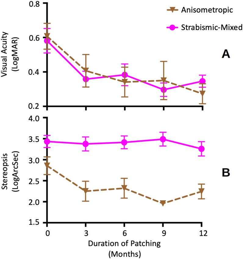
www.nature.com/scientificreports/
therapy can improve visual acuity in FMNS patients. Birch et al. have described an association between abnormal
stereo-acuity and patients with FMNS. They found that 67% of all children with abnormal stereo-acuity had
FMNS waveform, and nearly all children with nil stereo-acuity had FMNS w aveforms26. Bosworth and B irch31
have found that the risk for persistent amblyopia was greater among children with nil stereo-acuity than those
with measurable stereoacuity at treatment onset. The current standard of amblyopia treatment comprises of
part-time patching ranging from 2 to 6 h/day per the amblyopia severity as per the guidelines from the landmark
PEDIG Amblyopia Treatment Studies32–34. Little is known to date about the association between stereo-acuity
and part-time patching treatment in FMNS.
In the current manuscript, we measured fixation eye movements at the end of part-time patching treatment
and analyzed the rate of improvement of visual acuity and stereoacuity improvement in amblyopia patients with
and without FMNS. We hypothesize that the presence of FMNS, would be associated with a slower rate of visual
acuity improvement in amblyopic patients treated with patching therapy which requires monocular viewing
compared to patients without FMNS. We also hypothesize that the presence of FMNS will be associated with
minimal/no change in stereoacuity, as FMNS is a hallmark of binocular maldevelopment in early infancy. We
also analyzed the regression of amblyopia in patients after stopping part-time patching.
Results
Clinical type of amblyopia. The study subjects were categorized based on the clinical type (anisometropia:
n = 15, strabismic n = 3, mixed n = 22). The age of the start of patching treatment was similar in patients with
mixed/strabismic amblyopia compared to anisometropic amblyopia (anisometropic: 75 ± 17 months vs mixed/
strabismic: 64 ± 29 months, p = 0.36). Similarly, there was no difference in visual acuity at the time of diagnosis per
the clinical types of amblyopia (anisometropic = 0.64 ± 0.42 logMAR, strabismic/mixed = 0.53 ± 0.26 logMAR,
p = 0.54), while there was an expected significant difference in stereoacuity (anisometropic = 2.09 ± 0.66 logarc-
sec vs. strabismic/mixed = 2.82 ± 0.87 logarcsec, p = 0.002). Eight patients required strabismus surgery (Table 1).
There was no difference in age (surgery vs no surgery: 54 ± 33 months vs 66 ± 21 months, Mann Whitney U test
p = 0.47), visual acuity (surgery vs no surgery: 0.60 ± 0.30 log MARvs 0.58 ± 0.34 logMAR, Mann Whitney U Test
p = 0.48) and stereoacuity (surgery vs no surgery: 3.05 ± 0.85 logarcsec vs 2.45 ± 0.85 logarcsec, p = 0.693) at the
time of diagnosis between patients with strabismus requiring surgery versus those that did not require surgery.
Eye movement characteristics. The fixational eye movement traces obtained at the end of treatment
were evaluated, and amblyopic patients were classified based on the presence or absence of nystagmus (Fig. 1)35.
Patients without nystagmus (no nystagmus—Fig. 1A) exhibited alternating fixational saccades with inter-sac-
cadic drifts, similar to healthy subjects23,36,37. Patients with nystagmus were further classified into those with
FMNS (Fig. 1B) versus those that did not meet the criteria of FMNS (Fig. 1C). The presence of FMNS was
determined based on the classic reversal in the quick phase of nystagmus with linear/decreasing velocity nasally
directed slow phase observed during monocular viewing conditions28. Patients with nystagmus/nystagmus
like movements who did not exhibit the classic reversal in the direction of quick phases were characterized
as Nystagmus without FMNS (Nyst no FMN). These patients had jerk nystagmus with dynamic overshoots of
quick phases and differed from Infantile Nystagmus Syndrome patients in that their velocity was decreasing or
linear, unlike the increasing eye velocity characteristics seen in patients with Infantile Nystagmus Syndrome.
Also, patients with nystagmus but no FMNS did not have the Dissociated Vertical Deviation frequently seen
in FMNS patients. The fixational eye movements were evaluated, and amblyopic patients were classified into
those with no nystagmus (n = 18), those with FMNS (n = 8), and those with nystagmus but without the clas-
sic reversal in the quick phase of nystagmus seen in FMNS (n = 14). There was no difference in the age of the
start of patching per the fixational eye movement characteristics (No nystagmus = 68 ± 22 months, Nystagmus
no FMNS = 73 ± 19 months, FMNS = 60 ± 41 months, p = 0.38). Per the fixation eye movement characteristics,
there was no difference in visual acuity at the time of diagnosis (None = 0.52 ± 0.23 logMAR, Nystagmus no
FMNS = 0.62 ± 0.43 logMAR and FMNS = 0.50 ± 0.16 logMAR, p = 0.9), while there was a significant difference in
stereoacuity (None = 2.05 ± 0.56 logarcsec, Nystagmus no FMNS = 2.56 ± 0.84 logarcsec and FMNS = 3.49 ± 0.65
logarcsec, p = 0.001).
Improvement and type of amblyopia. Figure 2A,B plot the average and stdev of visual acuity and ste-
reo-acuity in the anisometropic and mixed/strabismic groups at baseline and within the first year after initiating
patching treatment. To determine statistical significance per different groups, we employed mixed-effect model
with random intercepts. We found that the visual acuity change within the first year of patching treatment was
found to be significantly different between the anisometropic versus strabismic/mixed amblyopia type (mixed
effect model: F = 5.9, p = 0.016). Both groups had improvement in visual acuity (indicated by negative values).
The average beta coefficient of visual acuity change ((log MAR visual acuity/3 months of patching treatment)
was greater in patients with anisometropia (− 0.025 ± 0.01) compared to the strabismic/mixed amblyopia group
(− 0.012 ± 0.02). We also found that the change of stereo-acuity within the first year of patching treatment was
significantly different between anisometropic versus strabismic/mixed groups (mixed effect model: F = 8.8,
p = 0.005). The average beta coefficient of stereo-acuity change (log arcsec/3 months of patching treatment) was
greater in the anisometropic group (− 0.05 ± 0.08) compared to strabismic/mixed amblyopia type (0.003 ± 0.04).
A positive value indicates a lack of improvement in the strabismic/mixed group.
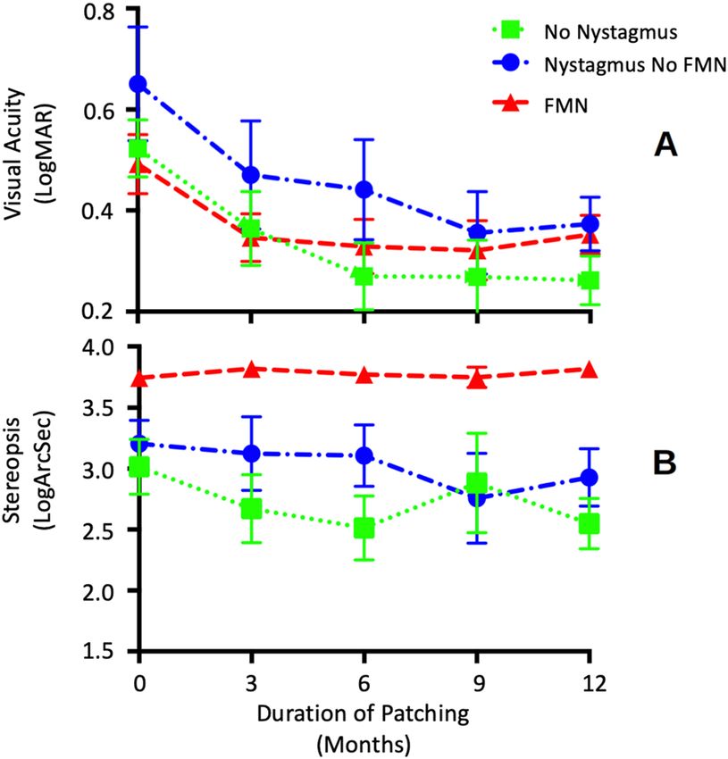
Improvement and fixational eye movements characteristics. Figure 3A,B plots the average and
stdev of visual acuity and stereo acuity for each subgroup at baseline and within the first year after initiating
patching treatment. To determine statistical significance between different groups, we employed a mixed-effect
Scientific Reports | (2021) 11:1217 | https://doi.org/10.1038/s41598-020-79077-5 2
Vol:.(1234567890)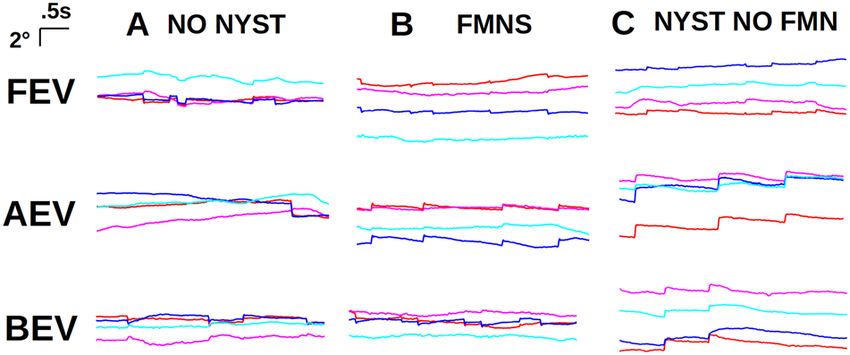
www.nature.com/scientificreports/
Age at patching Visual Acuity at Stereoacuity at Strabismus (near;
(duration) Category at time time of patching time of patching Eye movement Refraction (RE; distance) (prism Surgery and age
ID Gender (months) of patching (LogMAR) (arc second) characteristics LE) (diopters) diopters) (years)
Strabismic 1.4 Nil None + 6.5 ET 30 BMR
1 F 27 (30) REC
Severe 0.3 + 6.25 ET 30
Age 3
Mixed 0.5 200 None +5 ET 35 R&R RE
2 M 69 (26)
Moderate 0 +1 ET 35 Age 6
Mixed 0.4 100 None + 3.0 + 1.25 × 65 ET 12
3 F 82 (18)
Moderate 0.1 + 1.25 + 0.25 × 115 E(T) 4–6
Mixed 0.2 140 None + 2.5
4 M 44 (48) Ortho with glasses
Moderate 0.5 + 4.5
Mixed 0 100 None Plano + 0.50 × 95 XT 20
5 M 84 (9)
Moderate 0.4 − 0.75 + 3.5 × 85 XT 30
Mixed 0 140 None + 6.5 + 2.00 × 70
6 M 46 (75) Ortho with glasses
Severe 0.8 + 0.5 + 0.5 × 90
Mixed 0.1 50 None + 5.5 + 1.00 × 100
7 F 41 (22) Ortho with glasses
Moderate 0.3 + 6.5 + 1.0 × 80
Mixed 0 Nil None + 4.50 + 2.00 × 90 E(T) 8
8 F 77 (21)
Moderate 0.3 + 5.5 + 2.25 × 90 E(T) 10
Anisometropic 0 Nil None Plano + 0.75 × 95
9 M 79 (6) Ortho
Moderate 0.5 + 4.25 + 2.00 × 90
Anisometropic 0.5 80 None + 5 + 0.50 × 100
10 F 66 (14) Ortho
Moderate 0.1 + 3 + 0.50 × 80
Anisometropic 0.7 50 None + 7.5
11 F 60 (19) Ortho
Severe 0.1 + 5.0 + 0.50 × 180
Anisometropic 0.55 40 None + 4 + 0.50 × 10⁵
12 M 53 (15) Ortho
Moderate 0.2 + 0.5 + 0.5 × 85
Anisometropic 0 40 None − 0.25 + 0.5 × 90
13 F 117 (6) Ortho
Mild 0.2 Plano + 2 × 85
Anisometropic 0.4 60 None − 2.75 + 4.25 × 95
14 F 81 (6) Ortho
Moderate 0 + 1.5
Anisometropic 0.1 60 None + 0.5 + 1.00 × 90
15 F 53 (21) Ortho
Moderate 0.6 + 3.5 + 1.00 × 90
Anisometropic 0.4 40 None + 4.25 + 1.0 × 95
16 F 63 (6) Ortho
Moderate 0 + 1.75 + 0.25 × 80
Anisometropic 0.2 50 None Plano + 0.50 × 85
17 M 90 (30) Ortho
Moderate 0.5 + 5.25 + 2.00 × 10⁵
Anisometropic 1.2 140 None + 7.00 + 0.50 × 60
18 M 90 (22) Ortho
Severe 0 + 1.00 + 0.25 × 50
Strabismic 0 140 Nystagmus No + 2.75 + 0.50 × 180 ET 35 BMR
19 M 39 (21) REC
Moderate 0.3 FMNS + 2.75 + 0.50 × 180 ET 35
Age 3
Mixed 0.2 100 Nystagmus No − 1.75 + 3 × 85
20 F 83 (17) Ortho with glasses
Moderate 0.6 FMNS − 10.00 + 3.75 × 85
Mixed 0.3 Nil Nystagmus No + 2.25 + 0.75 × 80
21 F 66 (11) Ortho with glasses
Severe 0.7 FMNS + 3.5 + 0.5 × 135
Mixed 0.2 Nil Nystagmus No + 1.25 + 0.75 × 110
22 M 63 (27) Ortho with glasses
Mild 0 FMNS + 0.25 + 2.0 × 80
Mixed 0.4 200 Nystagmus No − 11.5 + 0.75 × 75 XT 14 RLR
23 F 95 (28) REC
Moderate 0.1 FMNS − 6.5 + 1.0 × 10⁵ XT 25
Age 8
Mixed 0.1 100 Nystagmus No + 6 + 2.0 × 90
24 M 102 (28) Ortho with glasses
Mild 0.2 FMNS + 7 + 1.75 × 90
Mixed 0.2 Nil Nystagmus No + 1.50 LE(T) 8
25 M 33 (31)
Severe 0.7 FMNS +4 LE(T) 10
Continued
Scientific Reports | (2021) 11:1217 | https://doi.org/10.1038/s41598-020-79077-5 3
Vol.:(0123456789)www.nature.com/scientificreports/
Age at patching Visual Acuity at Stereoacuity at Strabismus (near;
(duration) Category at time time of patching time of patching Eye movement Refraction (RE; distance) (prism Surgery and age
ID Gender (months) of patching (LogMAR) (arc second) characteristics LE) (diopters) diopters) (years)
Mixed 0.1 200 Nystagmus No − 0.75 + 0.5 × 75 XT 20 RLR
26 F 84 (20) REC
Moderate 0.4 FMNS + 1.5 + 1.00 × 90 XT 25
Age 8
Mixed 0 80 Nystagmus no + 1.25 + 1.5 × 100 Flick X(T)
27 M 85 (6)
Moderate 0.6 FMNS + 4.00 + 2.00 × 70 6 LX(T)
Anisometropic 0.1 140 Nystagmus No + 0.25 + 0.5 × 90
28 M 63 (34) Ortho
Severe 1.9 FMNS − 10.75 + 2.0 × 50
Anisometropic 0.2 100 Nystagmus No + 7.25 + 1.5 × 90
29 M 80 (39) Ortho
Moderate 0.4 FMNS + 8.25 + 1.5 × 100
Anisometropic 0.8 200 Nystagmus No + 6.75 + 3 × 90
30 M 75 (31) Ortho
Severe 0 FMNS + 0.5
Anisometropic 0.4 100 Nystagmus No + 4 + 1.25 × 85
31 F 71 (16) Ortho
Moderate 0 FMNS + 1.5 + 0.5 × 85
Anisometropic 0 80 Nystagmus No + 1.00 + 0.5 × 90
32 F 81 (25) Ortho
Moderate 0.5 FMNS + 3.75
Strabismic 0.7 Nil FMNS + 3.50 + 1.75 × 90 ET 45 BMR
33 F 14 (45) REC
Severe 0.2 + 3.50 + 1.75 × 90 ET 45
Age 1
Mixed 0.2 Nil FMNS + 5.00 + 0.50 × 90
34 M 72 (20) Ortho with glasses
Severe 0.7 + 6.25 + 1.00 × 95
Mixed 0.55 Nil FMNS − 9.5 + 2.5 × 165 ET 4
35 M 80 (6)
Moderate 0 plano + 0.75 × 45 ET 4
Mixed 0 Nil FMNS + 5 + 1.5 × 80
36 F 67 (38) Ortho with glasses
Moderate 0.6 + 6 + 1.5 × 95
Mixed 0.4 Nil FMNS − 6.75 + 3.75 × 90 XT 25 BLR
37 M 83 (25) REC
Severe 0.8 − 9.0 + 3.75 × 90 XT 45
Age 8
Mixed 0.3 Nil FMNS + 4.5 E(T) 30 BMR
38 M 18 (54) REC
Severe 0.2 + 3.5 ET 25
Age 3
Mixed 0.3 400 FMNS +4 XT 20
39 F 60 (38)
Moderate 0.1 + 2.25 XT 20
Mixed 0.5 200 FMNS + 8.00 + 1.5 × 90 ET 6–8
40 M 30 (79)
Moderate 0.3 + 7.25 + 0.5 × 90 ET 4
Table 1. Demographics, ophthalmic exam and strabismus surgery data of the enrolled subjects. BLR bilateral
lateral recti muscles, BMR bilateral medial recti muscles, CC with correction, ET esotropia, E(T) intermittent
esotropia, F female, FMNS fusion maldevelopment nystagmus syndrome, LE left eye, M male, RE right
eye, REC recession, R&R recession and resection, RLR right lateral rectus recession, XT exotropia, X(T)
intermittent exotropia.
Figure 1. Eye movement records. Eye positions vs time for patients without nystagmus (A), FMNS (B), and
with nystagmus but not FMNS (C), during fellow eye viewing (top row), amblyopic eye viewing (middle row),
and both eyes viewing (bottom row). Waveforms are plotted with a common scale indicated at upper left.
Red: right horizontal, blue: left horizontal, magenta: right vertical, cyan: left vertical. The positive vertical axis
corresponds to rightward and upward eye movements. Note the reversal in direction between FEV and AEV
in FMNS patients that is not present in nystagmus without FMN patient. Note also the greater intensity during
AEV and the absence of acceleration during slow phases in nystagmus patients both with and without FMNS.
Scientific Reports | (2021) 11:1217 | https://doi.org/10.1038/s41598-020-79077-5 4
Vol:.(1234567890)www.nature.com/scientificreports/
Figure 2. Visual acuity and stereoacuity per type of amblyopia. The mean and standard error of mean of
visual acuity (A) and stereoacuity (B) change sub-grouped by the type of amblyopia at baseline and during
the first year after initiating patching treatment. The lower scores indicate better visual acuity (log MAR)
and stereoacuity (log arc sec). visual acuity and stereoacuity improvement was greater in anisometropic than
strabismic/mixed group. Brown triangles—dashed line: anisometropic; magenta circles—full line: strabismic/
mixed.
Figure 3. Visual acuity and stereoacuity per fixational eye movements characteristcs. The mean and standard
error of mean of visual acuity (A) and stereoacuity (B) change sub-grouped by the fixation eye movement
characteristics at baseline and during the first year after initiating patching treatment. The lower scores indicate
better visual acuity (log MAR) and stereoacuity (log arc sec). Visual acuity and stereoacuity improvement was
greater in patients without nystagmus. No improvement of stereoacuity was recorded in FMNS group. Green
squares: no nystagmus; blue circles: nystagmus no FMNS; red triangles: FMNS.
Scientific Reports | (2021) 11:1217 | https://doi.org/10.1038/s41598-020-79077-5 5
Vol.:(0123456789)www.nature.com/scientificreports/
model with random intercepts. We found that the change of visual acuity within the first year of patching treat-
ment was found to be significantly different between no nystagmus, nystagmus without FMNS, and FMNS
groups (mixed effect model: F = 4.3, p = 0.04). All three groups had improvement in visual acuity (indicated by
negative values). The average beta coefficient of visual acuity change (log MAR visual acuity/3 months of patch-
ing treatment) was greater in patients with no nystagmus (− 0.025 ± 0.01) compared to nystagmus no FMNS (−
0.018 ± 0.02) and FMNS (− 0.01 ± 0.01) groups. We also found that the change of stereoacuity within the first year
of patching treatment was found to be significantly different between no nystagmus, nystagmus without FMNS,
and FMNS groups (mixed effect model: F = 5.8, p = 0.02). The average beta coefficient of stereoacuity change (log
arcsec/3 months of patching treatment) was greater in no nystagmus group (− 0.049 ± 0.09) compared to nys-
tagmus without FMNS (− 0.015 ± 0.03) group with no improvement in the FMNS group (0.02 ± 0.04). A positive
value indicates a lack of improvement in the FMNS group.
No improvement and regression. In our cohort, 17% of patients had regression with a decrease in visual
acuity after stopping patching treatment. All of the patients who experienced regression had strabismic/mixed
amblyopia and had received patching treatment for at least 6 months before the treatment was discontinued due
to a plateau in visual acuity or the resolution of amblyopia (defined as inter-ocular visual acuity difference of < 2
lines). Regression was noted in 2/18 patients without nystagmus, 1/14 patient with nystagmus without FMNS,
and 4/8 patients with FMNS. Of the two patients without nystagmus who experienced regression (Subject 3
and 4), both patients regained their visual acuity on restarting the patching treatment and had mild residual
amblyopia with some stereoacuity at the end of treatment. The patients with nystagmus without FMNS (Subject
22) and FMNS (Subjects 33, 38, 39, and 40) experienced regression and regained the visual acuity after restarting
treatment but did not have any stereoacuity at the end of treatment.
10% of our cohort had no improvement in visual acuity post patching treatment. These include one subject
with no nystagmus (Subjects 2), two with nystagmus without FMNS (Subject 23 and 27), and one with FMNS
(Subject 35). Patching was started after age 5 in all four subjects, and all of them had high anisometropia (2 aniso-
myopia, 2 with aniso-hyperopia). The two aniso-hyperopic patients opted for atropine penalization treatment.
Visual acuity improvement and treatment plateau. The improvement in visual acuity and stereoacu-
ity was analyzed between groups per the fixation eye movement characteristics and as a function of the clinical
type of amblyopia. Patients who did not have any improvement in visual acuity with patching treatment were
excluded from this analysis. Similar visual acuity improvement levels were found in patients with and with-
out FMNS (no nystagmus: 0.39 ± 0.28, nystagmus no FMNS: 0.33 ± 0.36, FMNS: 0.29 ± 0.16 LogMAR, F = 0.30,
p = 0.74). A similar analysis was performed as a function of the clinical type of amblyopia. The results were
comparable between anisometropic versus strabismic/mixed groups (visual acuity improvement: anisometropia:
0.41 ± 0.31 and strabismic/mixed: 0.29 ± 0.26 LogMAR, t-test p = 0.29). Children with no nystagmus plateaued
sooner in terms of visual acuity improvement compared to the no nystagmus group (no nystagmus: 9.3 ± 6.5;
nystagmus no FMNS: 14.2 ± 8.2; FMNS: 30.2 ± 23.2 months, one way ANOVA, F = 6.4, p = 0.005). Children with
anisometropic amblyopia plateaued sooner in terms of visual acuity improvement than strabismic/mixed ambly-
opia groups (anisometropia: 9.1 ± 6.8 months, strabismic/mixed: 22.1 ± 17 months, unpaired t test p = 0.01).
Stereoacuity improvement and treatment plateau. 21/40 patients had improvement in stereoacuity.
The majority of patients without nystagmus had improvement in stereoacuity (14/18) compared to nystagmus
no FMNS (6/14) and FMNS (1/8). The extent of stereoacuity improvement was analyzed in patients with and
without nystagmus—we excluded FMNS patients from this analysis as only 1 FMNS patient had improvement
in stereoacuity after 29 months of treatment. Of the patients that had improvement in stereoacuity, there was
a trend that no nystagmus had greater improvement (1.3 ± 0.76 log arcsec) compared to nystagmus no FMNS
group (0.96 ± 0.57 log arcsec, p = 0.15, Mann–Whitney U test). There was no difference in the time to reach the
best possible stereoacuity in patients without nystagmus (26 ± 19 months) versus those with nystagmus without
FMNS (20.5 ± 9.5 months, p = 0.5, Mann Whitney U test).
Of the 21 patients with improvement in stereoacuity, 14 had anisometropic amblyopia, and 7 had strabismic/
mixed amblyopia. The extent of stereoacuity improvement was similar irrespective of amblyopia type (aniso-
metropia: 1.2 ± 0.82 versus mixed/strabismic: 1.4 ± 0.30 log arcsec, Mann–Whitney U test p = 0.5). Like visual
acuity improvement, patients with anisometropic amblyopia (19.7 ± 12.5 months) reached the best possible
stereoacuity sooner with treatment than patients with strabismic/mixed amblyopia (34 ± 19.5 months, Mann
Whitney test p = 0.02).
Discussion
In this retrospective study, we characterized fixational eye movements at the end of treatment in amblyopic
patients treated with part-time patching therapy and evaluated the rate of improvement in visual acuity and
stereoacuity. We found that the presence of FMNS was associated with a slower rate of visual acuity improve-
ment and poor recovery of stereopsis. Amblyopic patients with nystagmus without the reversal in the direction
of the quick phase, as seen in FMNS, had a similar rate of improvement in visual acuity but less improvement
in stereoacuity compared to patients without nystagmus. The velocity waveform of nystagmus differs from that
seen in patients with idiopathic infantile nystagmus syndrome. Nystagmus that is not FMNS or INS, has been
previously reported in patients with monocular vision loss in early childhood38,39. Amblyopic patients have
increased drifts. Thus, reduced visual acuity due to amblyopia and the increased drifts interrupted by corrective
saccades could result in the development of nystagmus beats in the absence of FMNS/INS. We found that the
rate of visual acuity and stereoacuity improvement was faster in anisometropic amblyopia and plateaued sooner
Scientific Reports | (2021) 11:1217 | https://doi.org/10.1038/s41598-020-79077-5 6
Vol:.(1234567890)www.nature.com/scientificreports/
with part-time patching than strabismic/mixed amblyopia patients. Similarly, patients without nystagmus (both
anisometropic and strabismic, or mixed, amblyopia) plateaued sooner than those with nystagmus. The patients
who experienced regression had strabismic/mixed amblyopia and had received part-time patching treatment
for at least 6 months before the treatment was discontinued. The risk of regression was greater in FMNS patients
and required longer durations of treatment than amblyopic patients without FMNS.
Effect of patching on visual acuity. A few studies describe the visual acuity improvement as a function
of patching duration. Stewart et al. found an average visual acuity improvement of 0.35 logMAR from a cumula-
tive dose of patching for 150–250 h irrespective of the type of amblyopia and a flattening of the dose/response
curve after 400 h of t reatment40. ATS2A and A TS2B32,33 did not find any differences in the extent of visual acuity
improvement at 17 weeks in groups per the clinical type of amblyopia- however, they found that patients with
worse initial visual acuity and age at treatment with children < 5 years of age had greater visual acuity improve-
ment. In a large retrospective study of 877 patients recruited per the PEDIG ATS2 A and B inclusion c riteria34,
the authors found similar levels of visual acuity improvement, as reported in PEDIG studies. However, the treat-
ment duration was longer, probably due to the differences in motivation for compliance and follow-ups between
patients included in trials versus in real-world clinical practice. We have previously described the results of visual
acuity and stereoacuity measurements obtained at the end of part-time patching therapy per the clinical type
of amblyopia and per the presence of F MNS41. We found that anisometropic patients had less severe residual
amblyopia at the end of part-time patching treatment. We found that the visual acuity of patients with FMNS
improved with part-time patching but required longer treatment duration with poor stereoacuity at the end
of the treatment. In the current study, we found although visual acuity improved in patients with and without
FMNS within the first year of treatment, but the presence of FMNS was associated with a lower rate of visual
acuity improvement. The treatment plateau occurs sooner in patients without nystagmus (average 39 weeks)
compared to those with nystagmus without FMNS (60 weeks) and FMNS patients (> 2 years). We computed
the treatment duration, which included the time when patching had to resume patching due to regression of
the amblyopic eye visual acuity. We also had 4 (10%) children < 3 years of age. These could potentially result in
greater treatment durations reported in our study than other studies32–34.
In our cohort, four patients did not have any visual acuity improvement after part-time patching. All these
patients had mixed amblyopia with high anisometropia (> 4 diopters) and initiated patching treatment at age
5.5 years or older. These results agree with other studies that have reported high anisometropia and late age at
therapy as risk factors for no improvement in visual acuity post patching treatment26,34,42–47.
Effect of patching on stereoacuity. We analyzed the stereoacuity improvement in patients with and
without FMNS and per the clinical type of amblyopia. We found that patients without nystagmus and anisome-
tropic amblyopia had better stereoacuity at baseline, and both the visual acuity and stereoacuity improved with
treatment. On the other hand, we found that patients with FMNS typically had no stereoacuity improvement
despite the reduction of visual acuity deficit in the amblyopic eye with part-time patching treatment. This is in
agreement with an observational study by Birch et al., where they reported that none of the amblyopic patients
with normal stereoacuity had FMNS, whereas 67% of children with abnormal stereoacuity had FMNS. In con-
trast, all the children with nil stereoacuity had FMNS w aveforms26. Also, in patients without FMNS, the stereo-
acuity improvement rate was slower, and the plateau time was higher than that of visual acuity improvement.
This is likely due to the delayed development of fine stereoacuity, which is thought to still be immature at 5 years
of age and with adult levels reaching between 6 and 9 years of age48–51.
Regression. In our cohort, we found a regression risk of 17%. A few other studies have reported similar
regression rates17,18. The PEDIG study 2004 has reported regression of 24% following patching therapy, with
6% of patients patching for more than 8 h/day52. Another PEDIG study reported regression of 7% within the
first year of treatment cessation in older children between the ages of 7–12 years53. Other studies have found a
regression rate of 24–27% in children after full-time occlusion therapy with a gradual taper19 versus an abrupt
taper54. Studies have found that the risk of regression inversely correlates with the patient’s age at termination of
treatment19. Other factors reported to be associated with regression are better visual acuity at the time of cessa-
tion of patching, greater visual acuity improvement during treatment, or previous r egression55,56. In our cohort,
one of our patients had experienced previous regression. All the patients except one who experienced regression
were < 6 years of age. Overall we did not see a systematic trend between the risks of regression versus the level
of visual acuity improvement. The differences between regression rates between our and other studies could be
due to the varying ages of children in our cohort and the strictly part-time patching employed in our study. In
our cohort, we found a significantly higher proportion of regression in patients with FMNS (50%) than in other
groups (10%), with regression occurring only in those with strabismic/mixed form of amblyopia.
Increased risk of recurrence has been reported previously in patients with mixed amblyopia15,17. Nilsson
et al. have reported the presence of microstrabismus alone as a risk factor of recurrence18. Holmes et al. and
Rutstein and Fuhr found that excellent stereoacuity does not preclude the recurrence of amblyopia55,57. On the
other hand, Bosworth and Birch reported the risk for persistent amblyopia was 2.2 times greater among children
with nil s tereoacuity31. Birch et al. have also described a higher rate of persistent amblyopia in patients affected
by infantile esotropia (up to 60%) than accommodative e sotropia26. In our cohort, we found that patients with
strabismic or mixed amblyopia and FMNS both had greater chances to develop regression, whereas patients with
strabismus without FMNS had similar levels of visual acuity and stereoacuity improvement as anisometropic
amblyopes with a lower risk of regression.
Scientific Reports | (2021) 11:1217 | https://doi.org/10.1038/s41598-020-79077-5 7
Vol.:(0123456789)www.nature.com/scientificreports/
Animal model studies have shown that disruption of binocularity during infancy is invariably associated with
gaze instabilities, most often FMNS6,58. Tychsen and colleagues have shown in experiments that the prevalence
and severity of FMNS increases with the longer duration of binocular decorrelation with 100% prevalence of
FMNS in primates who are exposed to periods of binocular decorrelation that is equivalent to 3 months in
humans. Tychsen has proposed that the binocular maldevelopment of the striate cortex is passed on to down-
stream extrastriate regions, namely the medial superior temporal area that drive conjugate gaze. The disruption
results in a nasalward bias that is pathognomic of FMNS. Thus, animal model studies suggest that the develop-
ment of FMNS is strongly associated with abnormal visual experience in infancy and can be used as a surrogate
marker of the presence of amblyogenic risk factors/strabismus in the first year of l ife59.
Amblyopic patients both with and without nystagmus have fixation eye movement abnormalities compared
to controls23,37,60. We have found that patients without nystagmus have a reduced frequency of physiologic
microsaccades in the amblyopic eye compared to the fellow eye with increased inter-saccadic drifts in both the
fellow and amblyopic eye. Patients with nystagmus with and without FMNS have increased slow phase velocities
compared to inter-saccadic drift velocities in patients without nystagmus. We have also found that the slow phase
velocities of FMNS patients are greater compared to patients with nystagmus without FMNS60. In the current
paper, the analysis shows that FMNS is associated with a slower visual acuity rate of improvement, poor stereo-
acuity recovery, and higher regression rate with part-time patching treatment. We also found that patients with
nystagmus but not FMNS tended to respond better to patching treatment than those with FMNS, even though
eye movement abnormalities are still present. Thus, we speculate that patients with FMNS are likely to have early
onset of amblyopia than those without FMNS, resulting in differences between the treatment outcomes in this
cohort. The findings from our paper highlight the utility of fixational eye movement recordings in amblyopic
patients in order to advance our understanding and field of knowledge of residual/recurrent amblyopia and to
improve amblyopia therapy for specific types of amblyopia.
The study’s main limitations are that the eye movement recordings were obtained at the end of treatment and
that the treatment effect was determined based on a retrospective chart review. To reduce the impact of inaccurate
data and individual testing biases, we excluded patients with incomplete data or that were not interpreted the
same by at least two independent reviews. In our experience, these patients were noncompliant with treatment
and did not follow up as frequently in our office per the recommendations. When evaluating the efficacy of
patching treatment, it is essential to consider the effects of compliance. While we could not measure objective
compliance, we did extract data obtained from clinical history to determine subjective compliance and included
patients thought to be at least 50% compliant. Since the study is a longitudinal follow-up over a period of years,
the visual acuity testing method differs depending on the techniques judged appropriate for the child’s maturity.
Regression was determined by two separate measurements with the same testing method to reduce the bias. The
study reflects real-world scenario mimicking as encountered in clinical practice.
In summary, we examined the association between the presence of FMNS (confirmed on eye movement
recordings) and the rate of improvement of visual acuity and stereoacuity and regression in amblyopia patients
treated with part-time patching. We found that patients without nystagmus have a faster improvement of visual
acuity and stereoacuity and plateaued sooner to reach their best possible visual acuity. FMNS is seen in patients
with strabismic/mixed amblyopia, and the presence of FMNS was associated with a slower rate of improvement
in visual acuity with poor/absent recovery of stereoacuity and a higher risk of regression. Thus, these results col-
lectively highlight the link between the lack of binocular function and recurrent amblyopia. The current study’s
data suggest that eye movement characterization and quantification can play an essential role in amblyopia
management. Children with FMNS and amblyopia should be observed closely with long-term follow-up and
with a careful taper of the patching treatment. Future prospective studies, that measure FEMs at the time of
treatment initiation will allow us to directly probe and understand the association of severity of visual acuity
and stereoacuity deficits at the time of diagnosis and effects of different amblyopia treatments in patients with
FMNS and other FEM abnormalities.
Methods
Study participants. Eye movement recordings were obtained in 80 amblyopic patients without any struc-
tural anomalies of the eye or neurologic disorders. The Cleveland Clinic Institutional Review Board approved
the protocol and written informed consent was obtained from each participant or parent/legal guardian in
accordance with the Declaration of Helsinki. The clinical parameters were extracted from a retrospective chart
review for all the enrolled subjects. After review, we recruited 40 patients who had at least 12 months of follow
up after initiating patching treatment and three sets of measurements, first at baseline, the second measurements
between 3 and 6 months and third measurement between 9 and 12 months after initiating treatment, were
included. Patients deemed to be at least 50% compliant were included in the study.
We categorized them based on the clinical type of amblyopia61 and on the fixational eye movements waveform
characteristics (Table 1). Patients with manifest strabismus were treated according to the American Academy of
Ophthalmology Preferred Practice P attern62.
Eye movement recording and analysis. A high-resolution video-based eye tracker (EyeLink 1000®, SR
Research, Ontario, Canada) was used to measure binocular horizontal and vertical eye positions during bin-
ocular, fellow and amblyopic eye viewing conditions. All eye movement recordings were obtained at the end of
patching treatment. An infrared permissive filter that blocked the visible light but allowed eye movement meas-
urements of the non-viewing eye was used. Monocular calibration and validation were performed per the manu-
facturer’s guidelines. The subjects fixated their gaze on a circular target projected on the LCD screen on a white
background (luminance 144 cd/m2) in a completely dark room for 45 s. The eye position data was analyzed after
Scientific Reports | (2021) 11:1217 | https://doi.org/10.1038/s41598-020-79077-5 8
Vol:.(1234567890)www.nature.com/scientificreports/
removal of blinks. The eye position signal was differentiated using MatlabTM (Mathworks, Natick, MA, USA)
differential function and was further smoothened with the Savitzkey–Golay filter to measure eye velocity22,35.
Fixational saccades and quick phases of nystagmus were identified using an unsupervised clustering method35.
Drifts and slow phases were defined as epochs between fixational saccades and quick phases, respectively.
Measurement of visual acuity, stereoacuity and strabismus. The clinical parameters were extracted
from a retrospective chart review. The ages at the start of treatment and at follow up visits, visual acuity of the fel-
low and amblyopic eye and stereoacuity, cycloplegic refraction, strabismus angle measurements, and treatment
compliance was noted. Visual acuity was measured in each eye monocularly, starting from the right eye, using
the participant’s optimal spectacle correction with Snellen linear optotype. For patients younger than 6 years
of age, per the child’s ability to perform the test, crowding bars HOTV optotypes were preferred and used over
picture optotypes (Allen optotypes with crowding bars presented with commercially available computer-based
system Accomodata Stimuli™). Visual acuity was measured at 20 feet distance, and the value was considered
only if the patient could read all the letters (or symbols) of the line. Stereoacuity was measured with the Titmus
Stereo Test at 40 cm. For analyses, visual acuity scores were converted into logMAR values, and stereoacuity
scores in seconds of arc were converted to log arcsec values. For the purpose of analysis, subjects with no detect-
able (nil) stereoacuity were assigned a value of 7000″. There were only four patients that were diagnosed before
their ability to perform any optotype and stereo-testing—they all had manifest strabismus with strong fixation
preferencee (Table 1, patients n. 1, 25, 33, 38).
These four patients were all assigned as having severe amblyopia with absent stereoacuity. The strabismus
was assessed in the primary position at distance and near measured by alternate and simultaneous prism cover
tests and Hirschberg and Krimsky tests in younger patients. The clinical categorization of amblyopia subtype
and severity at the time of diagnosis was based on PEDIG s tudies32,33,47,63.
Amblyopia treatment and measurement. The treatment comprised of part-time occlusion (2–6 h/
day), prescribed per the severity of amblyopia32,64. Strabismic patients were diagnosed before other groups
(anisometropic vs strabismic vs mixed: 71.7 ± 15.9 vs 23.7 ± 13.4 vs 59.7 ± 15.1 months, Kruskal–Wallis Test
p = 0.016), while no differences in presentation time to start of patching treatment were observed grouped
per fixational eye movement characteristics (no nystagmus vs nystagmus no FMNS vs FMNS: 68.2 ± 21.1 vs
62 ± 23.9 vs 49.8 ± 28.1 months, Kruskal–Wallis Test p = 0.346). Investigators judged patching compliance to be
good (> 50%), fair (26–50%), or poor (≤ 25%), based on discussions with the parents documented in the chart
comparing the number of hours prescribed and the ones declared by the parents including the number of daily
and weekly hours of patching treatment65.
Visual acuity and stereoacuity from the start to the end of treatment were computed as a function of the
clinical type and fixation eye movements characteristics. We also calculate the rate of visual acuity and stereo-
acuity change within the first year. The rate of improvement was analyzed as a function of the clinical type of
amblyopia and fixational eye movements characteristics. Patients who did not have any improvement in visual
acuity on two consecutive visits were not included in this analysis as they were considered to be non-responsive
to patching treatment. These patients opted for either atropine penalization or stopped the treatment. The patch-
ing treatment was continued beyond the first year per the clinical management. Patching was discontinued if the
visual acuity had stabilized with no further improvement or deterioration ≥ 2 consecutive visits ≥ 6 weeks apart in
patients with at least 50% compliance. Patients who were patching 6 h/day were gradually weaned of treatment
as the visual acuity i mproved52. After the patching treatment was discontinued, the visual acuity and stereoacu-
ity measurements obtained at 3 months interval were recorded to detect regression. Regression was defined as
a drop in visual acuity by 2 lines as obtained by two separate measurements (on the same or different day) from
the previous visit, and treatment was restarted in these patients52. The duration of patching treatment (treatment
plateau in months) required to reach the best possible visual acuity and stereoacuity with no further improve-
ment or regression was analyzed as a function of clinical subtype and fixational eye movements characteristics.
Statistical analysis. All analyses were performed in SPSS and GraphPad Prism 7 (La Jolla, CA, USA). A t
test, Kruskal–Wallis Test, Mann Whitney U Test and one-way ANOVA were used to compare the demographics
and baseline characteristics amongst the groups.
Mixed-effects regression models with random intercepts to test the hypothesis of a difference in the rate
of change in the visual acuity and stereoacuity between patients per fixational eye movements characteristics
was used. The hypothesis was assessed by comparing the slope of change in the LogMAR visual acuity over the
12-month period per the fixational eye movement characteristics with a negative slope reflecting visual acuity
improvement. The slope of change in the log arc seconds stereoacuity over the 12-month period per the fixa-
tional eye movement characteristics was compared with a negative slope reflecting stereoacuity improvement.
We also performed linear regression separately for each patient (visual function versus treatment duration) and
extracted the beta coefficient (degree of visual function change for every 3 months of patching treatment). We also
report the average beta coefficient values for a given subgroup. A similar analysis per the different clinical types
of amblyopia: anisometropic and strabismic/mixed was done (patients with strabismus and mixed amblyopia
were pooled together as there were few strabismic patients n = 3). One-way ANOVA was used to compare the
total improvement of visual acuity and stereoacuity and treatment duration grouped per the fixational eye move-
ment characteristics. A t test was used to analyze these parameters as a function of the clinical type of amblyopia
(strabismic/mixed versus anisometropic patients).
Scientific Reports | (2021) 11:1217 | https://doi.org/10.1038/s41598-020-79077-5 9
Vol.:(0123456789)www.nature.com/scientificreports/
Received: 9 October 2020; Accepted: 3 December 2020
References
1. von Noorden, G. & Campos, E. Binocular Vision and Ocular Motility: Theory and Management of Strabismus (C.V. Mosby Co,
Maryland Heights, 2002).
2. Wong, A. M. F. New concepts concerning the neural mechanisms of amblyopia and their clinical implications. Can. J. Ophthalmol.
47, 399–409 (2012).
3. Löwel, S. & Singer, W. Selection of intrinsic horizontal connections in the visual cortex by correlated neuronal activity. Science
(80–) 255, 209–212 (1992).
4. Löwel, S. & Engelmann, R. Neuroanatomical and neurophysiological consequences of strabismus: Changes in the structural and
functional organization of the primary visual cortex in cats with alternating fixation and strabismic amblyopia. In Strabismus vol.
10 95–105 (Strabismus, 2002).
5. Trachtenberg, J. T. & Stryker, M. P. Rapid anatomical plasticity of horizontal connections in the developing visual cortex. J. Neurosci.
21, 3476–3482 (2001).
6. Tychsen, L. Fusion maldevelopment (Latent) nystagmus: How insights from nonhuman primate experiments have benefitted
clinical practice. In Advances in Translational Neuroscience of Eye Movement Disorders 255–270 (Springer, Berlin, 2019).
7. Tychsen, L. et al. The neural mechanism for Latent (fusion maldevelopment) nystagmus. J. Neuroophthalmol. 30, 276–283 (2010).
8. Tychsen, L. Causing and curing infantile esotropia in primates: The role of decorrelated binocular input (an American Ophthal-
mological Society thesis). Trans. Am. Ophthalmol. Soc. 105, 564–593 (2007).
9. Tychsen, L. Binocular vision. In Adler’s Physiology of the Eye; Clinical Application (Mosby, Maryland Heights, 1992).
10. Hasany, A., Wong, A., Foeller, P., Bradley, D. & Tychsen, L. Duration of binocular decorrelation in infancy predicts the severity of
nasotemporal pursuit asymmetries in strabismic macaque monkeys. Neuroscience 156, 403–411 (2008).
11. Committee, N. E. I./N. I. H. A Classification of Eye Movement Abnormalities and Strabismus (CEMAS). Report of a National Eye
Institute Sponsored Workshop. (2001).
12. Tusa, R. J., Mustari, M. J., Das, V. E. & Boothe, R. G. Animal models for visual deprivation-induced strabismus and nystagmus.
Ann. N. Y. Acad. Sci. 956, 346–360 (2002).
13. Repka, M. X. et al. Two-year follow-up of a 6-month randomized trial of atropine vs patching for treatment of moderate amblyopia
in children. Arch. Ophthalmol. 123, 149–157 (2005).
14. Holmes, J. M. et al. Risk of amblyopia recurrence after cessation of treatment. J. AAPOS 8, 420–428 (2004).
15. Levartovsky, S., Oliver, M., Gottesman, N. & Shimshoni, M. Factors affecting long term results of successfully treated amblyopia:
Initial visual acuity and type of amblyopia. Br. J. Ophthalmol. 79, 225–228 (1995).
16. Wallace, D. K. et al. Time course and predictors of amblyopia improvement with 2 hours of daily patching. JAMA Ophthalmol.
133, 606–609 (2015).
17. Tacagni, D. J., Stewart, C. E., Moseley, M. J. & Fielder, A. R. Factors affecting the stability of visual function following cessation of
occlusion therapy for amblyopia. Graefe’s Arch. Clin. Exp. Ophthalmol. 245, 811–816 (2007).
18. Nilsson, J., Baumann, M. & Sjöstrand, J. Strabismus might be a risk factor for amblyopia recurrence. J. AAPOS 11, 240–242 (2007).
19. Bhola, R., Keech, R. V., Kutschke, P., Pfeifer, W. & Scott, W. E. Recurrence of amblyopia after occlusion therapy. Ophthalmology
113, 2097–2100 (2006).
20. González, E. G., Wong, A. M. F., Niechwiej-Szwedo, E., Tarita-Nistor, L. & Steinbach, M. J. Eye position stability in amblyopia and
in normal binocular vision. Invest. Ophthalmol. Vis. Sci. 53, 5386–5394 (2012).
21. Subramanian, V., Jost, R. M. & Birch, E. E. A quantitative study of fixation stability in amblyopia. Invest. Ophthalmol. Vis. Sci. 54,
1998–2003 (2013).
22. Shaikh, A. G., Otero-Millan, J., Kumar, P. & Ghasia, F. F. Abnormal fixational eye movements in amblyopia. PLoS One 11, 20 (2016).
23. Kang, S. L., Beylergil, S. B., Otero-Millan, J., Shaikh, A. G. & Ghasia, F. Fixational eye movement waveforms in amblyopia: Char-
acteristics of fast and slow eye movements. J. Eye Mov. Res. (2019) (in press).
24. Chen, D., Otero-Millan, J., Kumar, P., Shaikh, A. G. & Ghasia, F. F. Visual search in amblyopia: Abnormal fixational eye movements
and suboptimal sampling strategies. Invest. Ophthalmol. Vis. Sci. 59, 4506–4517 (2018).
25. Birch, E. E., Subramanian, V. & Weakley, D. R. Fixation instability in anisometropic children with reduced stereopsis. J. AAPOS
17, 287–290 (2013).
26. Birch, E. E. Amblyopia and binocular vision. Prog. Retin. Eye Res. 33, 67–84 (2013).
27. Duke-Elder, S. & Wybar, K. C. Ocular motility and strabismus. In System of Ophthalmology 824 (Mosby CV, Maryland Heights,
1973).
28. Abadi, R. V. & Scallan, C. J. Waveform characteristics of manifest latent nystagmus. Invest. Ophthalmol. Vis. Sci. 41, 3805–3817
(2000).
29. von Noorden, G. K., Avilla, C., Sidikaro, Y. & LaRoche, R. Latent nystagmus and strabismic amblyopia. Am. J. Ophthalmol. 103,
87–89 (1987).
30. Simonsz, H. J. The effect of prolonged monocular occlusion on latent nystagmus in the treatment of amblyopia. Doc. Ophthalmol.
72, 375–384 (1989).
31. Bosworth, R. G. & Birch, E. E. Binocular function and optoype-grating acuity discrepancies in amblyopic children. Invest. Oph-
thalmol. Vis. Sci. 44, e3183 (2003).
32. Holmes, J. M. et al. A randomized trial of prescribed patching regimens for treatment of severe amblyopia in children. Ophthalmol-
ogy 110, 2075–2087 (2003).
33. Repka, M. X. et al. A randomized trial of patching regimens for treatment of moderate amblyopia in children. Arch. Ophthalmol.
121, 603 (2003).
34. Buckle, M., Billington, C., Shah, P. & Ferris, J. D. Treatment outcomes for amblyopia using PEDIG amblyopia protocols: A retro-
spective study of 877 cases. J. AAPOS 23(98), e1-98.e4 (2019).
35. Otero-Millan, J., Castro, J. L. A., Macknik, S. L. & Martinez-Conde, S. Unsupervised clustering method to detect microsaccades.
J. Vis. 14, 18–18 (2014).
36. Martinez-Conde, S. Fixational eye movements in normal and pathological vision. Prog. Brain Res. 154, 151–176 (2006).
37. Shaikh, A. G. & Ghasia, F. F. Fixational saccades are more disconjugate in adults than in children. PLoS One 12, 20 (2017).
38. Schneider, R. M. et al. Neurological basis for eye movements of the blind. PLoS One 8, e56556 (2013).
39. Felius, J. et al. Nystagmus and related fixation instabilities following extraction of unilateral infantile cataract in the Infant Aphakia
Treatment Study (IATS). Investig. Ophthalmol. Vis. Sci. 55, 5332–5337 (2014).
40. Stewart, C. E., Stephens, D. A., Fielder, A. R. & Moseley, M. J. Modeling dose-response in amblyopia: Toward a child-specific
treatment plan. Investig. Ophthalmol. Vis. Sci. 48, 2589–2594 (2007).
41. Scaramuzzi, M. et al. Fixation instability in amblyopia: Oculomotor disease biomarkers predictive of treatment effectiveness. In
Progress in Brain Research Vol. 249 235–248 (Elsevier, New York, 2019).
42. Chen, P. L. et al. Anisometropic amblyopia treated with spectacle correction alone: Possible factors predicting success and time
to start patching. Am. J. Ophthalmol. 143, 54–60 (2007).
Scientific Reports | (2021) 11:1217 | https://doi.org/10.1038/s41598-020-79077-5 10
Vol:.(1234567890)www.nature.com/scientificreports/
43. Holmes, J. M. et al. Effect of age on response to amblyopia treatment in children. Arch. Ophthalmol. (Chicago, Ill. 1990) 129,
1451–1457 (2011).
44. Pediatric Eye Disease Investigator Group. A comparison of atropine and patching treatments for moderate amblyopia by patient
age, cause of amblyopia, depth of amblyopia, and other factors. Ophthalmology 110, 1632–1638 (2003).
45. Weakley, D. R. The association between nonstrabismic anisometropia, amblyopia, and subnormal binocularity. Ophthalmology
108, 163–171 (2001).
46. Weakley, D. R. The association between anisometropia, amblyopia, and binocularity in the absence of strabismus. In Transactions
of the American Ophthalmological Society Vol. 97 987–1021 (American Ophthalmological Society, New York, 1999).
47. Cotter, S. A. et al. Treatment of anisometropic amblyopia in children with refractive correction. Ophthalmology 113, 895–903
(2006).
48. Giaschi, D., Narasimhan, S., Solski, A., Harrison, E. & Wilcox, L. M. On the typical development of stereopsis: Fine and coarse
processing. Vis. Res. 89, 65–71 (2013).
49. Parrish, E. E., Giaschi, D. E., Boden, C. & Dougherty, R. The maturation of form and motion perception in school age children.
Vis. Res. 45, 827–837 (2005).
50. Hayward, J., Truong, G., Partanen, M. & Giaschi, D. Effects of speed, age, and amblyopia on the perception of motion-defined
form. Vis. Res. 51, 2216–2223 (2011).
51. Hadad, B.-S., Maurer, D. & Lewis, T. L. Long trajectory for the development of sensitivity to global and biological motion. Dev.
Sci. 14, 1330–1339 (2011).
52. Pediatric Eye Disease Investigator Group. Risk of amblyopia recurrence after cessation of treatment. J. AAPOS 8, 420–428 (2004).
53. Hertle, R. W. et al. Stability of visual acuity improvement following discontinuation of amblyopia treatment in children aged 7 to
12 years. Arch. Ophthalmol. 125, 655–659 (2007).
54. Walsh, L. A., Hahn, E. K. & LaRoche, G. R. The method of treatment cessation and recurrence rate of amblyopia. Strabismus 17,
107–116 (2009).
55. Holmes, J. M., Melia, M., Bradfield, Y. S., Cruz, O. A. & Forbes, B. Factors associated with recurrence of amblyopia on cessation
of patching. Ophthalmology 114, 1427–1432 (2007).
56. Saxena, R. et al. Factors predicting recurrence in successfully treated cases of anisometropic amblyopia. Indian J. Ophthalmol. 61,
630–633 (2013).
57. Rutstein, R. P. & Fuhr, P. S. Efficacy and stability of amblyopia therapy. Optom. Vis. Sci. 69, 747–754 (1992).
58. Upadhyaya, S. et al. Fixational saccades and their relation to fixation instability in strabismic monkeys. Investig. Ophthalmol. Vis.
Sci. 58, 5743–5753 (2017).
59. Tychsen, L. Can ophthalmologists repair the brain in infantile esotropia? Early surgery, stereopsis, monofixation syndrome, and
the legacy of Marshall Parks. J. AAPOS 9, 510–521 (2005).
60. Scaramuzzi, M. et al. Part time patching treatment outcomes in children with amblyopia with and without fusion maldevelopment
nystagmus: An eye movement study. PLoS One 15, e0237346 (2020).
61. Manh, V. M. et al. A randomized trial of a binocular ipad game versus part-time patching in children aged 13 to 16 years with
amblyopia. Am. J. Ophthalmol. 186, 104–115 (2018).
62. American Academy of Ophthalmology. Pediatric Ophthalmology/Strabismus Summary Benchmarks. https: //www.aao.org/summa
ry-benchmark-detail/pediatric-ophthalmology-strabismus-summar y-benchma (2019).
63. Holmes, J. M. et al. A randomized trial of binocular dig rush game treatment for amblyopia in children aged 7 to 12 years. Oph-
thalmology 126, 456–466 (2019).
64. Repka, M. X. et al. Two-year follow-up of a 6-month randomized trial of atropine vs patching for treatment of moderate amblyopia
in children. Arch. Ophthalmol. (Chicago, Ill. 1960) 123, 149–157 (2005).
65. Pediatric Eye Disease Investigator Group. A randomized trial of atropine vs patching for treatment of moderate amblyopia in
children. Arch. Ophthalmol. 120, 268 (2002).
Acknowledgements
Supported by Grants from Blind Children’s Center, RPB Unrestricted Grant CCLCM-CWRU, CTSC Pilot Grant
Program, Research to Prevent Blindness Walt and Lilly Disney Award for Amblyopia Research and Cleveland
Clinic RPC Grant (FG) and Departmental NEI T32 Grant (JM).
Author contributions
M.S. has made substantial contributions in the acquisition, analysis, and interpretation of data, has drafted the
work, has approved the submitted version and has agreed both to be personally accountable for the author’s own
contributions and to ensure that questions related to the accuracy or integrity of any part of the work, even ones
in which the author was not personally involved, are appropriately investigated, resolved, and the resolution
documented in the literature. J.M. has made substantial contributions in the acquisition, analysis, and inter-
pretation of data, has approved the submitted version and has agreed both to be personally accountable for the
author’s own contributions and to ensure that questions related to the accuracy or integrity of any part of the
work, even ones in which the author was not personally involved, are appropriately investigated, resolved, and
the resolution documented in the literature. P.J. has revised the article, has approved the submitted version and
has agreed both to be personally accountable for the author’s own contributions and to ensure that questions
related to the accuracy or integrity of any part of the work, even ones in which the author was not personally
involved, are appropriately investigated, resolved, and the resolution documented in the literature. A.G.S. has
made substantial contributions to the conception or design of the work, in the creation of new software used in
the work, has approved the submitted version and has agreed both to be personally accountable for the author’s
own contributions and to ensure that questions related to the accuracy or integrity of any part of the work, even
ones in which the author was not personally involved, are appropriately investigated, resolved, and the resolu-
tion documented in the literature. F.F.G. has made substantial contributions to the conception or design of the
work, in the acquisition, analysis, or interpretation of data, in the creation of new software used in the work, has
drafted the work, has approved the submitted version and has agreed both to be personally accountable for the
author’s own contributions and to ensure that questions related to the accuracy or integrity of any part of the
work, even ones in which the author was not personally involved, are appropriately investigated, resolved, and
the resolution documented in the literature.
Scientific Reports | (2021) 11:1217 | https://doi.org/10.1038/s41598-020-79077-5 11
Vol.:(0123456789)You can also read