Safety and Efficacy of a Novel Shunt Surgery Combined with Foam Sclerotherapy of Varices for Prehepatic Portal Hypertension: A Pilot Study - SciELO
←
→
Page content transcription
If your browser does not render page correctly, please read the page content below
ORIGINAL ARTICLE

Safety and Efficacy of a Novel Shunt Surgery
Combined with Foam Sclerotherapy of Varices for
Prehepatic Portal Hypertension: A Pilot Study
Zhe Zhang0 0 -0 01-6965-362 ,I Xueming Chen0 0 -0 03-0439-4564 ,I Chenyu Li0 0 -0 02-642 -929 ,I Hai Feng0 0 -0 01-87 1-568X ,I Hongzhi Yu0 0 -0 01-6945-2919 ,I Renming Zhu0 0 -0 02-458 -0967 ,I
Tianyou Wang0 0 -0 02-15 9-61 3 II ,*
I
  Department of Vascular Surgery, Beijing Friendship Hospital, Capital Medical University, China. II Department of Thoracic Surgery, Beijing Friendship
Hospital, Capital Medical University, China.
Zhang Z, Chen X, Li C, Feng H, Yu H, Zhu R, et al. Safety and Efficacy of a Novel Shunt Surgery Combined with Foam Sclerotherapy of Varices for
Prehepatic Portal Hypertension: A Pilot Study. Clinics. 2019;74:e704
*Corresponding author. E-mail: wangtianyou2005@163.com

      OBJECTIVES: This pilot study investigated the safety and efficacy of a novel shunt surgery combined with foam
      sclerotherapy of varices in patients with prehepatic portal hypertension.
      METHODS: Twenty-seven patients who were diagnosed with prehepatic portal hypertension and underwent
      shunt surgeries were divided into three groups by surgery type: shunt surgery alone (Group A), shunt surgery
      and devascularization (Group B), and shunt surgery combined with foam sclerotherapy (Group C). Between-group
      differences in operation time, intraoperative blood loss, portal pressure decrease, postoperative complications,
      rebleeding rates, encephalopathy, mortality rates and remission of gastroesophageal varices were compared.
      RESULTS: Groups A, B and C had similar operation times, intraoperative bleeding, and portal pressure decrease.
      The remission rates of varices differed significantly (po0.001): one patient in Group A and 6 patients in Group B
      had partial response, and all 9 patients in Group C had remission (2 complete, 7 partial). Two Group A patients
      and one Group B patient developed recurrent gastrointestinal bleeding postoperatively within 12 months.
      No postoperative recurrence or bleeding was observed in Group C, and no sclerotherapy-related complications
      were observed.
      CONCLUSIONS: Shunt surgery combined with foam sclerotherapy obliterates varices more effectively than shunt
      surgery alone does, decreasing the risk of postoperative rebleeding from residual gastroesophageal varices. This
      novel surgery is safe and effective with good short-term outcomes.

      KEYWORDS: Prehepatic Portal Hypertension; Foam Sclerotherapy; Shunt Surgery; Gastroesophageal Varices.

’ INTRODUCTION                                                                                                 PPH is to control and prevent gastrointestinal bleeding.
                                                                                                               Vasoactive drugs (somatostatin and octreotide) can prevent
   Prehepatic portal hypertension (PPH) is elevated pressure                                                   bleeding, and propranolol is used to reduce portal pressure
of the portal vein system as a result of extrahepatic portal                                                   (6), but the effects of these drugs are limited.
venous obstruction (EHPVO) or presence of arterioportal                                                           Current treatments for PPH mainly include endoscopic
fistula. Stenosis, obstruction, or thrombosis of the extra-                                                    therapy, interventional therapy and surgery (5,7,8). Endo-
hepatic portal vein system may result in EHPVO (1), which                                                      scopic sclerotherapy (EST) or band ligation are commonly
is a major cause of noncirrhotic portal hypertension and
                                                                                                               used to treat gastroesophageal varices (7). However, although
variceal bleeding in children (2,3), as well as a common
                                                                                                               endoscopic treatment is less invasive, long-term results have
cause of upper gastrointestinal bleeding in adults (4).
                                                                                                               been reported to be poorer than those of other methods (9).
   The most severe and common complication of PPH is
gastrointestinal bleeding. Approximately 5% to 30% of                                                          Endoscopic therapy does not effectively reduce portal
patients will die of gastrointestinal bleeding if timely treat-                                                pressure, and bleeding typically recurs (10). Among the
ment is not administered (5). Thus, the goal of treatment for                                                  interventional options for treating PPH, placement of a
                                                                                                               transjugular intrahepatic portosystemic shunt (TIPS) is the
                                                                                                               standard treatment for bleeding due to esophageal varices
Copyright & 2019 CLINICS – This is an Open Access article distributed under the                                as a result of PPH that is refractory to endoscopic therapy (11).
terms of the Creative Commons License (http://creativecommons.org/licenses/by/                                    Surgery remains an effective strategy for the control and
4.0/) which permits unrestricted use, distribution, and reproduction in any
medium or format, provided the original work is properly cited.
                                                                                                               prevention of PPH-related gastrointestinal bleeding (12)
                                                                                                               and is especially applicable to patients for whom endoscopic
No potential conflict of interest was reported.
                                                                                                               therapy has failed (13). Surgical interventions include various
Received for publication on December 18, 2018. Accepted for publi-                                             shunt surgeries and devascularizations, and the selection is
cation on May 13, 2019                                                                                         determined by patients’ individual conditions. Portosystemic
DOI: 10.6061/clinics/2019/e704                                                                                 shunts are the most common approach, including mesocaval,

                                                                                                          1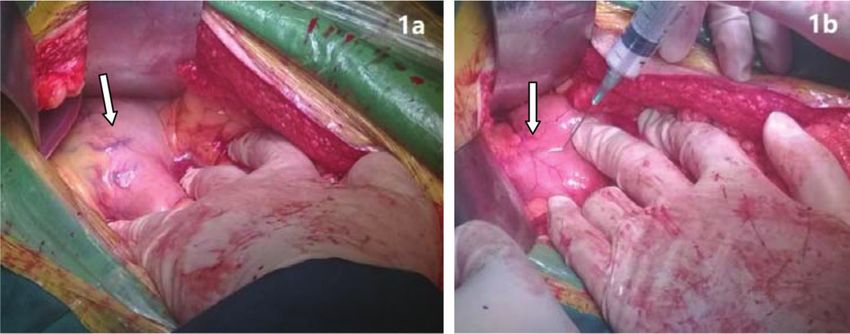
Treatments for prehepatic portal hypertension                                                                   CLINICS 2019;74:e704
Zhang Z et al.

splenorenal and portacaval shunts (14). Devascularization,            patients only, was administered during the procedure by the
mainly including pericardial devascularization, esophageal            surgeon after performing the shunt surgery.
transection and the Sugiura operation, directly blocks the
paradoxical blood flow between the azygos vein and portal                Mesocaval shunt surgery. Patients received general
vein, precisely controlling the acute bleeding of ruptured            anesthesia, and a midline incision was made for laparotomy.
esophageal varices and assuring blood flow to the liver (15).         Portal pressure was measured using the piezometer before
However, devascularization alone cannot reduce portal vein            shunting. The major superior mesenteric vein and its
pressure and may result in deterioration of portal hyperten-          branches were exposed after the mesenteric tissues were
sive gastropathy, causing thrombosis in the portal vein and           separated and ligated. Then, the inferior vena cava was
increasing the risk of rebleeding (15). Shunt surgery and             exposed through the posterior peritoneum. The superior
devascularization are also used together. For example, meso-          mesenteric vein and inferior vena cava were clamped, and
caval shunts or splenorenal shunts can be combined with               10-15 mm incisions were made on the vessel wall. Then,
pericardial or gastric devascularization (8). Shunt surgery           5-0 Prolene suture was used for side-to-side anastomosis.
effectively reduces portal vein pressure, and devascularization       If a side-to-side suture was impossible due to the long
removes the varicose veins, blocks blood circulation between          distance between the superior mesenteric vein and the
the azygos vein and portal vein and maintains blood flow to           inferior vena cava, an artificial (prosthetic) blood vessel
the liver, preventing variceal rebleeding and encephalopathy.         with a diameter of 8-12 mm (W.L. Gore & Associates, Newark,
   Surgeons in our hospital are still exploring surgical              DE, USA) was used to anastomose. After shunting, the pres-
approaches for PPH. Previously, we employed portosystemic             sure of the omentum vein was measured again and recorded,
shunts such as the mesocaval shunt and proximal splenor-              followed by wound closure.
enal shunt. For the past five years, we have used shunt
surgery combined with devascularization. In the last two
years, we have used shunt surgery combined with foam                    Splenorenal shunt surgery. After general anesthesia,
sclerotherapy of the varices as an alternative method to treat        a midline incision or an ‘‘L’’ type incision was made. Before
PPH, although the safety and efficacy of this operation               exploration, portal vein pressure was measured, followed
has not yet been reported. Therefore, this pilot study was            by splenectomy. Approximately 3-4 cm of the splenic
undertaken to compare the safety and efficacy of our novel            vein was reserved for further anastomosis, and the renal
surgical approach with that of conventional surgeries in              vein was exposed through the posterior peritoneum. After
obliterating esophagogastric varices and preventing post-             an incision was made on the renal vein, an end-to-side
operative rebleeding.                                                 anastomosis of the splenic and renal veins was established.
                                                                      A drainage tube was placed in the splenic fossa, and the
                                                                      portal vein pressure was measured again before wound
’ PATIENTS AND METHODS                                                closure.
Patient population
   In this retrospective, comparative pilot study, data from             Shunt surgery combined with devascularization.
33 patients who were diagnosed with PPH based on findings             After shunt surgery, the gastric fundus was exposed, and the
of abdominal ultrasonography and enhanced computer tomo-              proximal branches of gastroepiploic vessels and short gastric
graphy (CT) between January 2007 and March 2016 were                  veins were exposed along the greater and lesser curvature,
collected. The data of 4 patients who received conserva-              followed by ligation. Some varicose veins at the gastric body
tive treatment were excluded from the analysis. Of the                or fundus were sutured through the gastric wall.
29 patients who received surgical interventions, 1 received
exploratory laparotomy, 1 underwent splenectomy alone,
and 27 received shunt surgery or shunt surgery combined                  Shunt surgery combined with foam sclerotherapy.
with other surgical interventions. Data from the 27 shunt             After shunt surgery, the gastric varicose veins were exposed.
surgery patients were included for analysis and divided               Foam sclerosant was made up of 2 ml of 1% polidocanol
into 3 groups according to the type of surgery received:              (Aethoxysklerol, Kreussler, Wiesbaden, Germany) mixed
Group A (n=10) included patients who received shunt                   with 8 ml of air, as previously described by Tessari et al.
surgery alone; Group B (n=8) included patients who received           (16). Then, 2-5 ml of foam was injected into the major
shunt surgery combined with devascularization; and Group C            varicose vein. After injection, the puncture site vessel was
(n=9) included patients who received shunt surgery combined           ligated, followed by injection of the foam into other varicose
with foam sclerotherapy.                                              veins (Figure 1a, 1b). Foam sclerosant at different concentra-
   All included patients had portal obstruction due to any            tions was selected based on the size of the varicose veins;
cause, resulting in increased blood flow and symptoms of              generally, 1% foam sclerosant was used when the vessel was
portal hypertension. Ultrasonography and CT showed filling            smaller than 6 mm in diameter. However, if the varices
defects in the main portal vein, absence of a main portal vein        exceeded 6 mm in diameter, 3% polidocanol was applied. In
or portal cavernoma. The protocol for this retrospective study        one patient with rectal varices, the inferior mesenteric vein
was reviewed and approved by the Institutional Review                 was exposed, and a 4 Fr sheath was inserted, followed by a
Board of Beijing Friendship Hospital. Signed consent was not          guide wire (0.035 inch) and a 4 Fr catheter inserted into the
needed because the retrospective data were deidentified.              distal inferior mesenteric vein. Contrast agent was injected to
                                                                      visualize the distal inferior mesenteric vein and rectal
                                                                      varices. After withdrawal of the guide wire, 3% polidocanol
Surgical procedures                                                   foam (10 ml) was injected to occlude the rectal varices.
  All 27 patients in the three groups underwent surgery by            A second angiography showed the absence of varicose veins
the same team of surgeons. Foam sclerotherapy, for Group C            (Figure 2a, 2b).

                                                                  2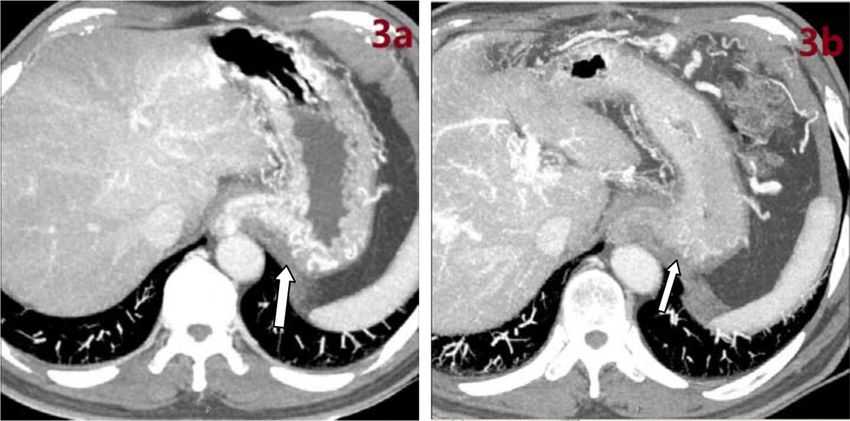
CLINICS 2019;74:e704                                                                     Treatments for prehepatic portal hypertension
                                                                                                                         Zhang Z et al.

Figure 1 - Varicose veins before and after treatment. a) Varicose veins at lesser gastric curvature. b) Occlusion of varicose veins after
injection of polidocanol foam.

Figure 2 - Rectal varices before and after treatment. a) Rectal varices on intraoperative angiography of the inferior mesenteric vein.
b) Occlusion of rectal varicose veins after injection of polidocanol foam.

Definitions and measures                                               of encephalopathy did not have ammonia determinations at
  The complete remission of gastroesophageal varices was               that time.
defined as an absence of any varicose vein in the esophagus               Patients were followed for 12 months after surgery, and
and gastric fundus on CT and/or gastric endoscopy; partial             the incidence of rebleeding (hematemesis or bloody stool due
remission was defined as the presence of but a reduction               to variceal bleeding), the incidence of hepatic encephalo-
in varicose veins in the esophagus and gastric fundus;                 pathy and the survival rates were compared among groups
no-remission was defined as unchanged varicose veins in the            at 12 months. Findings from abdominal CT or gastric endo-
esophagus and gastric fundus. The remission rate was cal-              scopy before surgery and within 12 months after surgery
culated as the proportion of patients with partial remission           were also compared. The remission rates of esophageal and
or complete remission to total patients.                               gastric varices were compared after surgery among the three
                                                                       groups. The possible causes of rebleeding were explored,
Intraoperative and postoperative observations and                      including the etiology (portal cavernoma or portal thrombo-
follow-up                                                              sis), reduction in portal pressure after shunt surgery and
   Operation time, intraoperative blood loss, reduction in             remission of gastroesophageal varices. Associations between
portal pressure after shunt surgery (difference between portal         these factors and rebleeding were further evaluated.
pressure before and portal pressure after shunt surgery),
incidence of postoperative complications, and mortality within         Statistical analysis
30 days after surgery were compared between groups. All                  Patient characteristics are presented as n (%) for categorical
patients were examined for patency of shunts before dis-               data and mean with range (min. to max.) for continuous data.
charge. All patients had ammonia determination before being            The characteristics during surgery, within 30 days and within
discharged and after receiving surgery; during the postopera-          one year of surgery are presented as the mean ± standard
tive follow-up after discharge, patients without symptoms              deviation (SD) for operation time, blood loss and reduction

                                                                   3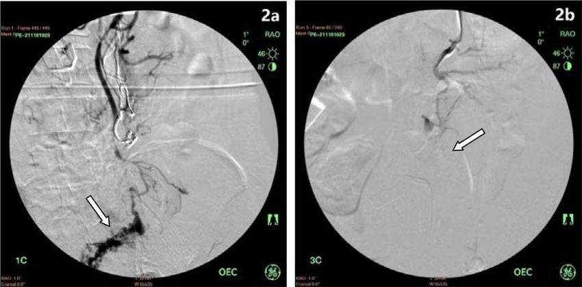
Treatments for prehepatic portal hypertension                                                                                 CLINICS 2019;74:e704
Zhang Z et al.

in portal pressure, and n (%) for other categorical data of                    splenocaval shunt using an artificial vessel in 1 patient.
safety and efficacy evaluations. Differences among groups                      The mean operation time was 241.0±92.0 minutes, and the
were compared using the Kruskal-Wallis test for continuous                     mean blood loss was 925.0±962.4 ml. The mean post-
data and Fisher’s exact test for categorical data. The Mann-                   operative reduction in portal pressure was 10.9±6.7 cm
Whitney U test was also applied for continuous data between                    H2O. After splenectomy, thrombocytosis was noted in one
two groups. All statistical analyses were carried out using                    patient who was treated with anti-platelet therapy; one
IBM SPSS statistical software version 22 for Windows (IBM                      patient developed pleural and peritoneal effusion, which
Corp., Armonk, NY, USA).                                                       was alleviated after supplementation with albumin and
                                                                               diuretic treatment.
’ RESULTS                                                                         In Group B, mesocaval shunt + devascularization was
                                                                               performed in 6 patients, splenectomy + mesocaval shunt +
Patient characteristics                                                        devascularization in 1 patient, and splenectomy + splenorenal
   In this retrospective pilot study, the data of 27 patients                  shunt + devascularization in 1 patient. The mean operation
with PPH (15 males and 12 females) with a mean age of                          time was 246.2±31.7 minutes, and the mean intraoperative
35.5 years (range: 8 to 62 years) were collected from January                  blood loss was 475.0±315.1 ml. The mean postoperative
2007 to March 2016. All patients were diagnosed via                            reduction in portal pressure was 9.2±4.6 cm H2O. After
enhanced abdominal CT screening, including 16 patients                         splenectomy, one patient developed a splenic fossa abscess
with portal cavernoma and 11 with chronic portal venous                        and an abdominal infection, which was corrected by splenic
thrombosis. Comorbidities included 27 patients with gastro-                    fossa drainage; one patient developed abdominal bleeding
esophageal varices, 20 with splenomegaly, 14 with hypers-                      within 5 hours after surgery, so a second exploratory
plenism, 11 with liver cirrhosis, 10 with ascites and one with                 laparotomy was performed, and the patients had a smooth
rectal varices. The medical histories revealed that 25 patients                recovery.
had gastrointestinal hemorrhage, and 2 had portal hyperten-                       In Group C, mesocaval shunt + foam sclerotherapy was
sive gastropathy. Eighteen patients had Child-Pugh class A                     performed in 8 patients, and a mesocaval shunt was placed
cirrhosis, and 9 had class B (Table 1).                                        using an artificial vessel + foam sclerotherapy in 1 patient.
                                                                               The median volume of polidocanol used in surgery was
Safety and efficacy                                                            16.9 ml. The mean operation time was 242.2±55.7 minutes,
  In Group A, a mesocaval shunt was placed in 5 patients,                      and the mean intraoperative blood loss was 511.1±493.6 ml.
a mesocaval shunt using an artificial vessel in 2 patients,                    The mean reduction in portal pressure was 9.8±3.4 cm
a splenectomy + splenorenal shunt in 2 patients, and a                         H2O. One patient developed upper gastrointestinal bleeding

Table 1 - Patient demographics and clinical characteristics (n=27).
                                            Total (n=27)         Group A (n=10)            Group B (n=8)          Group C (n=9)            p-value

Years                                                                                                                                        n/a
  2007                                          1                      1                        0                      0
  2008                                          3                      3                        0                      0
  2009                                          2                      2                        0                      0
  2010                                          4                      4                        0                      0
  2012                                          1                      0                        1                      0
  2013                                          2                      0                        2                      0
  2014                                          3                      0                        3                      0
  2015                                          8                      0                        2                      6
  2016                                          3                      0                        0                      3
Sex                                                                                                                                         0.560
  Males                                       15 (55.6)               6 (60)                    3 (37.5)               6 (66.7)
  Females                                     12 (44.4)               4 (40)                    5 (62.5)               3 (33.3)
Age, years                                  35.5 (8-62)            29.9 (11-62)              29.4 (8-60)            47.2 (18-61)            0.108
Etiology                                                                                                                                    1.000
  Portal cavernoma                            16 (59.3)               6 (60)                    5 (62.5)               5 (55.6)
  Chronic portal vein thrombosis              11 (40.7)               4 (40)                    3 (37.5)               4 (44.4)
Comorbidity
  Gastroesophageal varices                    27    (100)            10    (100)                8   (100)              9   (100)             n/a
  Splenomegaly                                20    (74.1)            7    (70)                 7   (87.5)             6   (66.7)           0.642
  Hypersplenism                               14    (51.9)            5    (50)                 5   (62.5)             4   (44.4)           0.794
  Liver cirrhosis                             11    (40.7)            4    (40)                 3   (37.5)             4   (44.4)           1.000
  Ascites                                     10    (37)              3    (20)                 5   (62.5)             2   (22.2)           0.247
  Rectal varices                               1    (3.7)             0    (0)                  0   (0)                1   (11.1)           0.631
Medical history                                                                                                                             0.512
  Gastrointestinal hemorrhage                 25 (92.6)              10 (100)                   7 (87.5)               8 (88.9)
  Portal hypertensive gastropathy              2 (7.4)                0 (0)                     1 (12.5)               1 (11.1)
Child-Pugh class                                                                                                                            0.098
  A                                           18 (66.7)               7 (70)                    3 (37.5)               8 (88.9)
  B                                            9 (33.3)               3 (30)                    5 (62.5)               1 (11.1)

Data are summarized as the number of patients by group for the years of patient enrollment; n (%) is shown for other categorical data; mean (range:
min.-max.) is shown for age.
n/a, not available.

                                                                           4
CLINICS 2019;74:e704 Treatments for prehepatic portal hypertension
Zhang Z et al.
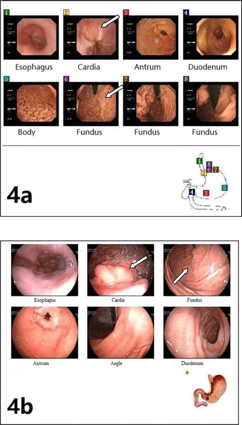
within 3 days after surgery, and endoscopy showed ulcer- complete remission (Figure 3a, 3b) and 7 with partial
related mucosal bleeding due to the shedding of tissue glue remission (Figure 4a, 4b) (1 had complete remission of rectal
that was administered before surgery; this patient recovered varices). No patients in any group died within 12 months
after blood transfusion and anti-acid treatment. One patient after surgery (Table 2).
developed persistent nasal bleeding due to thrombocytope- Table 3 presents associations between postoperative
nia, which resolved after focal hemostasis and plasma bleeding and other factors. No associations were found
transfusion. Sclerotherapy-related complications were not between the included factors (all p-value40.05) (Table 3).
observed in this group.
In Group A, 2 patients developed recurrent gastro-
’ DISCUSSION
intestinal bleeding at 4 and 10 months after surgery,
of whom 1 received endoscopic hemostasis and the other The results of this retrospective pilot study showed that
received conservative therapy. One patient developed hepatic the safety and efficacy of shunt surgery combined with foam
encephalopathy at 5 months after surgery but recovered after sclerotherapy were comparable to the safety and efficacy
corresponding treatment, and similar symptoms were not of shunt surgery plus devascularization for patients with
observed again. In Group B, recurrent bloody stool was PPH. Notably, both shunt surgery plus foam sclerotherapy
observed in 1 patient at 10.5 months after surgery but and shunt surgery plus devascularization were superior to
resolved after conservative therapy. In the follow-up shunt surgery alone in terms of preventing recurrence and
period, obstruction of the shunt was observed in 1 patient, rebleeding.
but without rebleeding, and no patients developed hepatic The goal of PPH treatment is the control and prevention
encephalopathy. In Group C, no postoperative rebleeding of gastrointestinal bleeding. Unlike in patients with hepatic
was observed; 1 patient developed hepatic encephalopathy cirrhosis-related portal hypertension, gastroesophageal varices
at 2 months after surgery, which resolved after correspond- in PPH are evidently causing more blood loss, and con-
ing treatment and was not observed thereafter. comitant esophageal and gastric varices are more common
Table 2 presents the operative parameters by group. (31%-44% vs. 22%). Some PPH patients have concomitant
During the operations, all three groups had similar operation rectal and anal varices. Isolated varicose veins are found in
times, intraoperative blood loss, and a reduction in portal approximately 6% of PPH patients, and ectopic or duodenal
pressure (all p-values40.05) (Table 2). No patients in any varices are also common (17,18).
group died within 30 days after surgery. A total of 6 patients, Surgery is the conventional treatment of choice for PPH
including 2 in Group A, 2 in Group B, and 2 in Group C, according to the guidelines of the American Association for
developed complications within 30 days after surgery. the Study of Liver Diseases (19), but with the development of
Within 12 months after surgery, a total of 3 patients had minimally invasive treatment, endoscopy and interventional
recurrent bleeding at 4 months, 10 months, and 10.5 months; procedures have become the preferred treatments. Evidence
2 patients had encephalopathy, and one experienced shunt shows that the efficacy of hemostasis by endoscopy is as high
occlusion. The remission rates of varices were significantly as 90% in the emergency department (4), and endoscopy is
different between the three groups (po0.001): one patient especially applicable in patients with acute bleeding who are
in Group A and 6 patients in Group B had partial remis- not candidates for open surgery. Endoscopy is also employed
sion; 9 patients in Group C had remission, including 2 with to prevent bleeding, but it cannot reduce portal pressure and
Table 2 - Comparisons of intraoperative and postoperative characteristics (n=27).
Total (n=27) Group A (n=10) Group B (n=8) Group C (n=9) p-value
Characteristics during surgery
Operation time (min) 243.0±64.5 241.0±92.0 246.2±31.7 242.2±55.7 0.986
Intraoperative blood loss (ml) 653.7±683.7 925.0±962.4 475.0±315.1 511.1±493.6 0.613
Preshunting portal pressure level (cm H2O) 42.4±6.8 46.1±6.7 40±7.9 40.3±4.1 0.085
Postshunting portal pressure level (cm H2O) 32.3±5.3 35.2±5.3 30.7±5.0 30.5±4.8 0.099
Reduction level of portal pressure (cm H2O) 10.1±5.0 10.9±6.7 9.2±4.6 9.8±3.4 0.931
Within 30 days after surgery
Complications 6 (22.2) 2 (20) 2 (25) 2 (22.2) 1.000
30-days mortality 0 (0) 0 (0) 0 (0) 0 (0) NA
Within 12 months after surgery
Recurrent bleeding 3 (11.1) 2 (20) 1 (12.5) 0 (0) 0.614
Encephalopathy 2 (7.4) 1 (10) 0 (0) 1 (11) NA
Shunt occlusion 1 (3.7) 1 (10) 0 (0) 0 (0) NA
Remission level of varices o0.001*
Completed remission 2 (7.4) 0 (0) 0 (0) 2 (22.2)
Partial remission 14 (51.9) 1 (10) 6 (75) 7 (77.8)
No remission 11 (40.7) 9 (90) 2 (25) 0 (0)
Remission rate 16 (59.3) 1 (10) 6 (75) 9 (100) o0.001*
Mortality rate 0 (0) 0 (0) 0 (0) 0 (0) NA
Data are summarized as the mean±SD for continuous data and as n(%) for categorical data. Differences among groups were compared using the Kruskal-
Wallis test for continuous data and Fisher’s exact test for categorical data.
* po0.05, indicates significant differences between groups.
NA, not available.
Three patients exhibited recurrent bleeding at 4 months, 10 months, and 10.5 months after the operation.
5Treatments for prehepatic portal hypertension CLINICS 2019;74:e704
Zhang Z et al.
Figure 3 - Perioperative enhanced CT images. a) Gastroesophageal varices on preoperative enhanced CT. b) Absence of varicose veins on
postoperative enhanced CT after foam sclerotherapy in the same patient.
may increase the risk for recurrent gastric varices and ectopic In the present study, all patients had portal obstruction, so
varices (20). Bleeding recurrence is common after endoscopy, the mesocaval shunt technique was employed. The blood
with a rate as high as 40%-70% (21,22). Therefore, regular flow shunted is less than that with the portocaval shunt,
follow-up and repeated treatments are needed for patients reducing the risk for postoperative hepatic encephalopathy.
who receive endoscopy alone, but this therapeutic pattern is Because the long distance between the superior mesenteric
often inapplicable in developing countries. Interventional vein and inferior vena cava may make direct anastomosis
procedures such as TIPS and percutaneous mesocaval shunt difficult, an artificial (prosthetic) vessel can be used for the
creation are minimally invasive and suitable for patients who shunt between these vessels. However, artificial vessels have
cannot undergo open surgery, although portal vein obstruc- a low long-term patency rate and cannot grow over time;
tion or chronic portal vein thrombosis remains a relative thus, the use of a prosthetic is inapplicable in children. For
contraindication to TIPS (11,23). Additionally, the technique patients with severe splenomegaly or hypersplenism, shunt
for percutaneous mesocaval shunt placement is a challenging surgery should be prepared during splenectomy, and a
and potentially precarious procedural approach requiring proximal splenorenal shunt is preferred for surgical inter-
transmesenteric, intraperitoneal vessel puncture and clinical vention. Splenectomy cannot effectively reduce the portal
experience is limited. PPH may also cause other complica- pressure or relieve esophageal and gastric varicosities but
tions, such as splenomegaly, hypersplenism, growth and can increase the susceptibility to splenic vein thrombosis,
development disorders, portal hypertensive gastropathy and which may risk involvement of the portal vein and superior
ectopic varices, which cannot be managed by endoscopy. mesenteric vein. This condition requires performing shunt
Thus, treatment must be individualized, and comprehensive surgery (28), and splenectomy alone should be avoided for
treatment is required for PPH patients. these patients.
To date, surgery remains an effective treatment for PPH, Although shunt surgery effectively reduces portal pres-
and the long-term survival rate of PPH is higher than 95% sure, recurrent bleeding is still possible after surgery due to
after shunt surgery (24). Its morbidity and mortality can be obstruction of shunt vessels and/or residual varicose veins
minimized by experienced surgeons. However, few studies (29). PPH patients may have large varicose veins, and even
have compared the efficacy between shunt surgery and though pressure is reduced, the dilated vessels cannot
endoscopy. A single-center, randomized, controlled study remodel, and thin-walled veins may rupture. In the present
indicated that the mortality was comparable between shunt study, although the absence of varices was not associated
surgery and endoscopy in PPH patients, but the rate of with postoperative recurrence of bleeding, three patients
bleeding recurrence in the endoscopy group (22.6%) was with recurrent bleeding had no improvement in varices after
significantly higher than that in the shunt surgery group surgery, indicating that residual varicose veins may increase
(3.3%), and a high rate of treatment failure was also found in the risk for bleeding recurrence. Therefore, in shunt surgery,
the endoscopy group (19.4% vs. 6.7%) (25). Surgery is also eliminating varices is necessary for concomitant devascular-
reported to improve pediatric patients’ growth and devel- ization or concomitant foam sclerotherapy.
opment, attenuate the development of varices, and relieve Theoretically, the combined use of shunt and devascular-
PPH-related biliary diseases (26,27). Accepted indications for ization not only reduces portal pressure but also removes
surgery for PPH include (1) acute gastrointestinal bleeding residual varicose veins, helping to prevent recurrent bleeding.
that is nonresponsive to endoscopic hemostasis; (2) gastric Feng et al. (30) achieved favorable efficacy using mesocaval
and ectopic varicose veins for which endoscopic treatment is artificial vessel shunts, ligation of gastric fundal veins and
too difficult to perform; (3) the presence of splenomegaly, coronary veins of the stomach, and partial splenectomy. In that
hypersplenism, growth and development disorders, portal study of 100 patients, no recurrence, bleeding or hepatic
hypertensive biliary tract disease or other diseases; and (4) encephalopathy were observed during follow-up. In the
when patients have difficulty making repeat hospital visits present study, among the eight patients (Group B) who
and are willing to receive one-time treatment (12). underwent surgical intervention with shunt creation and
6CLINICS 2019;74:e704 Treatments for prehepatic portal hypertension
Zhang Z et al.
Figure 4 - Endoscopy images. a) Preoperative endoscopy showing gastroesophageal varices. b) Endoscopy review of the same patient
showing obviously decreased gastric varices but residual esophageal varices postoperatively.
devascularization and received a postoperative CT scan, to shunt surgery alone. Therefore, we attempted to combine
six patients had remission of varices, indicating that the a shunt with foam sclerotherapy to replace conventional
combined use of a shunt and devascularization is effective shunt surgery, aiming to effectively remove the varicose
for attenuating varices. However, two patients still had no veins and simplify the surgery.
attenuation of varices, which might be ascribed to residual Foam sclerotherapy has been widely used in treating limb
varicose veins due to incomplete devascularization. In varices, endoscopic sclerotherapy and balloon-occluded
addition, devascularization sometimes requires expansion retrograde transvenous obliteration (BRTO), and can effec-
of the surgical field to expose the varicose veins completely, tively close varicose veins. However, direct injection of
which may increase surgical risk and difficulty compared sclerosant into fundal varicose veins during open surgery
7Treatments for prehepatic portal hypertension CLINICS 2019;74:e704
Zhang Z et al.
Table 3 - Associations between postoperative bleeding and clinical factors.
With postoperative bleeding (n=3a) Without postoperative bleeding (n=24) p-value
Operation time (min) 240±80 243.3±64.4 0.856
Intraoperative blood loss (ml) 750±1082.8 641.7±652.0 0.532
Reduction level of portal pressure (cm H2O) 7.7±0.6 10.4±5.3 0.532
Etiology
Portal cavernoma 2 (66.7) 14 (58.3) 1.000
Chronic portal vein thrombosis 1 (33.3) 10 (41.7) 1.000
Encephalopathy 1 (33.3) 1 (4.2) 0.214
Complications 1 (33.3) 5 (20.8) 0.545
Remission rate within 12 months after operation 0 (0) 16 (66.7) 0.056
a
Two patients in Group A and one patient in Group B had rebleeding within 12 months after operation.
Data are presented as n(%) and were analyzed using Mann-Whitney test for continuous data or Fishers’ exact test for categorical data.
No significant associations were derived.
has not been reported. Based on the classification of gastric are completely exposed, and 2-5 ml of polidocanol foam is
varices, some patients have both esophageal and gastric then injected into the target vessels. Before injection, with-
varices with connections between the varicose veins (GOV1 drawal of the syringe is needed to ensure that the needle
type and GOV2 type) (31). After injecting sclerosant into localizes in the vessel lumen. Stable injection may be difficult
fundal varicose veins, it may be distributed to the entire when the target vessels are small or curled or if the space is
varices system via communicating collaterals and even narrow. In such cases, an intravenous infusion needle with
further up to the esophageal veins. One-time injection may extended tubes can be used to inject the foam sclerosant,
cause the obstruction of a wide range varices, which increases increasing needle stability. The sclerosant dose should be
the efficacy of varicose vein removal and decreases surgical determined based on the severity of varices before and
trauma and difficulty. In addition, ectopic varicose veins during surgery. The sclerosant dose can usually reference
(e.g., rectal veins) that are difficult to expose anatomically that used in the treatment of limb varices, in which 2-5 ml
can also be managed with hybrid surgery. The catheter can of sclerosant is injected into each vessel, and the dose can be
be inserted into the distal lesion with the aid of digital increased for larger varicose veins. Polidocanol is available
subtraction angiography (DSA), and the sclerosant is then in concentrations of 0.5%, 1% and 3%; 1% polidocanol is
injected to obstruct the varicose veins, which may also most commonly used in treating fundal varices, while 3%
simplify this complex surgery. polidocanol is used if the diameter of varicose veins is larger
In the present study, we used polidocanol, a liquid than 6 mm. For fundal varices, pressurization is inapplicable
embolizing agent of the sclerosing class, as the foam sclerotic after injection of foam sclerosant, and unsuccessful obstruc-
reagent and found it to be safe and effective. This reagent tion of the target vessels may increase the possibility of
has been widely used in the treatment of limb varicosis and recirculation. Studies with long follow-up are needed to
venous malformation, as well as in endoscopic treatment determine whether this will affect long-term efficacy.
(32,33). Polidocanol is mixed with gas (1:4) to form the foam In the present study, although none of the patients in
sclerotic reagent. This reagent can damage vascular endothe- Group C who received shunt + foam sclerotherapy had
lial cells chemically, leading to thrombosis, fibrosis and final rebleeding, 20% of patients receiving shunt surgery alone
permanent occlusion. Foam sclerosant has several advan- (Group A) and 12.5% of patients receiving shunt surgery +
tages over liquid sclerosant: a lower dose of foam sclerosant devascularization (Group B) developed rebleeding within
can be used, increasing safety, and the distribution of foam 12 months after surgery. For short-term efficacy, shunt surgery
sclerosant is even greater in the vessels, increasing the area of combined with foam sclerotherapy prevented postopera-
sclerosant contacting vessel walls and elevating the efficacy tive recurrent bleeding more effectively, although signifi-
of sclerotherapy (34). Darke et al. (35) reported that poli- cant differences were not observed among the three groups,
docanol foam effectively induced occlusion of varicose so we cannot conclude that the new approach is superior to
veins of the lower limbs with 490% overall effectiveness. the conventional one. Further comparisons of findings from
The results of a multicenter study showed that the efficacy abdominal enhanced CT or gastroscopy before and after
of foam sclerotherapy (69%) was significantly higher than surgery showed decreased varices in more patients in
that of liquid sclerotherapy (27%) in patients with lower Groups B and C than in Group A due to the treatment of
extremity varices (36). Foam sclerosant is also used in fundal varicose veins in addition to shunt surgery in
BRTO. Several studies employed retrograde injection of Groups B and C. Postoperative imaging in Group C showed
polidocanol foam via a spontaneous gastrorenal shunt to that the esophageal and gastric varices decreased in all
obstruct fundal varicose veins, resulting in an obstruction patients within 12 months, with complete remission in two
rate as high as 91%-100%, and only one patient had bleed- patients and partial remission in seven patients, suggesting
ing recurrence during follow up (37-39). that obstruction of varicose veins with foam sclerosant has
Foam sclerotherapy is usually performed after shunt favorable efficacy. Notably, complete remission was not
surgery because the shunt reduces the pressure of the achieved in seven patients due to residual esophageal
esophageal and fundal veins and decreases the blood flow varices, which might be ascribed to the large vascular
from the varicose veins into systemic circulation. Therefore, volume and the insufficient amount of sclerosant applied.
injection of foam sclerosant after shunt surgery may not Injection of foam sclerosant into the fundal veins alone
induce systemic embolism. In shunt surgery, the varicose may not spread into esophageal veins, but a decrease in
veins at the greater curvature, lesser curvature and fundus fundal varices theoretically improves the prognosis of PPH
8CLINICS 2019;74:e704 Treatments for prehepatic portal hypertension
Zhang Z et al.
patients. When the pressure gradient between the portal embolism do arise, the incidence is low (51), and lower
and systemic vein is o12 mmHg after shunt or TIPS doses of sclerosant are better than giving the maximal dose.
placement, recurrent bleeding due to esophageal varices is In the present study, nine patients received 8-25 ml of 1%
rare, but fundal varices may still cause repeated bleeding polidocanol (median: 16.9 ml). Given a liquid to air ratio
(40). In addition, residual esophageal varices can also be of 1:4, the actual dose of polidocanol used was 16-50 mg.
managed by endoscopy, but the efficacy of endoscopy for Rectal varices were treated in one patient with 10 ml of 3%
fundal varices is inferior to that for esophageal varices (41). polidocanol, which was equivalent to 60 mg of polidocanol
Therefore, a decrease in fundal varices after foam scler- and was lower than the maximal dose, and no sclerosant-
otherapy is crucial for the control of postoperative recurrent related complications were observed, corroborating the safety
bleeding. of treating varices with polidocanol foam sclerosant.
Foam sclerotherapy is simpler to perform than devascu-
larization. Although no significant differences were found in Limitations
operation time or intraoperative blood loss in our patients The present pilot study still has several limitations. The
who received foam sclerotherapy, this technique only incidence of PPH was low, leading to a small sample size.
requires exposure of fundal varicose veins and subsequent Because conducting a single-center, prospective, randomized
injection of sclerosant without excessive exposure of the controlled study of a new approach is difficult, the new
surgical field. The hybrid surgery we used employs both approach can only be compared retrospectively to the conven-
interventional procedures and foam sclerotherapy, which can tional one. Additionally, only short-term efficacy was evaluated,
resolve difficulties common in conventional surgery (such as and it is not clear whether risk exists for long-term vascular
ectopic or rectal varices) with minimal invasion. In rectal and recanalization after foam sclerotherapy and whether significant
anal varices, redistribution of the portal vein may cause differences may be found in long-term recurrent bleeding and
increased pressure in the inferior mesenteric vein. Although survival rates among the surgical groups. Although no
the rate of relevant bleeding is low (0.5%-10%), severe or significant differences were found in patients’ baseline char-
even life-threatening consequences may still develop (42,43). acteristics (etiology, gender, age, complications and liver
One patient in Group C who had concomitant rectal varices function), study bias may still exist, emphasizing the need
had a history of repeated bloody stool and severe anemia for more multicenter, prospective, randomized controlled
before surgery. This lesion was hard to treat by conventional studies to confirm the results of the current trial.
surgery, and rectal resection might be needed. In the shunt
surgery, hybrid surgery was employed and involved catheter
insertion via the inferior mesenteric vein and injection of ’ CONCLUSION
sclerosant to obstruct the rectal varices. After the hybrid Shunt surgery combined with foam sclerotherapy has
procedure, the rectal varices were occluded, and the bloody similar safety and efficacy as shunt surgery with devascular-
stool was relieved. ization. This combined procedure provides more effective
Typically, PPH patients have normal liver function or mild variceal obliteration than shunt surgery alone does, and the
liver dysfunction, and the prognosis is good after surgery. In combined procedure may reduce the risk of postoperative
the present study, all patients had Child-Pugh class A or B recurrent bleeding caused by residual gastroesophageal varices.
cirrhosis and were tolerant of surgery with low surgical risk In addition, foam sclerotherapy is easy to perform, is safe and
compared to those with decompensated liver function. In has favorable short-term efficacy in combination with shunt
addition, no significant differences were found in 30-day surgery, but its long-term efficacy requires further study.
postoperative mortality, operation times or intraoperative
blood loss among the three groups, indicating similar safety
between the different procedures. In one patient in Group C ’ ACKNOWLEDGMENTS
who developed upper gastrointestinal bleeding, endoscopy The authors thank Lishan Lian for helping with collection of the clinical
showed ulcer formation rather than ruptured varices after documents.
endoscopic glue obturation. Other studies have reported
severe complications due to the shedding of tissue glue after
endoscopic therapy (44,45). During the 12-month follow-up,
’ AUTHOR CONTRIBUTIONS
all patients in the present study survived. Although hepatic Zhang Z was responsible for the study design, manuscript preparation,
encephalopathy was noted in one patient in Group A and clinical studies and data analysis. Chen X: was responsible for the study
one patient in Group C, it did not progress to hepatic coma concepts, study design, manuscript editing and clinical studies. Li C,
and resolved after conservative treatment. Feng H, Yu H, Zhu R were responsible for the clinical studies and data
Compared with other sclerosants, such as sodium tetra- acquisition. Wang T is the guarantor of integrity of the entire study and was
responsible for the manuscript review.
decyl sulfate, polidocanol foam is reported to be more stable
and has fewer side effects (45). The manufacturer’s instruc-
tions advise a maximal safe dose of 2 mg/kg/d, suggesting ’ REFERENCES
that polidocanol at o2 mg/kg/d is safe. Evaluation of the 1. Wani ZA, Bhat RA, Bhadoria AS, Malwall R. Extrahepatic portal vein
safety and efficacy of polidocanol in 16804 patients who obstruction and portal vein thrombosis in special situations: Need for a
received sclerotherapy showed no allergy or death, indicat- new classification. Saudi J Gastroenterol. 2015;21(3):129-38. https://doi.
org/10.4103/1319-3767.157550
ing favorable safety (47). However, excessive amounts of
2. Grimaldi C, de Ville de Goyet J, Nobili V. Portal hypertension in children.
polidocanol has been administered without sclerotherapy- Clin Res Hepatol Gastroenterol. 2012;36(3):260-1. https://doi.org/10.1016/
related complications (48,49). Studies also indicate that foam j.clinre.2012.03.016
sclerosant in the blood may not cause biological reactions, 3. Poddar U, Thapa BR, Rao KL, Singh K. Etiological spectrum of esopha-
geal varices due to portal hypertension in Indian children: is it different
except for a transient increase in D-dimer (50). Although from the West? J Gastroenterol Hepatol. 2008;23(9):1354–7. https://doi.
complications such as cardiac toxicity and pulmonary org/10.1111/j.1440-1746.2007.05102.x
9Treatments for prehepatic portal hypertension CLINICS 2019;74:e704
Zhang Z et al.
4. Sarin SK, Sollano JD, Chawla YK, Amarapurkar D, Hamid S, Hashizume 27. Thomas V, Jose T, Kumar S. Natural history of bleeding after esophageal
M, et al. Consensus on extra-hepatic portal vein obstruction. Liver Int. variceal eradication in patients with extrahepatic portal venous obstruction:
2006;26(5):512–9. https://doi.org/10.1111/j.1478-3231.2006.01269.x a 20-year follow-up. Indian J Gastroenterol. 2009;28(6):206–11. https://doi.
5. Mowat AP. Prevention of variceal bleeding. J Pediatr Gastroenterol Nutr. org/10.1007/s12664-009-0086-0
1986;5(5):679–81. https://doi.org/10.1097/00005176-198609000-00001 28. Wu JD, Wang ZG, Wang SH, Liu CW, Zeng R. Diagnosis and man-
6. Webster GJ, Burroughs AK, Riordan SM. Review article: portal vein agement for prehepatic portal hypertension: 9 years’ experience. J Clin
thrombosis—new insights into aetiology and management. Aliment Hepatol. 2011;27:134-5. Available at: http://new.oversea.cnki.net/
Pharmacol Ther. 2005;21(1):1-9. https://doi.org/10.1111/j.1365-2036.2004. KCMS/detail/detail.aspx?dbcode=CJFQ&dbname=CJFD2011&filename=
02301.x LCGD201102011&v=MDAwNDFyQ1VSTE9mWU9SbUZpbm1VNzNL
7. Yoshida H, Mamada Y, Taniai N, Yamamoto K, Kawano Y, Mizuguchi Y, S1M3TWFyRzRIOURNclk5RVpZUjhlWDFMdXhZUzdEaDFUM3FUc
et al. A randomized control trial of bi-monthly versus bi-weekly endo- ldNMUY=
scopic variceal ligation of esophageal varices. Am J Gastroenterol. 2005; 29. Mercado MA, Morales-Linares JC, Granados-García J, Gómez-Méndez TJ,
100(9):2005-9. https://doi.org/10.1111/j.1572-0241.2005.41864.x Chan C, Orozco H. Distal splenorenal shunt versus 10-mm low-diameter
8. Yoshida H, Mamada Y, Taniai N, Tajiri T. New trends in surgical treatment mesocaval shunt for variceal hemorrhage. Am J Surg. 1996;171(6):591-5.
for portal hypertension. Hepatol Res. 2009;39(10):1044-51. https://doi. https://doi.org/10.1016/S0002-9610(96)00038-4
org/10.1111/j.1872-034X.2009.00549.x 30. Feng LS, Li K, Peng QP, Ma XX, Zhao YF, Xu PQ, et al. Triplex opera-
9. Rikkers LF, Jin G, Burnett DA, Buchi KN, Cormier RA. Shunt surgery tion for portal hypertension with esophageal variceal bleeding: report of
versus endoscopic sclerotherapy for variceal hemorrhage: late results of a 140 cases. Hepatobiliary Pancreat Dis Int. 2004;3(4):534–7.
randomized trial. Am J Surg. 1993;165(1):27-32. https://doi.org/10.1016/ 31. Kapoor A, Dharel N, Sanyal AJ. Endoscopic Diagnosis and Therapy in
S0002-9610(05)80400-3 Gastroesophageal Variceal Bleeding. Gastrointest Endosc Clin N Am.
10. Sarin SK, Kumar A, Chawla YK, Baijal SS, Dhiman RK, Jafri W, et al. 2015;25(3):491–507. https://doi.org/10.1016/j.giec.2015.03.004
Noncirrhotic portal fibrosis/idiopathic portal hypertension: APASL 32. Duffy DM. Sclerosants: a comparative review. Dermatol Surg. 2010;
recommendations for diagnosis and treatment. Hepatol Int. 2007;1(3): 36(Suppl 2):1010-25. https://doi.org/10.1111/j.1524-4725.2009.01469.x
398–413. https://doi.org/10.1007/s12072-007-9010-9 33. Yamaki T, Hamahata A, Soejima K, Kono T, Nozaki M, Sakurai H. Pro-
11. Casadaban LC, Gaba RC. Percutaneous Portosystemic Shunts: TIPS and spective randomised comparative study of visual foam sclerotherapy
Beyond. Semin Intervent Radiol. 2014;31(3):227-34. https://doi.org/ alone or in combination with ultrasound-guided foam sclerotherapy
10.1055/s-0034-1382789 for treatment of superficial venous insufficiency: preliminary report.
12. Chaudhary N, Mehrotra S, Srivastava M, Nundy S. Management of Eur J Vasc Endovasc Surg. 2012;43(3):343-7. https://doi.org/10.1016/
bleeding in extrahepatic portal venous obstruction. Int J Hepatol. j.ejvs.2011.07.029
2013;2013:784842. https://doi.org/10.1155/2013/784842 34. Rabe E, Pannier F. Sclerotherapy in venous malformation. Phlebology.
13. Superina R, Shneider B, Emre S, Sarin S, de Ville de Goyet J. Surgical 2013;28 Suppl 1:188-91. https://doi.org/10.1177/0268355513477282
guidelines for the management of extra-hepatic portal vein obstruction. 35. Darke SG, Baker SJ. Ultrasound-guided foam sclerotherapy for the
Pediatr Transplant. 2006;10(8):908–13. https://doi.org/10.1111/j.1399- treatment of varicose veins. Br J Surg. 2006;93(8):969–74. https://doi.org/
3046.2006.00598.x 10.1002/bjs.5423
14. Zhang H, Zhang N, Li M, Jin W, Pan S. Surgical treatment of portal vein 36. Rabe E, Otto J, Schliephake D, Pannier F. Efficacy and safety of great
cavernous transformation. World J Surg. 2004;28(7):708-11. https://doi. saphenous vein sclerotherapy using standardised polidocanol foam (ESAF):
org/10.1007/s00268-004-7265-z a randomised controlled multicentre clinical trial. Eur J Vasc Endovasc Surg.
15. Sarin SK, Agarwal SR. Extrahepatic portal vein obstruction. Semin Liver 2008;35(2):238-45. https://doi.org/10.1016/j.ejvs.2007.09.006
Dis. 2002;22(1):43–58. https://doi.org/10.1055/s-2002-23206 37. Clements W, Cavanagh K, Ali F, Kavnoudias H, Kemp W, Roberts S, et al.
16. Tessari L, Cavezzi A, Frullini A. Preliminary experience with a new Variant treatment for gastric varices with polidocanol foam using balloon-
sclerosing foam in the treatment of varicose veins. Dermatol Surg. 2001; occluded retrograde transvenous obliteration: a pilot study. J Med
27(1):58–60. https://doi.org/10.1111/j.1524-4725.2001.00192.x Imaging Radiat Oncol. 2012;56(6):599–605. https://doi.org/10.1111/j.1754-
17. Noronha Ferreira C, Seijo S, Plessier A, Silva-Junior G, Turon F, Rautou 9485.2012.02453.x
PE, et al. Natural history and management of esophagogastric varices in 38. Koizumi J, Hashimoto T, Myojin K, Itou C, Kagawa T, Nishibe T, et al.
chronic noncirrhotic, nontumoral portal vein thrombosis. Hepatology. Balloon-occluded retrograde transvenous obliteration of gastric vari-
2016;63(5):1640-50. https://doi.org/10.1002/hep.28466 ces: use of CT-guided foam sclerotherapy to optimize technique. AJR
18. Sarin SK, Shahi HM, Jain M, Jain AK, Issar SK, Murthy NS. The natural Am J Roentgenol. 2012;199(1):200–7. https://doi.org/10.2214/AJR.11.
history of portal hypertensive gastropathy: influence of variceal eradica- 7002
tion. Am J Gastroenterol. 2000;95(10):2888–93. https://doi.org/10.1111/ 39. Choi SY, Won JY, Kim KA, Lee DY, Lee KH. Foam sclerotherapy using
j.1572-0241.2000.03200.x polidocanol for balloon-occluded retrograde transvenous obliteration
19. Garcia-Tsao G, Abraldes JG, Berzigotti A, Bosch J. Portal hyperten- (BRTO). Eur Radiol. 2011;21(1):122–9. https://doi.org/10.1007/s00330-
sive bleeding in cirrhosis: Risk stratification, diagnosis, and management: 010-1895-3
2016 practice guidance by the American Association for the study of liver 40. Siramolpiwat S. Transjugular intrahepatic portosystemic shunts and
diseases. Hepatology. 2017;65(1):310-35. https://doi.org/10.1002/hep. portal hypertension-related complications. World J Gastroenterol. 2014;
28906 20(45):16996-7010. https://doi.org/10.3748/wjg.v20.i45.16996
20. Poddar U, Thapa BR, Singh K. Frequency of gastropathy and gastric 41. Lo GH, Liang HL, Chen WC, Chen MH, Lai KH, Hsu PI, et al.
varices in children with extrahepatic portal venous obstruction treated A prospective, randomized controlled trial of transjugular intrahepatic
with sclerotherapy. J Gastroenterol Hepatol. 2004;19(11):1253–6. https:// portosystemic shunt versus cyanoacrylate injection in the prevention of
doi.org/10.1111/j.1440-1746.2004.03470.x gastric variceal rebleeding. Endoscopy. 2007;39(8):679–85. https://doi.
21. Itha S, Yachha SK. Endoscopic outcome beyond esophageal variceal era- org/10.1055/s-2007-966591
dication in children with extrahepatic portal venous obstruction. J Pediatr 42. Maslekar S, Toh EW, Adair R, Bate JP, Botterill I. Systematic review of
Gastroenterol Nutr. 2006;42(2):196–200. https://doi.org/10.1097/01.mpg. anorectal varices. Colorectal Dis. 2013;15(12):e702-10. https://doi.org/
0000189351.55666.45 10.1111/codi.12417
22. Oh SH, Kim SJ, Rhee KW, Kim KM. Endoscopic cyanoacrylate injection 43. Urrunaga NH, Rockey DC. Portal hypertensive gastropathy and colo-
for the treatment of gastric varices in children. World J Gastroenterol. pathy. Clin Liver Dis. 2014;18(2):389-406. https://doi.org/10.1016/j.cld.
2015;21(9):2719-24. https://doi.org/10.3748/wjg.v21.i9.2719 2014.01.008
23. Bercu ZL, Sheth SB, Noor A, Lookstein RA, Fischman AM, Nowakowski 44. Kobilica N, Flis V, Sojar V. Major complication after Histoacryl
FS, et al. Percutaneous Mesocaval Shunt Creation in a Patient with injection for endoscopic treatment of bleeding peptic ulcer. Endos-
Chronic Portal and Superior Mesenteric Vein Thrombosis. Cardiovasc copy. 2012;44(Suppl 2) UCTN:E204-5. https://doi.org/10.1055/s-0032-
Intervent Radiol. 2015;38(5):1316-9. https://doi.org/10.1007/s00270-014- 1308923
0989-8 45. Cheng LF, Wang ZQ, Li CZ, Lin W, Yeo AE, Jin B. Low incidence of
24. Poddar U, Borkar V. Management of extra hepatic portal venous complications from endoscopic gastric variceal obturation with butyl
obstruction (EHPVO): current strategies. Trop Gastroenterol. 2011;32(2): cyanoacrylate. Clin Gastroenterol Hepatol. 2010;8(9):760–6. https://doi.
94-102. org/10.1016/j.cgh.2010.05.019
25. Wani AH, Shah OJ, Zargar SA. Management of variceal hemorrhage in 46. McAree B, Ikponmwosa A, Brockbank K, Abbott C, Homer-Vannia-
children with extrahepatic portal venous obstruction-shunt surgery vs. sinkam S, Gough MJ. Comparative stability of sodium tetradecyl sulphate
endoscopic sclerotherapy. Indian J Surg. 2011;73(6):409–13. https://doi. (STD) and polidocanol foam: impact on vein damage in an in-vitro model.
org/10.1007/s12262-011-0345-z Eur J Vasc Endovasc Surg. 2012;43(6):721-5. https://doi.org/10.1016/
26. Pal S, Mangla V, Radhakrishna P, Sahni P, Pande GK, Acharya SK, et al. j.ejvs.2012.02.026
Surgery as primary prophylaxis from variceal bleeding in patients with 47. Conrad P, Malouf GM, Stacey MC. The Australian polidocanol (aethox-
extrahepatic portal venous obstruction. J Gastroenterol Hepatol. 2013; ysklerol) study. Results at 2 years. Dermatol Surg. 1995;21(4):334–6.
28(6):1010-4. https://doi.org/10.1111/jgh.12123 https://doi.org/10.1111/j.1524-4725.1995.tb00184.x
10CLINICS 2019;74:e704 Treatments for prehepatic portal hypertension
Zhang Z et al.
48. Arakaki Y, Murakami K, Takahashi K, Sato R, Okimoto T, Ishitobi H, 50. Connor DE, Joseph JE, Exner T, Ma DD, Parsi K. Infusion of foam sclerosants
et al. Clinical evaluation of combined endoscopic variceal ligation and results in a distance-dependent procoagulant activity, haemoconcentration
sclerotherapy of gastric varices in liver cirrhosis. Endoscopy 2003; and elevation of D-dimer levels. Phlebology. 2014;29(10):677-87. https://doi.
35(11):940–5. https://doi.org/10.1055/s-2003-43475 org/10.1177/0268355513502333
49. Mimura H, Fujiwara H, Hiraki T, Gobara H, Mukai T, Hyodo T, et al. 51. Sylvoz N, Villier C, Blaise S, Senturier C, Mallaret M. [Polidocanol
Polidocanol sclerotherapy for painful venous malformations: evaluation induced cardiotoxity: a case report and review of the literature].
of safety and efficacy in pain relief. Eur Radiol. 2009;19(10):2474–80. J Mal Vasc. 2008;33(4-5):234–8. https://doi.org/10.1016/j.jmv.2008.
https://doi.org/10.1007/s00330-009-1442-2 09.004
11You can also read