Biochemical and structural characterization of a recombinant fibrinogen related lectin from Penaeus monodon
←
→
Page content transcription
If your browser does not render page correctly, please read the page content below
www.nature.com/scientificreports
OPEN Biochemical and structural
characterization of a recombinant
fibrinogen‑related lectin
from Penaeus monodon
Nongnuch Singrang1,2, Sirasit Laophetsakunchai1, Bich Ngoc Tran3, Paul T. Matsudaira3,
Anchalee Tassanakajon1 & Kittikhun Wangkanont1,2*
Fibrinogen-related lectins are carbohydrate-binding proteins of the innate immune system that
recognize glycan structures on microbial surfaces. These innate immune lectins are crucial for
invertebrates as they do not rely on adaptive immunity for pathogen clearance. Here, we characterize
a recombinant fibrinogen-related lectin PmFREP from the black tiger shrimp Penaeus monodon
expressed in the Trichoplusia ni insect cell. Electron microscopy and cross-linking experiments
revealed that PmFREP is a disulfide-linked dimer of pentamers distinct from other fibrinogen-
related lectins. The full-length protein binds N-acetyl sugars in a Ca2+ ion-independent manner.
PmFREP recognized and agglutinated Pseudomonas aeruginosa. Weak binding was detected with
other bacteria, including Vibrio parahaemolyticus, but no agglutination activity was observed. The
biologically active PmFREP will not only be a crucial tool to elucidate the innate immune signaling in
P. monodon and other economically important species, but will also aid in detection and prevention of
shrimp bacterial infectious diseases.
The black tiger shrimp Penaeus monodon is an important economic animal of many countries, including
Thailand1. However, there are many infectious diseases devastating shrimp farming. Viral pathogen such as
white spot syndrome virus2, yellow head virus3, or shrimp baculovirus4 have costed severe economic loss and
their molecular properties have been extensively investigated. There are various test kits available and factors
in controlling infections are k nown5. Recently, bacterial pathogen such as Vibrio parahaemolyticus, causing the
acute hepatopancreatic necrosis disease (AHPND), has emerged as a major disruption in the shrimp farming
industry6. Very little is known about the interplay of the shrimp innate immune system and pathogenic bacteria.
One key player is likely the lectins of the innate immune system7,8.
Lectins, or carbohydrate binding proteins, play important roles in pathogen recognition, especially in inver-
tebrates where adaptive immunity is not as developed compared to vertebrates9. Lectins of the innate immune
system generally can distinguish self from non-self by recognition of carbohydrate residues or specific glycan
structure not present on the host cells. There are several families of animal lectins that are involved in the innate
immunity, such as C-type lectins10, ficolins11, and intelectins12,13 (X-type lectin). Ficolins and intelectins are
classified as fibrinogen-like lectins based on structural homology. In mammalian systems, ficolins are known to
activate the lectin complement system for pathogen c learance7. Ficolins may act as an opsonin for phagocytosis
as well14,15. However, the sequence of signaling events after bacteria recognition by lectins in invertebrate is
largely unknown. Thus, we are interested in investigating the structure and function of fibrinogen-related lectin
in shrimp, as it may have applications in bacterial disease prevention and treatment.
Fibrinogen-related lectins are widely distributed in the animal kingdom and they have diverse molecular
structure (Fig. 1). Ficolins, found in both vertebrates and invertebrates, contain a cysteine-rich N-terminal region,
a collagen-like region, and the carbohydrate recognition domain (CRD) at the C terminus (Fig. 1A). Because of
the collagen-like region that is capable of forming a triple helix, ficolins can trimerize. The cysteine-rich region
then mediates disulfide-linked oligomerization of the trimer into higher order oligomers, resulting in a fan- or
1
Center of Excellence for Molecular Biology and Genomics of Shrimp, Department of Biochemistry, Faculty
of Science, Chulalongkorn University, Bangkok, Thailand. 2Molecular Crop Research Unit, Department of
Biochemistry, Faculty of Science, Chulalongkorn University, Bangkok, Thailand. 3Department of Biological
Sciences, Faculty of Science, Centre for BioImaging Sciences, National University of Singapore, Singapore,
Singapore. *email: kittikhun.w@chula.ac.th
Scientific Reports | (2021) 11:2934 | https://doi.org/10.1038/s41598-021-82301-5 1
Vol.:(0123456789)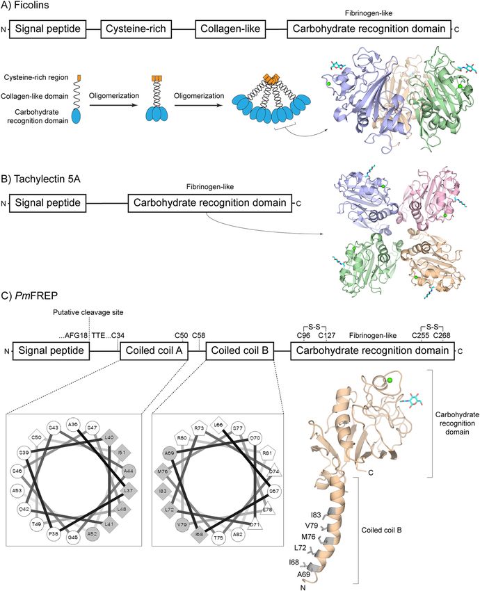
www.nature.com/scientificreports/
Figure 1. Comparison of fibrinogen-like lectin structures. (A) Ficolins. Each polypeptide chain can trimerize
and oligomerize into a flower bouquet-like structure. The structure represents a trimeric CRD. (B) Tachylectin
5A. The structure represents the tetrameric CRD. and (C) PmFREP. The coiled coil regions are also represented
with helical wheel diagrams showing the predicted amphipathic helices. The structure shown is a homology
model of the CRD and coiled coil B built with human fibrinogen (PDB ID 2HPC) as the template.
flower bouquet-shaped molecular complex16,17. Ficolins binds N-acetyl glucosamine (GlcNAc)-containing glycan
and subsequent activate the innate immune system, such as the lectin complement pathway in mammals18,19.
However, the signaling event in invertebrate is not well-studied. Another group of invertebrate fibrinogen-like
lectin is tachylectin 5A (Fig. 1B). The protein has a simple primary structure of only the CRD. The protein is
tetrameric and binds G lcNAc20,21. The protein is proposed to be involved in bacteria sensing and hemolymph
clotting to seal the bacteria-exposed wound, but the molecular mechanism following ligand recognition remains
unknown22. Recently, a unique fibrinogen-like lectin from P. monodon was described. The protein has been cloned
by various investigators, and named PmFREP23 or PL5-124. PmFREP binds bacterial p eptidoglycan23, thus likely
to bind GlcNAc as observed with ficolins and tachylectin 5A. PmFREP contains three cysteines outside of the
CRD (Fig. 1C). Because of the odd number of cysteines, they are likely to participate in intermolecular disulfide
bond formation. In contrast to ficolins, PmFREP does not contain the collagen-like sequence, but contains two
coiled coil regions that likely for amphipathic helices and mediate higher order oligomerization. Thus, we predict
that PmFREP has a novel molecular assembly unique among fibrinogen-like lectins.
PmFREP and related proteins have been purified from P. monodon hemolymph23,24. Because of the poten-
tial contamination of other GlcNAc-binding fibrinogen-related lectins, it is difficult to study the structure and
Scientific Reports | (2021) 11:2934 | https://doi.org/10.1038/s41598-021-82301-5 2
Vol:.(1234567890)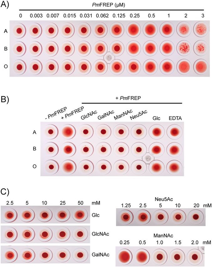
www.nature.com/scientificreports/
Figure 2. Western blot of His6 PmFREP and His6 PmFREP CRD under reducing (R) and non-reducing (NR)
conditions. Anti-His6 antibody was used as the primary antibody.
function of PmFREP using the protein purified from the hemolymph. In addition, the complex architecture of
PmFREP may cause difficulties in recombinant protein expression. To date investigators that have cloned and
studied PmFREP have reported recombinant expression of PmFREP-related proteins, such as PL5-2, in Escheri-
chia coli, but not PmFREP (PL5-1) i tself24. Given the potential intermolecular disulfide bonds and higher-order
oligomerization, it is unlikely that E. coli and in vitro refolding of inclusion bodies can produce functional
PmFREP, or any PmFREP-related proteins. The problem of protein quality is a bottleneck in investigating lectin
structure, function, and signaling in P. monodon. Therefore, we aim to produce functional recombinant PmFREP
to investigate its structure and function.
In this study, we produced recombinant PmFREP for biochemical and structural characterization. Due to
the complex architectures of fibrinogen-related lectins, high level of protein expression was successful in insect
cells. The dimer of pentamer structure of PmFREP was revealed by both biochemical methods and electron
microscopy. Binding to N-acetyl sugars and bacteria agglutination were also demonstrated. These results will
not only be crucial for further investigation of immune signaling in shrimp, but will also help in combating
bacterial infectious diseases in shrimp.
Results
PmFREP was cloned into various expression vectors for expression in bacteria (Escherichia coli), mammalian cells
(HEK293T), and insect cells (Trichoplusia ni). In the E. coli system, no signal peptide was included in the coding
sequences and both the N- and C-terminal hexahistidine (His6) tag was explored. Despite exploration of host
strains and IPTG concentrations, no protein expression was detected both by SDS-PAGE and western blot against
the His6 tag (data not shown). Thus, protein expression and secretion were further explored in mammalian and
insect cells (Figure S1). Protein expression was barely observable for PmFREP with its native signal peptide and
a C-terminal H is6 tag (Native SP PmFREP His6) when expressed and secreted from insect cells. Another expres-
sion construct examined is PmFREP with the Xenopus laevis embryonic epidermal lectin signal peptide and an
N-terminal His6 tag (XEEL SP His6 PmFREP, Figure S2A). This expression construct yielded higher amount of
protein in the insect system compared to the mammalian system. Truncation of PmFREP to only the CRD with
the XEEL signal peptide and a N-terminal H is6 PmFREP, Figure S2B) when expressed in insect
is6 tag (XEEL SP H
cells yielded high amount of protein in insect culture media, visible in SDS-PAGE with Coomassie Blue stain.
Therefore, the insect expression system was used to express the constructs XEEL SP His6 PmFREP and XEEL
SP His6 PmFREP CRD, yielding His6 PmFREP and His6 PmFREP CRD respectively. The protein was purified by
Ni–NTA affinity chromatography for subsequent experiments (Figure S3). Correct cleavage of the signal peptide
for both H is6 PmFREP and H is6 PmFREP CRD was verified with N-terminal protein sequencing (Figure S4).
With the ability to produce recombinant PmFREP, we next explored the disulfide linked oligomeric states of
PmFREP by examining the apparent molecular weight under reducing (with DTT in sample buffer) and non-
reducing conditions (no DTT in sample buffer) (Fig. 2). His6 PmFREP ran as a single species under reducing
conditions, but appear to assemble into large disulfide-linked oligomers of more than 315 kDa. On the other
hand, His6 PmFREP CRD appeared monomeric both in reducing and non-reducing conditions, indicating that
there are no intermolecular disulfide bonds in the CRD.
To further examine the quaternary structure of PmFREP, both His6 PmFREP and His6 PmFREP CRD were
examined by transmission electron microscopy (TEM) with negative staining (Fig. 3). His6 PmFREP appeared
Scientific Reports | (2021) 11:2934 | https://doi.org/10.1038/s41598-021-82301-5 3
Vol.:(0123456789)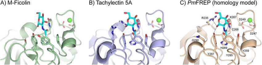
www.nature.com/scientificreports/
Figure 3. TEM micrograph of (A) His6 PmFREP, (B) His6 PmFREP CRD, and (C) 2D class averages of His6
PmFREP.
Figure 4. Crosslinking of PmFREP CRD with bis(sulfosuccinimidyl)suberate (BS3) at various concentrations.
PmFREP was detected by western blot probed with an anti-His6 antibody.
as dumbbell-shaped molecules with the dimension of roughly 100 × 200 Å. Two-dimensional class averaging
suggested that the molecule is a dimer of pentamer with a flexible linker in between. This oligomeric structure
is distinct from other fibrinogen-related lectins, such as mammalian ficolins, which are flower bouquet-shaped
is6 PmFREP CRD
consisting of oligomers of trimers, or intelectins which are also trimer or oligomer of trimers. H
appeared as 100 Å particles, but we were not able to obtain consistent 2D class average. Dynamic light scattering
(DLS) experiments also confirmed the sizes of H is6 PmFREP (215 ± 36 Å) and H is6 PmFREP CRD (97 ± 30 Å)
is6 PmFREP CRD might still be able to oligomerize in solution, but the
(Figure S5). These results suggested that H
oligomeric conformation may not be as stable as the full length His6 PmFREP protein. To examine whether His6
PmFREP CRD can oligomerize in solution, chemical cross-linking was performed (Fig. 4). As the concentration
of the cross-linker was increased, species with the molecular weight consistent with dimers and pentamers were
observed. These results suggested that His6 PmFREP CRD can self-associate in solution and were consistent
with the TEM and DLS results.
To explore the ligand binding properties of PmFREP, we performed competitive elution assays (Fig. 5A). The
proteins were bound to carbohydrate affinity resins and eluted with the corresponding monosaccharide or EDTA.
His6 PmFREP can bind GlcNAc as expected for fibrinogen-like lectin. However, EDTA was not able to eluted
His6 PmFREP from the GlcNAc resin. In contrast to the full length H is6 PmFREP, His6 PmFREP CRD failed to
bind any carbohydrate affinity resin. To further explore carbohydrate specificity of His6 PmFREP, His6 PmFREP
was bound to the GlcNAc resin and eluted with various carbohydrates (Fig. 5B). GlcNAc, N-acetylgalactosamine
(GalNAc), N-acetylmannosamine (ManNAc), and N-acetylneuraminic acid (Neu5Ac) were able to elute H is6
PmFREP from the GlcNAc resin. However, competitive elution was not observed with glycerol (Gro), ribose
Scientific Reports | (2021) 11:2934 | https://doi.org/10.1038/s41598-021-82301-5 4
Vol:.(1234567890)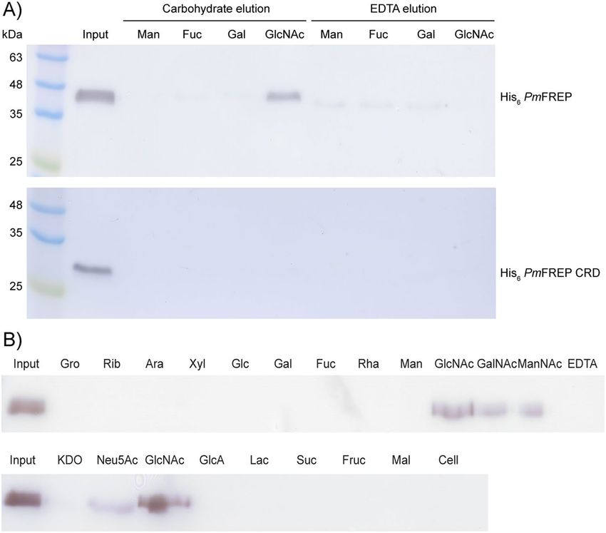
www.nature.com/scientificreports/
Figure 5. (A) Western blot analysis of H
is6 PmFREP (top panel) and H is6 PmFREP CRD (bottom panel) bound
to different carbohydrate resin and eluted with either the respective monosaccharide or EDTA. (B) Competitive
elution of His6 PmFREP bound to GlcNAc resin by different soluble carbohydrates. Anti-His6 antibody was used
as the primary antibody.
Figure 6. Comparison of the ligand binding site of (A) M-ficolin (crystal structure, PDB ID 2JHK), (B)
Tachylectin 5A (crystal structure, PDB ID 1JC9), and (C) PmFREP (homology model using SWISS-MODEL44,
PDB ID 1JC9 as the template). The GlcNAc ligand is shown in cyan and the structural calcium ion is shown in
green.
(Rib), arabinose (Ara), xylose (Xyl), glucose (Glc), galactose (Gal), fucose (Fuc), rhamnose (Rha), mannose
(Man), 3-deoxy-D-manno-2-octulosonic acid (KDO), glucoronic acid (GlcA), lactose (Lac), sucrose (Suc),
fructose (Fruc), maltose (Mal), and cellobiose (Cell). These results suggested that His6 PmFREP was specific for
acetyl group-containing carbohydrates, and while the ligand binding site and the calcium ion-binding residues
were conserved (Fig. 6), the calcium ion is not required for ligand binding.
To investigate recognition of carbohydrates in cellular context, we examined agglutination of red blood cells
by His6 PmFREP. Because of the dimer of pentamer arrangement, H is6 PmFREP is expected to agglutinate cells
displaying its ligand. His6 PmFREP agglutinates red blood cells of A-, B- and O-type starting at concentration
around 0.031 µM (Fig. 7A). Agglutination was inhibited by GlcNAc, GalNAc, ManNAc, and Neu5Ac (Fig. 7B).
However, glucose and EDTA could not inhibit agglutination. GlcNAc and GalNAc could inhibit agglutination
Scientific Reports | (2021) 11:2934 | https://doi.org/10.1038/s41598-021-82301-5 5
Vol.:(0123456789)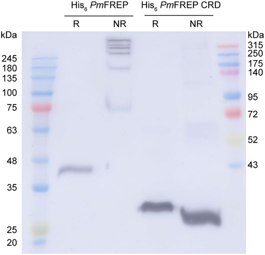
www.nature.com/scientificreports/
Figure 7. Agglutination of human red blood cells with His6 PmFREP. (A) Agglutination of different ABO blood
type by His6 PmFREP at various concentrations. (B) Inhibition of ABO blood type agglutination by different
carbohydrates (20 mM for Neu5Ac and 50 mM for other carbohydrates) and EDTA. (C) Inhibition of A blood
type agglutination by different carbohydrates at various concentrations.
at around 10 mM (Fig. 7C). Inhibition of agglutination was observed at 5 and 1 mM for Neu5Ac and ManNAc,
respectively.
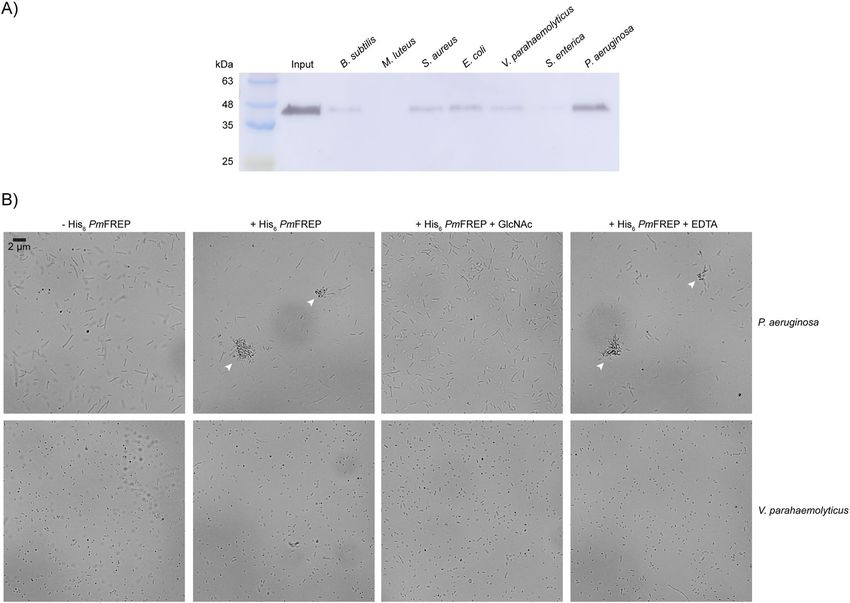
Because of its proposed role in P. monodon innate immune system, we next examined the ability of H is6
PmFREP to recognize bacteria (Fig. 8A). His6 PmFREP was bound to bacteria pellet and eluted with GlcNAc. The
eluted protein solutions were then examined by western blot. H is6 PmFREP strongly recognized Pseudomonas
aeruginosa. Noticeable binding was also observed with Bacillus subtilis, Staphylococcus aureus, Escherichia coli,
and Vibrio parahaemolyticus. Very little to no binding was observed toward Micrococcus luteus and Salmonella
enterica. Because the dimer of pentamer molecular architecture of PmFREP suggests that the protein can engage
two bacteria simultaneously and cause agglutination, bacteria agglutination activity of PmFREP was explored
(Fig. 8B). P. aeruginosa and V. parahaemolyticus were used in the agglutination because of the strong signal in
the bacteria binding assay and the importance as a shrimp pathogen respectively. At the highest concentration of
His6 PmFREP shown to agglutinate red blood cells (3 µM), His6 PmFREP agglutinated S. aureus and this activ-
ity is inhibited by addition of GlcNAc. However, sequestration of C a2+ ion by addition of EDTA did not inhibit
agglutination. In contrast, V. parahaemolyticus was not agglutinated by His6 PmFREP.
Discussion
Various investigators have purified PmFREP and its homologs from P. monodon hemolymph23–25. However,
the degree of contamination by other homologs is unknown, which complicates further analysis of PmFREP
structure and function. Moreover, in contrast to other PmFREP homologs in shrimp, there is no report of
expression or characterization of functional recombinant PmFREP23–25. We speculate that other investigators
have encounter issues with bacterial production of PmFREP. In our hands, we did not observe protein expression
in E. coli whether the His6 tag was placed at the N- or C-terminus. Thus, we conclude that bacterial expression
system might not be suitable for PmFREP. Some protein expression is detected in mammalian cells, but scaling
up protein production would be cost prohibitive, especially with transient transfection. Appreciable yield was
observed when PmFREP was expressed in insect cells for both H is6 PmFREP and H is6 PmFREP CRD. There-
fore, the insect cell expression is the system of choice for PmFREP. XEEL signal peptide was used because of the
observation that XEEL, a fibrinogen-related lectin of the intelectin family, is highly expressed in insect cells12.
Cleavage of the signal peptide occurred at the expected site as confirmed by N-terminal protein sequencing. We
reasoned that the placement of the His6 tag at the N-terminus is more suitable because the C-terminal carboxyl
group of other fibrinogen-related lectins, such as H-ficolin26 or intelectins12,13, form a salt bridge with another
amino acid residue. Placement of a C-terminal tag may disrupt this interaction and destabilize PmFREP. In this
Scientific Reports | (2021) 11:2934 | https://doi.org/10.1038/s41598-021-82301-5 6
Vol:.(1234567890)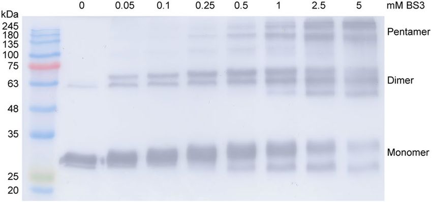
www.nature.com/scientificreports/
Figure 8. (A) Western blot of H is6 PmFREP eluted from bacteria pellet with 100 mM GlcNAc. Anti-His6
antibody was used as the primary antibody. (B) Agglutination of bacteria with His6 PmFREP (3 µM). Clumps of
bacteria are indicated with arrows.
case of human intelectin and XEEL, placement of the C-terminal tag drastically reduced the protein expression
yield12,13. Utilization of the His6 tag is ubiquitous in biochemistry. The tag is placed at a protein terminus, which
is not likely involved in interaction interfaces or any functional s ite27. Another advantage of the H is6 tag is its
function as an epitope tag. Thus, raising a specific antibody to PmFREP is not required. Moreover, having a tag
separated from the protein sequence allow better control over future immunoprecipitation experiments because
antibody binding to the tag will be less likely to interfere with interaction of PmFREP and other binding partners.
Because most lectins are oligomeric and the N-terminus of PmFREP contains an odd number of 3 cysteines
(C34, C50, and C58), we reason that PmFREP might be able to form intermolecular disulfide bonds. SDS-PAGE
analysis of His6 PmFREP and His6 PmFREP CRD under reducing and non-reducing conditions revealed that His6
PmFREP is a disulfide-linked oligomer. The cysteines in the CRD are conserved among other fibrinogen-related
lectins and are only involved in intramolecular disulfide bond f ormation21,26. This is consistent with the results
that His6 PmFREP CRD is monomeric both in reducing and non-reducing conditions. Because of this intermo-
lecular disulfide bonds, it is unlikely that recombinant PmFREP produced in E. coli, even if expressed, will be
fully functional. Further examination of His6 PmFREP quaternary structure by TEM revealed dumbbell-shaped
molecules. Two-dimensional class averaging suggested that His6 PmFREP is a dimer of pentamer with a flexible
linker. The molecular weight of the dimer of pentamer, or decamer, would be around 345 kDa, which is consistent
with the observation that His6 PmFREP ran at more than 315 kDa in SDS-PAGE under non-reducing conditions.
DLS also confirmed the particle size observed in TEM. Examination of H is6 PmFREP CRD by TEM revealed
protein molecules about the size of the pentamer in the full length H is6 PmFREP. However, 2D class averaging
did not yield sensible solutions, suggesting that H is6 PmFREP CRD might be able to oligomerize in solution,
but the structure is rather inhomogeneous. Chemical cross-linking of H is6 PmFREP CRD in solution and DLS
also indicate that the pentameric assembly of H is6 PmFREP CRD likely exists in solution. A similar molecular
property is observed in XEEL, a fibrinogen-related lectin of the intelectin family12. XEEL is a dimer of trimer,
and the CRD is capable of trimerization in solution even in the absence of the intermolecular disulfide bonds.
His6 PmFREP recognized GlcNAc, as expected from structural homology to other fibrinogen-related lectins
(Fig. 5). Most residues in the binding sites that interact directly with GlcNAc (R235, NH of C268, H269, Y285,
and Y297) are conserved between PmFREP and tachylectin 5 A21. The residues recognizing the N-acetyl group
Scientific Reports | (2021) 11:2934 | https://doi.org/10.1038/s41598-021-82301-5 7
Vol.:(0123456789)www.nature.com/scientificreports/
(NH of C268, H269, Y285, and Y297) are conserved in human M-ficolin as well28. PmFREP could also bind
GalNAc, ManNAc, and Neu5Ac. The red blood cell agglutination experiments suggested that PmFREP have the
highest affinity towards ManNAc. However, the lack of experimental structural data, which we will continue to
investigate, do not currently allow us to comments on specific interactions. It is also not clear whether ManNAc
is a biologically relevant epitope since there is limited information on glycobiology of shrimp diseases. However,
because PmFREP was first identified as a peptidoglycan-binding lectin23, the GlcNAc-binding activity might
still be biologically relevant.
The calcium ion binding residues (D247 and D249) are conserved between PmFREP, tachylectin 5A, and
M-ficolin, suggesting that PmFREP possesses C a2+ ion binding activity. However, our results showed that C a2+
ion was not required for GlcNAc binding. Because the C a2+ ion is not directly participating in ligand binding,
it is possible that the C a2+ ion may only have structural and stability role for PmFREP. There are reports that
other shrimp fibrinogen-related lectins require Ca2+ ion for ligand binding29–33. However, several ficolins are also
capable of C a2+-independent ligand binding19,34–36. The role of Ca2+ ion in PmFREP ligand binding modulated
by protein stability need to be further investigated. In contrast to the full length H is6 PmFREP, His6 PmFREP
CRD did not bind the GlcNAc affinity resin. The reduction in ligand binding affinity could be due to the lack
of intermolecular disulfide bonds that may stabilize the structure. This observation is consistent with the TEM
results which indicates that His6 PmFREP CRD has relatively low structural homogeneity and may not be stable
enough to bind GlcNAc with high affinity. In addition to potential reduction in structural stability, truncation of
the full length protein to merely the CRD certainly reduced the multivalent binding capability of H is6 PmFREP.
Multivalent binding event is well documented and is utilized ubiquitously in nature to increase apparent bind-
ing affinity, or avidity, especially in immune proteins and signaling e vents37–39. Therefore, reduction in binding
avidity is expected for any lectin when the oligomeric state is reduced.
His6 PmFREP clearly recognized P. aeruginosa. Binding is less strong towards B. subtilis, S. aureus, E. coli,
and V. parahaemolyticus. Because H is6 PmFREP is a dimer of pentamer, the molecule should be able to engage
two bacteria simultaneously and cause agglutination. Indeed, His6 PmFREP agglutinated P. aeruginosa and
this activity is inhibited by the presence of GlcNAc. Addition of EDTA to sequester C a2+ ion did not inhibit
agglutination. These results are consistent with the ligand binding and red blood cell agglutination experiments
which suggested that Ca2+ is not required for ligand binding. In contrast to P. aeruginosa, His6 PmFREP did not
agglutinate V. parahaemolyticus, which is consistent with the weaker binding affinity of His6 PmFREP towards
V. parahaemolyticus in the ligand binding assay.
In conclusion, we have characterized the recombinant fibrinogen-like lectin PmFREP from P. monodon
expressed in insect cells. The protein is a dimer of pentamer with intermolecular disulfide bonds at the N-ter-
minus. This dimer of pentamer molecular architecture is novel among fibrinogen-related lectins. PmFREP binds
GlcNAc like other fibrinogen-related lectins, such as tachylectin 5A and ficolins. In addition, binding to GalNAc,
ManNAc, and Neu5Ac was observed. C a2+ is not required for ligand binding, but may have structural roles as
the calcium binding residues are conserved. The CRD itself is capable of oligomerization in solution, but cannot
bind the GlcNAc ligand. PmFREP recognizes and agglutinates P. aeruginosa in a C a2+-independent manner, but
cannot agglutinate the shrimp pathogen V. parahaemolyticus although weak binding is observed in the bacteria
binding assay. The information obtained and His6 PmFREP produced in the study will be useful for further
biochemical investigation and the signaling pathway of innate immune lectins in shrimp that may help prevent
and treat bacterial infectious disease in shrimp in the future.
Methods
Expression plasmids and protein expression. For expression in insect cells, the open reading frame
for PmFREP (GenBank accession number AIE45535) was amplified from P. monodon hemocyte cDNA by the
primers 5′-GCGCGGATCCATGGCGCTCTTGCACAAGTTCATG-3′ and 5′-ATGCGGTACCTCATTAGAA
TGCCGGCCTTATCATCATTGTTG-3′, and cloned into the BamHI and KpnI sites of pFastBac1 (pFastBac1
PmFREP). After sequencing, G184D substitution was noted in all the 3 clones sequenced and was thus assumed
to be a natural variation. The plasmid for expression of PmFREP His6 was made similarly, but with 5′-ATGC
GGTACCTCATTAATGGTGATGGTGGTGATGGAATGCCGGCCTTATCATCATTGTTG-3′ as the reverse
primer (pFastBac1 PmFREP His6). To create the expression construct for His6 PmFREP with Xenopus laevis
embryonic epidermal lectin signal peptide (XEEL SP), pFastBac1 PmFREP was used as a template to amplify
with the primer 5′-CCAGCAGGGCACGCTGGTTCACATCACCACCATCACCACAGCGGTACAACAGAA
CGAACAGATACCGCGG-3′ and the same reverse primer used to make pFastBac1 PmFREP. The PCR prod-
uct was amplified again with the primers 5′-ATGCGGTACCATGTTGTCATATAGCCTGTTGCTTTTTGCA
CTTGCATTTCCAGCAGGGCACGCTGGTTCA-3′ and the same reverse primer. The PCR product was then
cloned into the BamHI and KpnI sites of pFastBac1 (pFastBac1 XEEL SP His6 PmFREP). To create the expres-
sion construct for the carbohydrate recognition domain of PmFREP with the XEEL signal peptide (XEEL SP
His6 PmFREP CRD), pFastBac1 PmFREP was used as a template to amplify with the primer 5′- CCAGCAGGG
CACGCTGGTTCACATCACCACCATCACCACGGTTCACGGCCGAGGCACTGCCGCGACCTGC-3′ and
the same reverse primer used to make pFastBac1 PmFREP. The PCR product was re-amplified and cloned into
pFastBac1 in the same manner as pFastBac1 XEEL SP H is6 PmFREP (pFastBac1 XEEL SP H is6 PmFREP CRD).
Insect cell transfection, baculovirus production in Sf21, and protein production in Trichoplusia ni were carried
out as described previously12.
The bacterial expression vectors were constructed using pFastBac1 PmFREP His6 as a template for PCR. For
the expression plasmid of PmFREP His6, the open reading frame was amplified using the primers 5′-TGGCCA
TGGGGACAACAGAACGAACAGATACC-3′ and 5′-TATACTCGAGGAATGCCGGCCTTATCATCATTG-3′,
and then cloned into the NcoI and XhoI sites of pET28a (pET28a PmFREP His6). For the expression plasmid
Scientific Reports | (2021) 11:2934 | https://doi.org/10.1038/s41598-021-82301-5 8
Vol:.(1234567890)www.nature.com/scientificreports/
of His6 PmFREP, the open reading frame was amplified using the primers 5′-ATACCATGGGCCATCATCATC
ATCATCACGGGACAACAGAACGAACAGATACCG-3′ and 5′- CTCGGATCCTCAT TAGAATGCCGGCCT
TATCATC -3′, and then cloned into the NcoI and BamHI sites of pET28a (pET28a His6 PmFREP). For protein
expression in Escherichia coli, the plasmids were each transformed into Tuner(DE3) and Rosetta(DE3). After
growing at 37 °C until OD600 reached 0.6, protein expression was induced by 0, 0.5, and 1 mM IPTG for 6 h.
Cells were collected by centrifugation and lysed by sonication. The soluble and insoluble fractions were analyzed
by SDS-PAGE and western blot.
The mammalian expression plasmid for H is6 PmFREP with the XEEL signal peptide was constructed by PCR
amplification of the XEEL SP H is6 PmFREP open reading frame from pFastBac1 XEEL SP H is6 PmFREP using
the primers 5′-ATGCGGTACCATGT TGTCATATAGCCTGT TGCTTT TTGCACTTGCAT TTCCAGCAGGG
CACGCTGGTTCA-3′ and 5′-CTCGGATCCTCATTAATGGTGATGGTGGTGATGG-3′, and then cloned into
the KpnI and BamHI sites of pcDNA4 myc His A (pcDNA4 myc His A XEEL SP His6 PmFREP). For protein
expression in mammalian cells, HEK293T was cultured in Dulbecco’s Modified Eagle Medium (DMEM) sup-
plemented with 10% fetal bovine serum and 100 U/mL of penicillin–streptomycin (Gibco). Prior to transfection,
cells were plated at 106 cells/well in a 6-well plate and incubated overnight. The transfection mixture contains 2 μg
plasmid and 6 μg PEI (linear MW 25,000, Polysciences) in 200 μL of Opti-MEM (Gibco). After 30 min incuba-
tion, Opti-MEM was added to the total volume of 1 mL. The culture media was aspirated from the adherent
cells and replaced with the transfection mixture. After 4 h, SFM4HKE293 (1.5 mL, HyClone) was added. Protein
secretion was allowed to proceed for 48 h before the culture media was collected for analysis by western blot.
Purification of His6 PmFREP and His6 PmFREP CRD. Insect culture media containing secreted protein
were dialyzed against 20 mM Bis–Tris pH 6.5 and 150 mM NaCl to reduce media component precipitation and
subsequently dialyzed against 20 mM HEPES pH 7.5, 150 mM NaCl, and 25 mM imidazole (loading buffer).
The dialyzed insect culture media was then applied a Ni–NTA column equilibrated with the loading buffer. The
column was washed with the loading buffer and the protein eluted with 20 mM HEPES pH 7.5, 150 mM NaCl,
and 250 mM imidazole. Buffer was exchanged to 20 mM HEPES pH 7.5, 150 mM NaCl, and 10 mM C aCl2
by dialysis. The purity of the protein was examined by SDS-PAGE and the presence of the His6 tag verified by
western blot. Protein concentrations were determined using spectrophotometry at 280 nm. For His6 PmFREP
(34.5 kDa) the extinction coefficient is 63,745 M−1 cm−1 or 1 absorbance unit = 1.849 mg/mL. For His6 PmFREP
CRD (26.6 kDa) the extinction coefficient is 63,620 M−1 cm−1 or 1 absorbance unit = 2.396 mg/mL. N-terminal
sequencing of the purified proteins was performed with ABI 494 Protein Sequencer (Tufts University Core Facil-
ity, Tufts Medical School). Dynamic light scattering data were collected on Malvern Zetasizer Nano ZS using
microcuvettes (40 µL).
Negative stain and transmission electron microscopy. Samples were diluted in 5 mM HEPES pH
7.5, 1 mM C aCl2 to concentration of 5 µg/mL. Negative stain was carried out as previously described40. TEM data
acquisition was performed on FEI Tecnai T12 electron microscope operating at 120 kV equipped with 4 k x 4 k
CCD camera (Gatan Ultrascan). Images were taken at magnifications of 110,000 (0.7 Å/pixel, defocus − 0.5 μm).
Images format conversion was performed with EMAN241. Image processing and 2D class averaging were per-
formed with cisTEM42.
His6 PmFREP CRD crosslinking. Bis(sulfosuccinimidyl)suberate (BS3, Pierce) was dissolved in water and
added to 90 µL solutions of H is6 PmFREP CRD (80 µg/mL in 20 mM HEPES pH 7.5, 150 mM NaCl, and 10 mM
CaCl2) to achieve the final concentrations of 0, 0.05, 0.1, 0.25, 0.5, 1, 2.5, and 5 mM with the total volume of
100 µL. The reaction mixture was incubated at room temperature for 30 min and the quenched by addition of
1 M Tris pH 7.5 to the final concentration of 20 mM. The product was then analyzed by western blot.
Carbohydrate binding assay. Affinity resins containing different carbohydrate ligands were prepared as
is6 PmFREP or H
previously described43. Purified H is6 PmFREP CRD in 20 mM HEPES pH 7.5, 150 mM NaCl,
and 10 mM C aCl2 (binding buffer). The protein solution (20 µg/mL, 100 µL) was then applied to 100 µL of affin-
ity resin in Bio-Rad Micro Bio-Spin column that had previously been washed with water and pre-equilibrated
with the binding buffer. After centrifugation, the resin was washed with 3 × 250 µL of the binding buffer. The
bound protein was then eluted with 2 × 100 µL of the elution buffer and analyzed by western blot with anti-His6
antibody as the primary antibody. For competitive elution with monosaccharide, the elution buffer is 20 mM
HEPES pH 7.5, 150 mM NaCl, 10 mM C aCl2, and 100 mM monosaccharide. For EDTA elution, the elution
buffer is 20 mM HEPES pH 7.5, 150 mM NaCl, and 10 mM EDTA. The eluted samples were precipitated in cold
acetone (4 volume) at − 20 °C for 2 h. The solution was centrifuged at 14,000×g at 4 °C for 30 min. The superna-
tant was removed and the pellet dried at room temperature. The precipitate was dissolves in SDS-PAGE sample
buffer and subjected to Western blot using anti-His6 antibody.
Red blood cell agglutination assay. Human red blood cells were purchased from Thai Red Cross and
washed with the binding buffer before use. His6 PmFREP was incubated with a suspension of 3% (v/v) human
red blood cells (A, B, and O) in 72-well Terasaki plates. In the inhibition experiments, various sugars were incu-
bated with 0.5 μM His6 PmFREP and 3% (v/v) human red blood cells was then added, agglutination activity was
observed as diffused cells that do not settle to the bottom of the well compared to untreated cells.
Scientific Reports | (2021) 11:2934 | https://doi.org/10.1038/s41598-021-82301-5 9
Vol.:(0123456789)www.nature.com/scientificreports/
Bacteria binding and agglutination assay. All bacteria were cultured in nutrient broth, except V. para-
haemolyticus that was cultured in nutrient broth with 3% NaCl. Bacteria (50 mg) were pelleted and washed
with 250 µL of the binding buffer. The pellets were then suspended in His6 PmFREP (20 µg/mL, 250 µL) then
incubated on ice for 10 min. The pellets were washed with 3 × 250 µL of the binding buffer. The protein was then
eluted from each pellet by 100 µL of 20 mM HEPES pH 7.5, 150 mM NaCl, 10 mM CaCl2, and 100 mM mono-
saccharide. The eluted protein was analyzed by western blot with anti-His6 antibody as the primary antibody.
For the agglutination assay, the bacteria were grown, washed, and resuspended in the binding buffer as men-
tioned above. The bacteria solution (50 µL) was mixed with H is6 PmFREP (100 µg/mL or 3 µM, 10 µL). The total
volume was then adjusted to 70 µL with the binding buffer with or without the addition of GlcNAc or EDTA to
the final concentration of 10 or 25 mM, respectively. A 20 µL sample of each mixture was then photographed
under an Olympus CX31 light microscope equipped with a Canon EOS 650D digital camera.
Received: 9 August 2020; Accepted: 19 January 2021
References
1. Sampantamit, T. et al. Aquaculture production and its environmental sustainability in Thailand: Challenges and potential solutions.
Sustainability 12, 2010 (2020).
2. Sanchez-Paz, A. White spot syndrome virus: An overview on an emergent concern. Vet. Res. 41, 43. https://doi.org/10.1051/vetre
s/2010015 (2010).
3. Munro, J. & Owens, L. Yellow head-like viruses affecting the penaeid aquaculture industry: A review. Aquac. Res. 38, 893–908.
https://doi.org/10.1111/j.1365-2109.2007.01735.x (2007).
4. Rajendran, K. V., Makesh, M. & Karunasagar, I. Monodon baculovirus of shrimp. Indian J. Virol. 23, 149–160. https://doi.
org/10.1007/s13337-012-0086-z (2012).
5. Chaivisuthangkura, P., Longyant, S. & Sithigorngul, P. Immunological-based assays for specific detection of shrimp viruses. World
J. Virol. 3, 1–10. https://doi.org/10.5501/wjv.v3.i1.1 (2014).
6. Tang, K. F. J. & Bondad-Reantaso, M. G. Impacts of acute hepatopancreatic necrosis disease on commercial shrimp aquaculture.
Revue scientifique et technique (International Office of Epizootics) 38, 477–490. https://doi.org/10.20506/rst.38.2.2999 (2019).
7. Fujita, T., Matsushita, M. & Endo, Y. The lectin-complement pathway—its role in innate immunity and evolution. Immunol. Rev.
198, 185–202 (2004).
8. Suresh, R. & Mosser, D. M. Pattern recognition receptors in innate immunity, host defense, and immunopathology. Adv. Physiol.
Educ. 37, 284–291. https://doi.org/10.1152/advan.00058.2013 (2013).
9. Loker, E. S., Adema, C. M., Zhang, S. M. & Kepler, T. B. Invertebrate immune systems—not homogeneous, not simple, not well
understood. Immunol. Rev. 198, 10–24 (2004).
10. Brown, G. D., Willment, J. A. & Whitehead, L. C-type lectins in immunity and homeostasis. Nat. Rev. Immunol. 18, 374–389. https
://doi.org/10.1038/s41577-018-0004-8 (2018).
11. Endo, Y., Matsushita, M. & Fujita, T. The role of ficolins in the lectin pathway of innate immunity. Int. J. Biochem. Cell Biol. 43,
705–712. https://doi.org/10.1016/j.biocel.2011.02.003 (2011).
12. Wangkanont, K., Wesener, D. A., Vidani, J. A., Kiessling, L. L. & Forest, K. T. Structures of xenopus embryonic epidermal lec-
tin reveal a conserved mechanism of microbial glycan recognition. J. Biol. Chem. 291, 5596–5610. https://doi.org/10.1074/jbc.
M115.709212 (2016).
13. Wesener, D. A. et al. Recognition of microbial glycans by human intelectin-1. Nat. Struct. Mol. Biol. 22, 603–610 (2015).
14. Bidula, S., Sexton, D. W. & Schelenz, S. Serum opsonin ficolin-A enhances host-fungal interactions and modulates cytokine expres-
sion from human monocyte-derived macrophages and neutrophils following Aspergillus fumigatus challenge. Med. Microbiol.
Immunol. 205, 133–142. https://doi.org/10.1007/s00430-015-0435-9 (2016).
15. Endo, Y., Matsushita, M. & Fujita, T. New insights into the role of ficolins in the lectin pathway of innate immunity. Int. Rev. Cell
Mol. Biol. 316, 49–110. https://doi.org/10.1016/bs.ircmb.2015.01.003 (2015).
16. Ohashi, T. & Erickson, H. P. The disulfide bonding pattern in ficolin multimers. J. Biol. Chem. 279, 6534–6539. https://doi.
org/10.1074/jbc.M310555200 (2004).
17. Lacroix, M. et al. Residue Lys57 in the collagen-like region of human L-ficolin and its counterpart Lys47 in H-ficolin play a key role
in the interaction with the mannan-binding lectin-associated serine proteases and the collectin receptor calreticulin. J. Immunol.
182, 456–465. https://doi.org/10.4049/jimmunol.182.1.456 (2009).
18. Matsushita, M. et al. A novel human serum lectin with collagen- and fibrinogen-like domains that functions as an opsonin. J. Biol.
Chem. 271, 2448–2454. https://doi.org/10.1074/jbc.271.5.2448 (1996).
19. Ohashi, T. & Erickson, H. P. Two oligomeric forms of plasma ficolin have differential lectin activity. J. Biol. Chem. 272, 14220–14226.
https://doi.org/10.1074/jbc.272.22.14220 (1997).
20. Gokudan, S. et al. Horseshoe crab acetyl group-recognizing lectins involved in innate immunity are structurally related to fibrino-
gen. Proc. Natl. Acad. Sci. U.S.A. 96, 10086–10091 (1999).
21. Kairies, N. et al. The 2.0-A crystal structure of tachylectin 5A provides evidence for the common origin of the innate immunity
and the blood coagulation systems. Proc. Natl. Acad. Sci. U.S.A. 98, 13519–13524. https://doi.org/10.1073/pnas.201523798 (2001).
22. Kawabata, S. & Tsuda, R. Molecular basis of non-self recognition by the horseshoe crab tachylectins. Biochem. Biophys. Acta. 1572,
414–421. https://doi.org/10.1016/s0304-4165(02)00322-7 (2002).
23. Udompetcharaporn, A. et al. Identification and characterization of a QM protein as a possible peptidoglycan recognition pro-
tein (PGRP) from the giant tiger shrimp Penaeus monodon. Dev. Comp. Immunol. 46, 146–154. https://doi.org/10.1016/j.
dci.2014.04.003 (2014).
24. Angthong, P., Roytrakul, S., Jarayabhand, P. & Jiravanichpaisal, P. Characterization and function of a tachylectin 5-like immune
molecule in Penaeus monodon. Dev. Comp. Immunol. 76, 120–131. https://doi.org/10.1016/j.dci.2017.05.023 (2017).
25. Angthong, P., Roytrakul, S., Jarayabhand, P. & Jiravanichpaisal, P. Involvement of a tachylectin-like gene and its protein in patho-
genesis of acute hepatopancreatic necrosis disease (AHPND) in the shrimp, Penaeus monodon. Dev. Comp. Immunol. 76, 229–237.
https://doi.org/10.1016/j.dci.2017.06.011 (2017).
26. Garlatti, V. et al. Structural insights into the innate immune recognition specificities of L- and H-ficolins. EMBO J. 26, 623–633.
https://doi.org/10.1038/sj.emboj.7601500 (2007).
27. Carson, M., Johnson, D. H., McDonald, H., Brouillette, C. & Delucas, L. J. His-tag impact on structure. Acta Crystallogr. Sect. D
Biol. Crystallogr. 63, 295–301. https://doi.org/10.1107/s0907444906052024 (2007).
28. Garlatti, V. et al. Structural basis for innate immune sensing by M-ficolin and its control by a pH-dependent conformational switch.
J. Biol. Chem. 282, 35814–35820. https://doi.org/10.1074/jbc.M705741200 (2007).
Scientific Reports | (2021) 11:2934 | https://doi.org/10.1038/s41598-021-82301-5 10
Vol:.(1234567890)www.nature.com/scientificreports/
29. Senghoi, W., Thongsoi, R., Yu, X. Q., Runsaeng, P. & Utarabhand, P. A unique lectin composing of fibrinogen-like domain from
Fenneropenaeus merguiensis contributed in shrimp immune defense and firstly found to mediate encapsulation. Fish Shellfish
Immunol. 92, 276–287. https://doi.org/10.1016/j.fsi.2019.06.009 (2019).
30. Zhang, X. W. et al. Cloning and characterization of two different ficolins from the giant freshwater prawn Macrobrachium rosen-
bergii. Dev. Comp. Immunol. 44, 359–369. https://doi.org/10.1016/j.dci.2014.01.009 (2014).
31. Chai, Y. M., Zhu, Q., Yu, S. S., Zhao, X. F. & Wang, J. X. A novel protein with a fibrinogen-like domain involved in the innate
immune response of Marsupenaeus japonicus. Fish Shellfish Immunol. 32, 307–315. https: //doi.org/10.1016/j.fsi.2011.11.020 (2012).
32. Sun, J. J. et al. A fibrinogen-related protein (FREP) is involved in the antibacterial immunity of Marsupenaeus japonicus. Fish
Shellfish Immunol. 39, 296–304. https://doi.org/10.1016/j.fsi.2014.05.005 (2014).
33. Han, K. et al. Novel fibrinogen-related protein with single FReD contributes to the innate immunity of Macrobrachium rosenbergii.
Fish Shellfish Immunol. 82, 350–360. https://doi.org/10.1016/j.fsi.2018.08.036 (2018).
34. Sugimoto, R. et al. Cloning and characterization of the Hakata antigen, a member of the ficolin/opsonin p35 lectin family. J. Biol.
Chem. 273, 20721–20727. https://doi.org/10.1074/jbc.273.33.20721 (1998).
35. Le, Y., Tan, S. M., Lee, S. H., Kon, O. L. & Lu, J. Purification and binding properties of a human ficolin-like protein. J. Immunol.
Methods 204, 43–49. https://doi.org/10.1016/s0022-1759(97)00029-x (1997).
36. Adler Sorensen, C., Rosbjerg, A., Hebbelstrup Jensen, B., Krogfelt, K. A. & Garred, P. The lectin complement pathway is involved in
protection against enteroaggregative Escherichia coli infection. Front. Immunol. 9, 1153. https: //doi.org/10.3389/fimmu. 2018.01153
(2018).
37. Lee, R. T. & Lee, Y. C. Affinity enhancement by multivalent lectin-carbohydrate interaction. Glycoconj. J. 17, 543–551. https://doi.
org/10.1023/a:1011070425430 (2000).
38. Sacchettini, J. C., Baum, L. G. & Brewer, C. F. Multivalent protein–carbohydrate interactions. A new paradigm for supermolecular
assembly and signal transduction. Biochemistry 40, 3009–3015. https://doi.org/10.1021/bi002544j (2001).
39. Kiessling, L. L., Young, T., Gruber, T. D. & Mortell, K. H. In Glycoscience: Chemistry and Chemical Biology (eds Fraser-Reid, B. O.
et al.) 2483–2523 (Springer, Berlin, 2008).
40. Tran, B. N. et al. Higher order structures of Adalimumab, Infliximab and their complexes with TNFalpha revealed by electron
microscopy. Protein Sci. 26, 2392–2398. https://doi.org/10.1002/pro.3306 (2017).
41. Tang, G. et al. EMAN2: An extensible image processing suite for electron microscopy. J. Struct. Biol. 157, 38–46. https://doi.
org/10.1016/j.jsb.2006.05.009 (2007).
42. Grant, T., Rohou, A. & Grigorieff, N. cisTEM, user-friendly software for single-particle image processing. eLife https://doi.
org/10.7554/eLife.35383 (2018).
43. Fornstedt, N. & Porath, J. Characterization studies on a new lectin found in seeds of Vicia ervilia. FEBS Lett. 57, 187–191. https://
doi.org/10.1016/0014-5793(75)80713-7 (1975).
44. Waterhouse, A. et al. SWISS-MODEL: Homology modelling of protein structures and complexes. Nucleic Acids Res. 46, W296-w303.
https://doi.org/10.1093/nar/gky427 (2018).
Acknowledgements
This work is supported by the Thailand Research Fund and Office of the Higher Education Commission, Ministry
of Education Research Grant for New Scholar number MRG6180012 (KW). This work is also partially supported
by the Chulalongkorn University grant to the Center of Excellence for Molecular Biology and Genomics of
Shrimp (KW and AT) and Molecular Crop Research Unit (KW). NS and KW is supported by the Second Century
Fund (C2F), Chulalongkorn University. We would like to thank Prof. Alisa Vangnai for the use of microscope
and camera, and DKSH (Thailand) Limited for access to a dynamic light scattering equipment.
Author contributions
K.W. and A.T. designed the study and analyzed the results. K.W. and S.L. performed protein expression. K.W.
and N.S. purified the protein, examined the biochemical properties, performed ligand binding, bacteria bind-
ing, and agglutination assays. K.W. acquired light scattering data. B.N.T., K.W. and P.T.M. acquired the electron
microscopy data. K.W. wrote the manuscript with input from other authors.
Competing interests
The authors declare no competing interests.
Additional information
Supplementary Information The online version contains supplementary material available at https://doi.
org/10.1038/s41598-021-82301-5.
Correspondence and requests for materials should be addressed to K.W.
Reprints and permissions information is available at www.nature.com/reprints.
Publisher’s note Springer Nature remains neutral with regard to jurisdictional claims in published maps and
institutional affiliations.
Open Access This article is licensed under a Creative Commons Attribution 4.0 International
License, which permits use, sharing, adaptation, distribution and reproduction in any medium or
format, as long as you give appropriate credit to the original author(s) and the source, provide a link to the
Creative Commons licence, and indicate if changes were made. The images or other third party material in this
article are included in the article’s Creative Commons licence, unless indicated otherwise in a credit line to the
material. If material is not included in the article’s Creative Commons licence and your intended use is not
permitted by statutory regulation or exceeds the permitted use, you will need to obtain permission directly from
the copyright holder. To view a copy of this licence, visit http://creativecommons.org/licenses/by/4.0/.
© The Author(s) 2021
Scientific Reports | (2021) 11:2934 | https://doi.org/10.1038/s41598-021-82301-5 11
Vol.:(0123456789)You can also read