Morphological, cytochemical and ultrastructural aspects of blood cells in freshwater stingray species in the middle Rio Negro basin of Amazonian ...
←
→
Page content transcription
If your browser does not render page correctly, please read the page content below
www.nature.com/scientificreports
OPEN Morphological, cytochemical
and ultrastructural aspects of blood
cells in freshwater stingray species
in the middle Rio Negro basin
of Amazonian Brazil
Adriano Teixeira de Oliveira 1*, Jefferson Raphael Gonzaga de Lemos2, Marcio Quara
de Carvalho Santos3, Jackson Pantoja‑Lima4, Paulo Henrique Rocha Aride1, Maria Lúcia Góes
de Araújo5, Marcos Tavares‑Dias6 & Jaydione Luiz Marcon7
In the present work, we examined the morphology, dimensions, cytochemical staining reactions
and ultrastructure of blood cells from three freshwater stingray species, Potamotrygon wallacei,
Potamotrygon motoro and Paratrygon aiereba, living in the waters of the middle Rio Negro basin
(Barcelos, Amazonas, Brazil). We identified erythrocytes, erythroblasts, thrombocytes and four types
of leukocytes (basophils, heterophils, lymphocytes and monocytes) in the blood of these stingray
species. In all the freshwater stingray species studied, the shapes and dimensions of these cells
were similar to those of marine elasmobranchs. Positive PAS staining occurred in heterophils and
thrombocytes, and weak staining occurred in lymphocytes and monocytes, while metachromasia
only occurred in basophils. Positive Sudan Black B staining was observed in thrombocytes and
lymphocytes, and weak staining occurred in heterophils. Basophils and heterophils were the only cells
with positive bromophenol blue staining, while no peroxidase staining was observed in any of the four
leukocyte types. This is the first study to establish the dimensions and cytochemical staining profiles
of blood cells in Amazonian stingray species. Because these elasmobranch species are exported as
ornamental fish to countries worldwide, this study can contribute to establishing standards for blood
constituents that may be helpful in assessing the health and welfare of these fish in artificial systems.
The subfamily Potamotrygoninae is a unique elasmobranch group composed of freshwater stingray species
distributed along most of the great fluvial systems of South America ending at the Atlantic Ocean or Caribbean
Sea1–3. There are four genera of freshwater stingrays: Plesiotrygon, Paratrygon, Potamotrygon4 and Heliotrygon5.
However, great effort and research investment are needed to achieve a better understanding of the diversity and
taxonomic status of this family6.
Freshwater stingrays are an important component of Amazonian biodiversity. They have great socioeconomic
importance, especially because of their use in the international ornamental fish trade and because they represent
an alternative source of income for riverine communities living along the tributaries of the middle Rio Negro
basin7. There is a relationship between freshwater stingrays and fishermen, especially because stingray stingers
1
Instituto Federal de Educação, Ciência e Tecnologia do Amazonas (IFAM), Campus Manaus Centro (CMC), Avenida
Sete de Setembro, 1975. Centro, Manaus, AM 69020‑120, Brazil. 2Faculdade Estácio do Amazonas, Avenida
Constantino Nery, 3693. Chapada, Manaus, AM 69050‑001, Brazil. 3Programa de Pós‑Graduação em Ciências
Pesqueiras nos Trópicos (PPG‑CIPET), Universidade Federal do Amazonas (UFAM), Avenida General Rodrigo
Octávio Jordão Ramos, 3000. Coroado I, Manaus, AM 69077‑000, Brazil. 4Instituto Federal de Educação, Ciência
e Tecnologia do Amazonas (IFAM), Campus Presidente Figueiredo, Avenida Onça Pintada, 1308. Galo da Serra,
Presidente Figueiredo, AM 69735‑000, Brazil. 5Universidade Federal de Sergipe (UFS), Avenida Marechal Rondon,
sn. Jardim Rosa Elze, São Cristovão, SE 49100‑000, Brazil. 6Empresa Brasileira de Pesquisas Agropecuárias
(EMBRAPA), Macapá, Rodovia Juscelino Kubitschek km 5, Macapá, AP 68903‑419, Brazil. 7Laboratório de Ciências
Fisiológicas, Universidade Federal do Amazonas (UFAM), Avenida General Rodrigo Octávio Jordão Ramos,
3000. Coroado I, Manaus, AM 69077‑000, Brazil. *email: adriano.oliveira@ifam.edu.br
Scientific Reports | (2021) 11:15685 | https://doi.org/10.1038/s41598-021-95183-4 1
Vol.:(0123456789)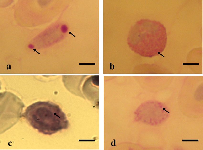
www.nature.com/scientificreports/
can cause a ccidents8,9. Four valid stingray species are found in the black waters of the Rio Negro basin: Potamo-
trygon motoro (Müller & Henle 1841), P. orbignyi (Castelnau 1855), P. schroederi (Fernández-Yépez 1958) and
Paratrygon aiereba (Müller & Henle 1841). In addition, a new species known as P. wallacei (cururu stingray)
(Carvalho et al. 2016) is currently being identified and scientifically characterized. This species is probably
endemic to this region, with a hotspot concentrated in the Mariuá archipelago near the municipality of Barcelos
(Amazonas, Brazil)10.
Investigations on the blood constituents of several marine elasmobranch fish species have been conducted,
especially in sharks11–16. Nevertheless, only a few studies have addressed freshwater elasmobranchs15,17–23. The
presence of erythrocytes, thrombocytes, lymphocytes, monocytes, heterophils and basophils in freshwater
stingrays of the Potamotrygonidae family has been r eported21. However, these authors did not investigate the
cytochemical features of these cell types to confirm their identities by examining traditional morphology from
cytochemical markings.
Hematological evaluations are becoming routine practice for assessing the health of fish and other
animals19,21,24–33. Studies on blood leukocytes can reveal the characteristics of the immune systems of different
fish species34,35, including free-living Amazonian stingrays. Hematological investigations have relied on classical
Romanowsky staining with the Leishman, Wright, May, Grünwald and Giemsa used to identify l eukocytes36–38,
but cell-based classifications of stingray leukocyte cells are not always reliable using classical staining methods
because the staining procedures vary, which can lead to errors in the identification of a cell type. Thus, cyto-
chemical staining of leukocytes in blood may be particularly useful for identifying cell lineages and may indicate
cell function.
This study aimed to investigate the morphology, dimensions, cytochemical staining reactivity and ultrastruc-
ture of blood cells from three freshwater stingray species, P. wallacei (cururu stingray), P. motoro and P. aiereba,
living in the black waters of the middle Rio Negro basin (Barcelos, Amazonas, Brazil). Because Brazil and other
Amazonian countries export these species as ornamental fish to consumers around the world, these results will
contribute to establishing standards for blood constituents that may be helpful in assessing the health and welfare
of these fish in artificial systems, especially in relation to the ornamental fish trade.
Materials and methods
Study area and specimen collection. Specimens of the Amazonian stingrays Potamotrygon wallacei
(cururu stingray; n = 53), Potamotrygon motoro (n = 55) and Paratrygon aiereba (n = 32) were collected from
the Mariuá archipelago (license: 15116-1 IBAMA). This is the largest complex of islands that exists in conti-
nental waters (more than 700 islands), and it is located in the black waters of the middle Rio Negro basin near
the municipality of Barcelos (Amazonas, Brazil). The fish were caught at different sites within the archipelago,
including beaches, lakes, small streams (igarapés), and areas of flooded forest (igapós), between January 2006
and October 2019. Professional fisherman caught the fish at night (19:00 to 03:00) through active searching with
the aid of a head flashlight, paddle and typical hand net (rapiché). We immediately anesthetized the captured
stingrays with eugenol (0.2 g L−1) and withdrew a blood sample (1.0–1.5 mL) from the gill arterial v essel18 using
10% EDTA as an a nticoagulant20. After these procedures, we measured the total length (TL, cm), disc width
(DW, cm) and body weight (BW, kg) of each specimen. All the stingrays sampled recovered from the anesthetic
and were safely returned to their respective capture sites.
For cytochemical staining and ultrastructural examination of different blood cell types, ten P. wallacei, P.
motoro and P. aiereba stingrays were caught by professional fishermen near the Daracuá community within the
Mariuá archipelago. These stingrays were transported by boat (journey of 24 h) to the Laboratory for Physiology
Applied to Aquaculture (LAFAP) at the National Amazon Research Institute (Instituto Nacional de Pesquisas da
Amazônia, INPA) in Manaus (Amazonas, Brazil). At the laboratory, the stingrays were acclimatized in 5000-L
tanks for 48 h, with constant water changes and oxygenation so that they would recover from the stress of being
captured and transported. After this period, a blood sample (1.0 mL) was collected from the gill arterial vessel
using 10% EDTA as an a nticoagulant20. Then, the biometric parameters were determined (TL, DW and BW).
Morphological and morphometric measurements and quantification of blood cells. Fresh
blood samples were collected from P. wallacei (n = 43), P. motoro (n = 45) and P. aiereba (n = 32). We stained these
blood smears with a combination of May-Grünwald-Giemsa-Wright stains to identify cells and make morpho-
metric measurements (µm) of 100 samples of each cell type found (erythrocytes, leukocytes and thrombocytes),
with the aid of an optical microscope and a millimeter ruler for determination of the largest and smallest cells.
Subsequently, the blood samples were used for leukocyte and total thrombocyte counts37 and for differential
leukocyte counts, which were based on the counts of 200 l eukocytes21.
Cytochemical staining. We collected fresh blood samples from 10 specimens of each stingray species. The
presence and intensity of glycogen deposits inside blood cells was confirmed by using the periodic acid-Schiff
(PAS) method. Controls for this reaction were obtained through smears exposed to salivary amylase digestion
for 60 min.
The peroxidase reaction was carried out by using the ortho-toluidine method in the presence or absence of
hydrogen peroxide. The reaction products were subjected to nuclear staining using Harris h ematoxylin37.
Reactions for metachromasia were tested in blood smears fixed in 1% lead subacetate for 10 min and sub-
sequently stained with 0.2% toluidine blue for 50 min37. The presence of lipids in different blood cell types was
confirmed in blood smears previously fixed with 70% ethanol for 5 s and then stained with 0.3% Sudan Black
B solution26.
Scientific Reports | (2021) 11:15685 | https://doi.org/10.1038/s41598-021-95183-4 2
Vol:.(1234567890)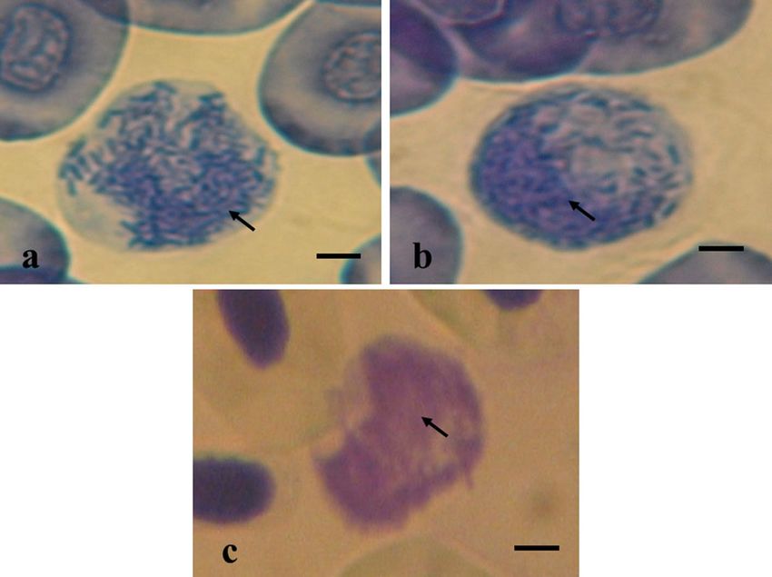
www.nature.com/scientificreports/
Species Total length (cm) Disk width (cm) Body weight (g)
P. wallacei 19.1 ± 2.5 17.4 ± 1.1 226.0 ± 48.5
P. motoro 25.1 ± 3.1 20.4 ± 1.8 351.0 ± 65.0
P. aiereba 44.8 ± 13.9 29.3 ± 10.8 966.5 ± 856.9
Table 1. Means (cm) ± standard deviations of the biometric variables for P. wallacei, P. motoro and P. aiereba in
the Middle Rio Negro of the Amazon are shown.
To identify total protein, blood smears were fixed in formalin, stained with bromophenol blue for 15 min,
immersed in 0.5% acetic acid, washed in phosphate buffer and finally dehydrated in butyl alcohol. Reticulocytes
were identified using a solution of brilliant cresyl blue and blood (1:1), which was homogenized, kept in a water
bath for 20 min at 37 °C and stained with a combination of May-Grünwald-Giemsa-Wright stains37. The results
from the cytochemical staining were expressed qualitatively, according to the intensity of staining observed for
each blood cell type, i.e., negative staining (–), weakly positive staining (+) and positive staining (++).
Ultrastructural analysis. The blood cell types were characterized ultrastructurally in four of the ten sting-
rays from each species that had been acclimatized for cytochemical studies. Blood samples were taken from
the gill vessel18 and centrifuged at 750g for 15 min to obtain pellets containing erythrocytes, thrombocytes and
leukocytes. We immediately fixed these pellets in 0.1 M sodium cacodylate solution (pH 7.4) containing 2.5%
glutaraldehyde and 2.0% paraformaldehyde at 4 °C for 2.5 h. We then immersed these samples in a 0.2 M sodium
cacodylate solution (pH 7.4) containing 1% osmium tetroxide at 4 °C for one hour. After these procedures, the
samples were dehydrated and embedded in Araldite resin (Sigma-Aldrich, USA), and sections were cut using a
Reichert OM-U3 ultratome, mounted on copper grids (200 mesh) and stained with 0.2% uranyl acetate solution
and lead citrate solution for 15 min. The sections were analyzed using a transmission electron microscope at the
Microscopy Center of the Institute of Biosciences at São Paulo State University (Universidade Estadual Paulista
Julio de Mesquita Filho, UNESP) in Botucatu of São Paulo, Brazil.
Ethical approval. The study was conducted at the Department of Physiological Sciences, Federal University
Amazonas, in accordance with the Brazilian guidelines for animal experiments and was approved by the govern-
ment of Amazonas (license: 15116-1), Brazil. All experiments were conducted according to local and ARRIVE
guidelines39.
Research involving human participants and/or animals. A total of 140 rays were captured in the
natural environment, 130 of which were returned, and 10 were processed and registered in the collection.
Results
The mean values for total length, disc width and body mass of P. wallacei, P. aiereba and P. motoro specimens
are shown in Table 1.
Morphological and morphometric measurements and quantification of blood cells. Blood
smears from P. wallacei, P. motoro and P. aiereba revealed erythroblasts, mature erythrocytes, thrombocytes,
lymphocytes, monocytes, heterophils and basophils of similar sizes among the species.
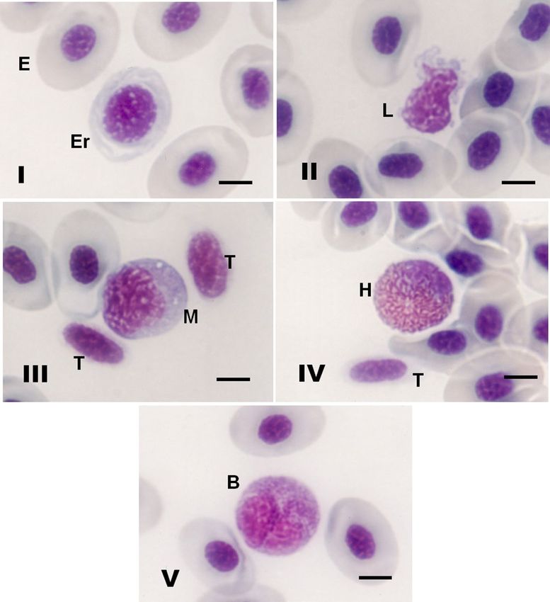
Mature erythrocytes were very similar in shape and size in the three Amazonian stingray species. Under an
optical microscope, mature erythrocytes presented as elliptical cells with abundant hyaline in the cytoplasm, and
the nucleus was usually centered and condensed with a shape that resembled the cell shape (Fig. 1-I). Erythro-
blasts were rounded cells and were easily distinguished from mature erythrocytes by their pale appearance or
by hyaline in the cytoplasm (Fig. 1-I).
Lymphocytes presented different sizes and irregular shapes, which were mostly elliptical and rarely oval, with
a nucleus occupying a large part of the basophilic cytoplasm. Lymphocytes presented cytoplasmic projections
without visible granulations and sometimes presented vacuoles (Fig. 1-II). Thrombocytes were generally fusiform,
with hyaline in the cytoplasm; the nucleus occupied almost the entire cell and resembled the cell shape (Fig. 1-
III). Monocytes were predominantly oval, with a nucleus morphology similar to that of thrombocytes (Fig. 1-III).
Heterophils were predominantly oval, with a large amount of heterophilic coarse granules and a nucleus that was
generally eccentric (Fig. 1-IV). Basophils were also predominantly oval, with basophilic granules and a nucleus
that was eccentric and generally bilobulated (Fig. 1-V).
In comparison with the other leukocyte cell types, monocytes were the largest cells in the three elasmobranch
species (Table 2). Basophils and lymphocytes were the smallest cell types in the blood of the three freshwater
stingray species investigated herein (Table 2).
Leukocyte and thrombocyte cell counts showed that lymphocytes and monocytes were the predominant
blood cells, while heterophils and basophils were the least abundant blood cells in the three freshwater stingray
species investigated (Table 3).
Cytochemical staining. Thrombocytes and leukocytes did not show any differences in cytochemical stain-
ing when comparing between the three stingray species (Table 4). Glycogen marking was observed in thrombo-
Scientific Reports | (2021) 11:15685 | https://doi.org/10.1038/s41598-021-95183-4 3
Vol.:(0123456789)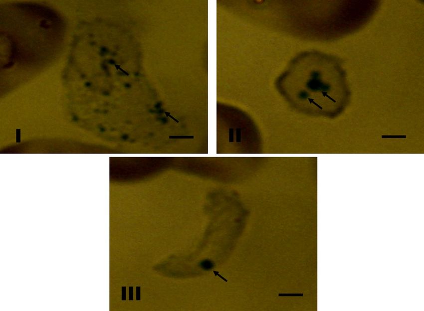
www.nature.com/scientificreports/

                                            Figure 1.  (I–V) Morphology of blood cells of three freshwater stingray species stained with May Grunwald-
                                            Giemsa-Wright stains. (I) (E) Erythrocytes and (Er) erythroblasts of P. wallacei; (II) (L) lymphocytes of P.
                                            wallacei; (III) (T) thrombocytes and (M) monocytes of P. wallacei; (IV) (H) heterophils and (T) thrombocytes
                                            of P. wallacei; (V) (B) basophils of P. wallacei. Bar = 8 µm.

                                             Cells                P. wallacei               P. motoro                 P. aiereba
                                             Erythrocytes (µm)    20.1 ± 0.7 × 14.1 ± 0.6   20.2 ± 0.8 × 14.1 ± 0.7   20.0 ± 0.8 × 14.0 ± 0.8
                                             Erythroblasts (µm)   19.0 ± 0.9 × 14.8 ± 0.4   19.0 ± 0.8 × 14.7 ± 0.5   19.1 ± 0.7 × 14.8 ± 0.5
                                             Thrombocytes (µm)    14.7 ± 1.4 × 9.6 ± 0.5    14.6 ± 1.5 × 9.5 ± 0.6    14.6 ± 1.3 × 9.6 ± 0.4
                                             Lymphocytes (µm)     14.4 ± 1.8 × 12.4 ± 2.7   14.7 ± 1.7 × 12.8 ± 3.1   14.8 ± 2.1 × 12.7 ± 2.9
                                             Monocytes (µm)       21.4 ± 1.1 × 21.4 ± 1.1   21.3 ± 1.2 × 21.3 ± 1.2   21.5 ± 1.0 × 21.5 ± 1.0
                                             Heterophils (µm)     14.5 ± 0.5 × 14.5 ± 0.5   14.4 ± 0.4 × 14.4 ± 0.4   14.4 ± 0.5 × 14.4 ± 0.5
                                             Basophils (µm)       13.5 ± 0.5 × 13.5 ± 0.5   13.4 ± 0.6 × 13.4 ± 0.6   13.6 ± 0.6 × 13.6 ± 0.6

                                            Table 2.  Mean diameters (µm ± SD) of the largest and smallest axes of different blood cells (n = 50) from three
                                            freshwater stingray species living in the middle Rio Negro basin in Amazonas, Brazil are shown.

                                            cytes (Fig. 2-I) and heterophils (Fig. 2-II), and there was weak positive staining in lymphocytes (Fig. 2-III) and
                                            monocytes (Fig. 2-IV).
                                                Weak positive staining with Sudan black was observed in heterophils (Fig. 3-I), lymphocytes (Fig. 3-II) and
                                            thrombocytes (Fig. 3-III). Positive identification of proteins using bromophenol blue occurred only in granules
                                            of heterophils (Fig. 4-I) and basophils (Fig. 4-II). The presence of reticulocytes was observed in erythrocytes,
                                            thus indicating the presence of crosslinking material fragments that were not stained with traditional dyes. There

          Scientific Reports |   (2021) 11:15685 |                https://doi.org/10.1038/s41598-021-95183-4                                                4

Vol:.(1234567890)
www.nature.com/scientificreports/
Species P. wallacei P. motoro P. aiereba
Leukocytes (µL) 3629 ± 2001 2998 ± 1107 3297 ± 1469
Thrombocytes (µL) 890 ± 498 826 ± 601 690 ± 468
Lymphocytes (%) 46.1 ± 15.8 45.6 ± 10.9 43.6 ± 14.5
Lymphocytes (µL) 1673 ± 316 1367 ± 121 1437 ± 213
Monocytes (%) 30.7 ± 14.7 26.2 ± 4.3 28.5 ± 12.3
Monocytes (µL) 1114 ± 294 785 ± 51 939 ± 180
Heterophils (%) 20.2 ± 10.7 25.1 ± 15.5 24.9 ± 14.0
Heterophils (µL) 733 ± 214 752 ± 171 820 ± 205
Basophils (%) 3.0 ± 2.0 2.7 ± 0.6 3.0 ± 1.8
Basophils (µL) 109 ± 40 81 ± 7 99 ± 26
Table 3. Leukocyte and thrombocyte counts of P. wallacei, P. motoro and P. aiereba from the Middle Rio
Negro of the Amazon are listed.
Bromophenol
PAS Peroxidase Toluidine blue Sudan Black B blue
Cells 1 2 3 1 2 3 1 2 3 1 2 3 1 2 3
Thrombocytes ++ ++ ++ − − − − − − ++ ++ ++ − − −
Lymphocytes + + + − − − − − − ++ ++ ++ − − −
Monocytes + + + − − − − − − − − − − − −
Heterophils ++ ++ ++ − − − − − − + + + ++ ++ ++
Basophils − − − − − − ++ ++ ++ − − − ++ ++ ++
Table 4. Cytochemical staining results of the blood cells of stingrays P. wallacei, P. motoro and P. aiereba
from the middle Rio Negro of the Amazon are shown. (1) P. wallacei; (2) P. motoro; (3) P. aiereba. −
Negative; + weakly positive; ++ positive.
Figure 2. (I–IV) PAS staining for detection of glycogen in blood cells of freshwater stingrays in central
Amazonia. (I) Thrombocytes of P. aiereba; (II) Heterophils of P. aiereba; (III) Lymphocytes of P. wallacei; (IV)
Monocytes of P. motoro. Bar = 8 µm.
Scientific Reports | (2021) 11:15685 | https://doi.org/10.1038/s41598-021-95183-4 5
Vol.:(0123456789)www.nature.com/scientificreports/
Figure 3. (I–III) Cytochemical staining of lipids with Sudan Black B was performed in blood cells of freshwater
stingrays in central Amazonia. (I) Heterophils of P. wallacei; (II) Lymphocytes of P. aiereba; (III) Thrombocytes
of P. aiereba. Bar = 8 µm.
Figure 4. (I–III) Cytochemical staining of total protein and metachromasia in blood cells of freshwater
stingrays in central Amazonia. (I) Total protein staining in heterophils of P. motoro; (II) Total protein staining in
basophils of P. wallacei; (III) Metachromasia in basophils of P. wallacei. Bar = 10 µm.
Scientific Reports | (2021) 11:15685 | https://doi.org/10.1038/s41598-021-95183-4 6
Vol:.(1234567890)www.nature.com/scientificreports/
Figure 5. (I,II) Ultrastructural analysis of blood cells from freshwater stingrays in central Amazonia. (I)
Thrombocytes of P. motoro; (II) Lymphocytes of P. wallacei. Increase 4000 x.
was no positive peroxidase staining, although metachromasia was observed (Fig. 4-III). This was characterized
using a blue reagent that reacted with the red-colored blood of the freshwater stingrays.
Ultrastructural analysis. Thrombocytes were generally round and spindle-shaped. In the cytoplasm, a
canalicular system with various-sized vesicles and canaliculi was occasionally observed, along with glycogen
pellets, granules and numerous mitochondria (Fig. 5-I). Lymphocytes presented amorphous forms, with sparse
cytoplasm. The presence of vacuoles and few mitochondria was observed, and the nucleus occupied almost the
entire cell, with dense chromatin in the periphery and no evident nucleolus (Fig. 5-II). Monocytes presented
nuclei with peripheral heterochromatin and cytoplasm with mitochondria, secretion vesicles, secretion granules
and endoplasmic reticulum. Because basophils were scarce in the blood, they could not be found in the potamo-
trygonids in this study. Staining of heterophils revealed the presence of heterochromatin, and there were large
numbers of granules that might have been glycogen, lipids and/or proteins, but they could not be distinguished.
Discussion
Morphological and morphometric measurements and quantification of blood cells. Most ver-
tebrates have seven blood cell types: erythrocytes, thrombocytes, lymphocytes, eosinophils, basophils, mono-
cytes and n eutrophils40,41. The morphology of each cell type appears to be similar, except for neutrophils. In some
cases, neutrophils are replaced by heterophils, which present the same immunological function40,42,43. It was
reported that erythrocytes, thrombocytes, lymphocytes, monocytes, neutrophils and eosinophils were present
in freshwater potamotrygonids17.
In contrast, no eosinophils were observed in blood from Amazon stingrays, thus suggesting that heterophils
have some importance in the immune defense of these potamotrygonids. In addition, there is a lack of standardi-
zation in the staining procedures adopted and in the classification of blood cell types. In the present study, eryth-
roblasts, mature erythrocytes, thrombocytes, lymphocytes, monocytes, heterophils and basophils were observed.
In the potamotrygonids of this study, reticulocytes were identified by the presence of ribonucleoproteins
inside some erythrocytes. High amounts of ribonucleoproteins indicate premature release of erythrocytes into
the bloodstream37. Therefore, quantification of the number of circulating reticulocytes can provide information
about erythropoietic activity and therefore about the animal health status.
The morphological features of freshwater stingray erythrocytes are similar to those of marine elasmobranchs,
such as the Dasyatis sabina (Lesueur 1824), Raja eglanteria (Bosc 1800)13,16, Raja microocellata (Montagu 1818),
R. brachyura (Lafont 1871) and Raja sp.44 stingrays and the Squalus acanthias (Linnaeus 1758)45, Schroederich-
thyes chilensis (Guichenot 1848)12, Ginglymostoma cirratum (Bonnaterre 1788) and Carcharhinus limbatus sharks
(Müller & Henle 1839).
In addition, in the C. coelolepis shark, immature erythrocytes (erythroblasts) may be smaller than mature
erythrocytes46, and this characteristic was also observed in the three potamotrygonid stingray species.
Thrombocytes in elasmobranchs are blood cells with functions analogous to mammalian platelets, which
play a role in homeostasis13,16. In dogfish (S. canicular), it was demonstrated that blood thrombocytes remove
antigenic substances, such as colloidal charcoal particles47. The cell sizes and morphological characteristics of
freshwater stingray thrombocytes were similar to those reported in the S. chilensis12 and C. leucas13 sharks and
different from those of C. plumbeus, which presented cytoplasmic g ranules11. Moreover, in the blood of the C.
coelolepis shark, the form known as "drop" (with fingerlike cytoplasmic projections) was observed46, but this was
not detected in the Amazonian stingrays herein.
In blood smears from marine elasmobranchs, leukocytes at different stages of maturation are frequently
observed. This can cause incorrect identification13, thereby contributing to the confusing terminology of
Scientific Reports | (2021) 11:15685 | https://doi.org/10.1038/s41598-021-95183-4 7
Vol.:(0123456789)www.nature.com/scientificreports/
elasmobranch leukocytes13 and causing errors in identifying small monocytes and large lymphocytes48. In the
present study, lymphocytes presented shapes ranging from round to amorphous, and this has also been observed
among lymphocytes in C. coelolepis46, S. chilensis12, G. cirratum12,13, C. plumbeus11, R. microocellata, R. brachyura,
R. sp.44, O. maculatus, O. ornatus, Orectolobus sp.49 and R. typus15. The size of the lymphocytes in the three Ama-
zonian stingray species was slightly smaller than that of the C. coelolepis shark46.
The lymphocyte morphological characteristics were similar to those observed in other
elasmobranchs11–13,15,44,49. Granulocytes have been reported in several elasmobranch species, but they are difficult
to identify and classify because of the great variations in shape and size and the poor staining of the c ells12. In
the present study, in the blood of freshwater stingrays, two types of granulocytes were detected: heterophils and
basophils. It was reported that the most common granulocytes in the blood of elasmobranchs were heterophils,
while basophils were rare in b lood12. It was reported that neutrophils and eosinophils were present in the blood
of potamotrygonids17. The identification of neutrophils and eosinophils in potamotrygonids can be correlated
with the extreme difficulty of the methods for staining smears and/or with incorrect classification of the differ-
ent types of leukocytes. Heterophils and basophils with the same morphological features observed in the three
Amazonian stingrays were also observed in C. coelolepis (dogfish shark)46, S. chilensis (catshark)12, C. limbatus
(blacktip shark)13 and R. typus (whale shark)15.
Erythrocytes are generally larger in lower orders, and variations in size may occur within species of the same
order40. The freshwater stingray erythrocytes were smaller than those of the Centroscymnus coelolepis shark
(Barbosa du Bocage & de Brito Capello 1864)46 and approximately two times larger than those in freshwater and
marine teleosts50 and in the Dicentrarchus labrax L. fish51.
The number of total leukocytes and thrombocytes observed in the present study was similar to the number
reported for freshwater stingrays P. falkneri, P. motoro, P. orbignyi, and P. scobina52, in addition to P. schroederi and
P. orbignyi19, as well as P. motoro, P. wallacei and P. aiereba21. For the differential leukocyte count, lymphocytes
were the predominant cells. Oliveira et al.20 and Oliveira et al.21 also reported the same results; however, Brito
et al.52 reported that neutrophils and leukocytes were predominant.
Cytochemical staining. The existence of neutrophils and eosinophils in the blood of an individual P.
motoro stingray was reported, and it was difficult to distinguish neutrophils from heterophils19. In the present
study, no neutrophils were detected. Instead, there were heterophilic granulocytes with morphological features
that were distinct from those of neutrophils. However, the granulocytes had heterophilic functions resembling
phagocytosis, which is also seen among neutrophils, as indicated by the presence of glycogen, lipids and proteins
in P. wallacei, P. motoro and P. aiereba. Glycogen is an important source of cellular energy reserves for the innate
defense mechanisms that occur, especially during phagocytosis37,53.
In the class Chondrichthyes, the cytochemical characteristics of leukocyte chimeras in the species Callorhyn-
chus milii (Bory de Saint-Vincent 1823), Chimaera phantasma (Jordan & Snyder 1900), Hydrolagus novaezea-
landiae (Fowler 1911), Hydrolagus sp., Harriotta raleighana (Goode & Bean 1895) and Rhinochimaera pacifica
(Mitsukuri 1895)54 were studied. It was reported that the esterase enzyme in the Holocephali subclass was very
different from that in elasmobranchs. However, the present study was the first aimed at determining the functions
of blood cell types in potamotrygonid species. Positive PAS staining was observed in thrombocytes of P. wallacei,
P. motoro and P. aiereba, but the staining in lymphocytes and monocytes was weak. Thrombocytes aid in blood
coagulation55, but they also play an important role in the immune activity of elasmobranchs13.
There was no peroxidase reaction in any of the blood cells of P. wallacei, P. motoro or P. aiereba. Peroxidase
is an important lysosomal enzyme involved in intracellular digestion, and one of its main features is that it
indicates the absence of eosinophilic and neutrophilic granulocytes in the species investigated here26. However,
this lack of peroxidase may be accompanied by compensatory development of other antibacterial components,
such as cationic p roteins13,38.
Though basophil leukocytes were rarely observed in the blood of P. wallacei, P. motoro and P. aiereba, their
existence was confirmed through metachromasia staining. In addition, the three potamotrygonids demon-
strated the presence of lipids in thrombocytes and lymphocytes but to a lesser degree than that in heterophils.
Similarly, in Xiphophorus helleri (Heckel 1848), a Sudan black reaction was also demonstrated in monocytes
and lymphocytes56. However, in other teleosts, this reaction has been observed in neutrophil granules36. Phago-
cytic leukocytes can use lipids as an energy source, thereby degrading these constituents through the action of
cytoplasmic enzymes.
The proteins in leukocyte granules are involved in host defense and microorganism death36. The heterophils
and basophils of P. wallacei, P. motoro and P. aiereba were positive for bromophenol blue staining, similar to
what had previously been found in eosinophils from S. brasiliensis36 in Amazonian t urtles26. Positive staining
was observed in basophils, eosinophils and neutrophils from P. motoro19. Therefore, these results indicate that
these proteins play an important role in the innate defense of animals, which is possibly performed by heterophil
and basophil granulocytes.
Ultrastructural analysis. The ultrastructural analyses of leukocytes from P. wallacei, P. motoro and P. aier-
eba were similar to each other and comparable with the findings from the sharks G. cirratum57 and S. canicula47.
The morphology and sizes of the different cell types were similar to those of marine rays and sharks. It is very
important to characterize the types of stingray leukocytes to provide basic information about these cells and
make correlations with health conditions. In this manner, leukocytes can be quantified in these stingrays, which
are extremely important for the aquarium industry. The cytochemical characteristics of heterophils indicates
that these major granulocytes are important in the immune defense of Amazonian potamotrygonids. The blood
Scientific Reports | (2021) 11:15685 | https://doi.org/10.1038/s41598-021-95183-4 8
Vol:.(1234567890)www.nature.com/scientificreports/
cell features of wild native stingrays may be useful for making diagnoses and comparisons among these same
species under controlled conditions.
Data availability
Data supporting the findings of this manuscript are available from the corresponding author upon reasonable
request.
Received: 2 January 2021; Accepted: 29 June 2021
References
1. Carvalho, M. R. Neotropical stingrays: family Potamotrygonidae. In Rays of The World (eds Last, P. R. et al.) 619–655 (Cornell
University Press, 2016).
2. Compagno, L. J. V. & Cook, S. F. The exploitation and conservation of freshwater elasmobrachs: Status of taxa and prospects for
the future. J. Aquat. Sci. 7, 62–90 (1995).
3. Lovejoy, N. R. Systematics of myliobatoid elasmobranchs: With emphasis on the phylogeny and historical biogeography of neotropi-
cal freshwater stingrays (Potamotrygonidae: Rajiformes). Zool. J. Linn. Soc. 117, 207–257. https://doi.org/10.1111/j.1096-3642.
1996.tb02189.x (1996).
4. Carvalho, M. R., Lovejoy, N. N. & Rosa, R. S. Family Potamotrygonidae (river stingrays). In Check List of the Freshwater Fishes of
South and Central América (eds Reis, R. E. et al.) 22–29 (Edipucrs, 2003).
5. Carvalho, M. R. & Lovejoy, N. R. Morphology and phylogenetic relationships of a remarkable new genus and two new species of
Neotropical freshwater stingrays from the Amazon basin (Chondrichthyes: Potamotrygonidae). Zootaxa 2776, 13–48 (2011).
6. Rosa, R. S., Charvet-Almeida, P. & Quijada, C. C. D. Biology of the South American potamotrygonid stingrays. In Sharks and their
Relatives II: Biodiversity, Adaptive Physiology, and Conservation (eds Carrier, J. F. et al.) 241–286 (CRC Press, 2010).
7. Chao, N. L., Petry, P., Prang, G., Sonneschien, L. & Tlusty, M. Conservation and Management of Ornamental Fish Resources of the
Rio Negro Basin, Amazonia, Brazil—Project Piaba (EDUA Manaus, 2001).
8. Oliveira, A. T. et al. Relação entre as populações naturais de arraias de água doce (Myliobatiformes: Potamotrygonidae) e pescadores
no baixo rio Juruá, estado do Amazonas, Brasil. Biota Amazônia. 5, 108–111. https://doi.org/10.18561/2179-5746/biotaamazonia.
v5n3p108-111 (2015).
9. Oliveira, A. T. et al. Conhecimento tradicional de pescadores de arraias de água doce da região Amazônica. Revista Ibero-americana
de Ciências Ambientais. 11, 128–135. https://doi.org/10.6008/CBPC2179-6858.2020.002.0015 (2020).
10. Duncan, W. P., Shibuya, A., Araújo, M. L. G. & Zuanon, J. Biologia e história natural de Potamotrygon wallacei (Carvalho, Rosa
and Araújo 2016) na bacia do Rio Negro, Amazônia Central, Brasil. In Rayas de agua dulce (Potamotrygonidae) de Suramérica.
Parte II: Colombia, Brasil, Perú, Bolivia, Paraguay, Uruguay y Argentina 1st edn (eds Lasso, C. A. et al.) 289–302 (Instituto de
Investigación de Recursos Biológicos Alexander von Humboldt, 2016).
11. Arnold, J. E. Hematology of the sandbar shark, Carcharhinus plumbeus: Standardization of complete blood count techniques for
elasmobranchs. Vet. Clin. Pathol. 34, 115–123. https://doi.org/10.1111/j.1939-165x.2005.tb00023.x (2005).
12. Valenzuela, A., Oyarzún, C. & Silva, V. Células sanguíneas de Schroederichthys chilensis (Guichenot 1848) (Elasmobranchii, Scylio-
rhinidae): la série blanca. Gayana 67, 130–136. https://doi.org/10.4067/S0717-65382003000100018 (2003).
13. Luer, C. A., Walsh, C. J. & Bodine, A. B. The Immune System of sharks, skates, and rays. In Biology of Sharks and their Relatives
(eds Carrier, J. C. et al.) 369–389 (CRC Marine Biology, 2004).
14. Cain, D. K., Harms, C. A. & Segars, A. Plasma biochemistry reference values of wild caught southern stingrays (Dasyatis americana).
J. Zoo Wildl. Med. 35, 471–476. https://doi.org/10.1638/03-107 (2004).
15. Dove, A. D. M., Arnold, J. & Clauss, T. M. Blood cells and serum chemistry in the world’s largest fish: The whale shark Rhincodon
typus. Aquat. Biol. 9, 177–183. https://doi.org/10.3354/ab00252 (2010).
16. Walsh, C. J. & Luer, C. A. Elasmobranch hematology: identification of cell types and practical applications. In The Elasmobranch
Husbandry Manual: Captive Care of Sharks, Rays and their Relatives (eds Smith, M. et al.) 307–323 (Ohio Biological Survey, 2004).
17. Griffith, R. W., Pang, P. K. T., Srivastava, A. K. & Pickford, G. E. Serum composition of freshwater stingrays (Potamotrygonidae)
adapted to fresh and diluted sea water. Biol. Bull. 144, 304–320 (1973).
18. Oliveira, A. T. et al. Procedimentos de manuseio e de colheita de sangue em arraias de água doce. Documentos Embrapa. 77, 1–18.
https://doi.org/10.13140/RG.2.2.19792.25605 (2012).
19. Oliveira, A. T., Pantoja-Lima, J., Aride, P. H. R., Tavares-Dias, M. & Marcon, J. L. Fisiologia de arraias de água doce: subsidios para
aplicabilidade na aquicultura. In Aquicultura no Brasil: novas perspectivas 1st edn, Vol. 1 (eds Tavares-Dias, M. & Mariano, W. S.)
45–74 (Pedro & João, 2015).
20. Oliveira, A. T., Santos, M. Q. C., Lemos, J. R. G., Tavares-Dias, M. & Marcon, J. L. Comparison of the effects of anticoagulants used
in blood collection for the determination of blood parameters of free-living stingrays of the Potamotrygon genus (Elasmobranchii:
Potamotrygonidae). Biota Amazônia. 5, 55–58. https://doi.org/10.18561/2179-5746/biotaamazonia.v5n3p55-58 (2015).
21. Oliveira, A. T. et al. Hematological parameters of three freshwater stingray species (Chondrichthyes: Potamotrygonidae) in the
middle Rio Negro, Amazonas state. Biochem. Syst. Ecol. 69, 33–40. https://doi.org/10.1016/j.bse.2016.07.002 (2016).
22. Oliveira, A. T. et al. Cyrilia sp. (Apicomplexa: Haemogregarinidae) in the Amazonian freshwater stingray Potamotrygon wallacei
(cururu stingray) in different hydrological phases of the Rio Negro. Braz. J. Biol. 77, 413–416. https://doi.org/10.1590/1519-6984.
00416 (2017).
23. Magro, N. M., Oliveira, A. T., Davies, A. & Odwyer, L. H. First report and description of a Cyrilia sp. (Apicomplexa: Haemogre-
garinidae) from a freshwater cururu stingray Potamotrygon cf. histrix (Elasmobranchii: Potamotrygonidae), from the Amazon
Region, Brazil. J. Fish Dis. 38, 1–5. https://doi.org/10.1111/jfd.12425 (2015).
24. Anselmo, N. P. et al. Hematological parameters of captive big-headed Amazon river turtles, Peltocephalus dumerilianus (Testudines:
Podocnemididae). Acta Biológica Colombiana. 26, 207–213. https://doi.org/10.15446/abc.v26n2.80616 (2021).
25. Polese, M. F. et al. Zootechnical parameters of Corn feed particle size in performance, digestibility and rate of passage in juvenile
tambaqui Colossoma macropomum. Braz. J. Biol. 82, 1–13. https://doi.org/10.1590/1519-6984.232612 (2022).
26. Oliveira, A. T. et al. Morphological and cytochemical characterization of thrombocytes and leukocytes in hatchings of three species
of Amazonian freshwater turtles. Veterinarski Arhiv. 81(5), 657–670 (2011).
27. Aride, P. H. R. et al. Growth and hematological responses of tambaqui, Colossoma macropomum fed different levels of rice, Oryza
spp. Braz. J. Biol. 81, 1–7. https://doi.org/10.1590/1519-6984.232560 (2020).
28. Aride, P. H. R. et al. Changes on physiological parameters of tambaqui (Colossoma macropomum) fed with diets supplemented
with Amazonian fruit Camu camu (Myrciaria dubia). Braz. J. Biol. 78, 360–367. https://d oi.o
rg/1 0.1 590/1 519-6 984.1 69442 (2018).
29. Aride, P. H. R. et al. Growth and hematological responses of tambaqui fed different amounts of cassava (Manihot esculenta). Arquivo
Brasileiro de Medicina Veterinária e Zootecnia 68, 1697–1704. https://doi.org/10.1590/1678-4162-8704 (2016).
Scientific Reports | (2021) 11:15685 | https://doi.org/10.1038/s41598-021-95183-4 9
Vol.:(0123456789)www.nature.com/scientificreports/
30. Castro, P. D. S. et al. Ecophysiological interactions in species of peacock bass Cichla spp. from the Amazon. Revista Ibero-americana
de Ciências Ambientais. 11, 594–599. https://doi.org/10.6008/CBPC2179-6858.2020.005.0053 (2020).
31. Castro, P. D. S. et al. Hematological parameters of three species of tucunarés (Cichla spp.) from Lake Balbina, Presidente Figueiredo,
Amazonas. Braz. J. Biol. 80, 1–7. https://doi.org/10.1590/1519-6984.219409 (2020).
32. Nascimento, G. B. et al. Parâmetros hematológicos do matrinxã Brycon amazonicus (Characidae: Bryconinae) criados em cativeiro
na região Amazônica. Braz. J. Dev. 6, 3303–3315. https://doi.org/10.34117/bjdv6n1-238 (2020).
33. Nascimento, M. S. et al. Supplementation of citric acid in plant protein-based diets for juvenile tambaqui. J. World Aquac. Soc.
https://doi.org/10.1111/jwas.12735 (2020).
34. Pavlidis, M., Futter, W. C., Kathario, P. & Divanach, P. Blood cells of six Mediterranean mariculture fish species. J. Appl. Ichthyol.
23, 70–73. https://doi.org/10.1111/j.1439-0426.2006.00771.x (2007).
35. Tavares-Dias, M. & Moraes, F. R. Leukocyte and thrombocyte reference values for channel catfish (Ictalurus punctatus Raf), with
an assessment of morphologic, cytochemical, and ultrastructural features. Vet. Clin. Pathol. 36, 49–54. https://doi.org/10.1111/j.
1939-165X.2007.tb00181.x (2007).
36. Tavares-Dias, M. A morphological and cytochemical study of erythrocytes, thrombocytes and leukocytes in four freshwater teleosts.
J. Fish Biol. 68, 1822–1833. https://doi.org/10.1111/j.1095-8649.2006.01089.x (2006).
37. Tavares-Dias, M. & Moraes, F. R. Morphological, cytochemical, and ultrastructural study of thrombocytes and leukocytes in
neotropical fish, Brycon orbignyanus Valenciones, 1850 (Characidae, Byconinae). J. Submicrosc. Cytol. Pathol. 38, 209–215 (2006).
38. Veiga, M. L., Egami, M. I., Ranzani-Paiva, M. J. T. & Rodrigues, E. L. Aspectos morfológicos y citoquímicos de las células sanguíneas
de Salminus maxilosus Valenciennes, 1840 (Characiformes, Characidae). Rev. Chil. Anat. 18(2), 245–250. https://doi.org/10.4067/
S0716-98682000000200005 (2000).
39. Percie du Sert, N. et al. The ARRIVE guidelines 2.0: Updated guidelines for reporting animal research. PLoS Biol. 18(7), e3000410.
https://doi.org/10.1371/journal.pbio.3000410 (2020).
40. Canfield, P. J. Comparative cell morphology in the peripheral blood film from exotic and native animals. Aust. Vet. J. 76, 793–800.
https://doi.org/10.1111/j.1751-0813.1998.tb12328.x (1998).
41. Tavares-Dias, M. & Moraes, F. R. Características hematológicas da Tilapia rendalli Boulenger, 1896 (Osteichthyes: Cichlidae)
capturada em “Pesque-Pague” de Franca, São Paulo, Brasil. Biosci. J. 19, 103–110 (2003).
42. Davis, A. K., Maney, D. L. & Maerz, J. C. The use of leukocyte profiles to measure stress in vertebrates: A review for ecologists.
Funct. Ecol. 22, 760–772. https://doi.org/10.1111/j.1365-2435.2008.01467.x (2008).
43. Hawkey, C. M. & Dennett, T. B. Color Atlas of Comparative Veterinary Hematology (Iowa State University Press, 1989).
44. Aragort, W., Alvarez, M. F., Leiro, J. L. & Sanmartín, M. L. Blood protozoans in elasmobranchs of the family Rajidae from Galicia
(NW Spain). Dis. Aquat. Org. 65, 63–68. https://doi.org/10.3354/dao065063 (2005).
45. Clewley, A., Kocan, R. M. & Kocan, A. A. An intraerythrocytic parasite from the spiny dogfish, Squalus acanthias L., from the
Pacific Northwest. J. Fish Dis. 25, 693–696. https://doi.org/10.1046/j.1365-2761.2002.00417.x (2002).
46. Sherburne, S. Cell types, differential cell counts, and blood cell measurements of a Portuguese shark, Centroscymnus coelolepis,
captured at 700 fathoms. Fish. Bull. 71, 435–439 (1973).
47. Morrow, W. J. W. & Pulsford, A. Identification of peripheral blood leukocytes of the dogfish (Scyliorhinus canicula L.) by electron
microscopy. J. Fish Biol. 17, 461–475 (1980).
48. DaMatta, R. A., Ribeiro, M. L. S., Carvalho, T. M. U. & Nascimento, J. L. M. Caracterização morfológica e funcional de leucócitos
de peixes. In Manejo e sanidade de peixes em cultivo (ed. Tavares-Dias, M.) 314–329 (Embrapa, 2009).
49. Old, J. M. & Huveneers, C. Morphology of the blood cells from three species of wobbegong sharks (Orectolobus species) on the
east coast of New South Wales. Zoo Biol. 25, 73–82. https://doi.org/10.1002/zoo.20079 (2006).
50. Vázquez, G. R. & Guerrero, G. A. Characterization of blood cells and hematological parameters in Cichlasona dimerus (Teleostei,
Perciformes). Tissue Cell 39, 151–160. https://doi.org/10.1016/j.tice.2007.02.004 (2007).
51. Esteban, M. A., Munoz, J. & Mesenguer, J. Blood cells of sea bass (Dicentrarchus labrax L.). Flow cytometric and microscopic
studies. Anat. Rec. 258, 80–89. https://doi.org/10.1002/(SICI)1097-0185(20000101)258:1%3c80::AID-AR9%3e3.0.CO;2-I (2000).
52. Brito, F. M. M. et al. Hematology, biochemical profile and thyroid hormones of four species of freshwater stingrays of the genus
Potamotrygon. Braz. J. Vet. Res. Anim. Sci. 52, 249–256. https://doi.org/10.11606/issn.1678-4456.v52i3p249-256 (2015).
53. Ueda, I. K., Egami, M. I., Sasso, W. S. & Matushima, E. R. Cytochemical aspects of the peripheral blood cells of Oreochromis (Tilapia)
niloticus (Linnaeus, 1758) (Cichlidae, Teleostei). Part II. Braz. J. Vet. Anim. Sci. 38, 273–277. https://doi.org/10.1590/S1413-95962
001000600005 (2001).
54. Hine, P. M. & Wain, J. M. The enzyme cytochemistry of leucocytes in blood and haematopoietic tissues of holocephalans (Chon-
drichthyes: Chimaeriformes). NZ J. Mar. Freshwater Res. 22, 57–62 (1988).
55. Hayhoe, F. G. J. & Qualigno, D. Haematological Cytochemistry (Churchill Livingstone, 1994).
56. Schütt, D. A., Lehmann, J., Goerlich, R. & Hamers, R. Haematology of swordtail, Xiphophorus helleri. I: Blood parameters and
light microscopy of blood cells. J. Appl. Ichthyol. 14, 83–89. https://doi.org/10.1111/j.1439-0426.1997.tb00106.x (1997).
57. Hyder, S. L., Cayer, M. L. & Pettey, C. L. Cell types in peripheral blood of the nurse shark: An approach to structure and function.
Tissue Cell 15(3), 437–4553. https://doi.org/10.1016/0040-8166(83)90075-7 (1983).
Acknowledgements
Dr. Elizabeth Gusmão Affonso for allowing physical space for the maintenance of freshwater stingrays and Pro-
fessor Dr. Irani Quagio-Grassiotto for helping with the analysis of electronic microscopy.
Author contributions
A.T.D.O. and J.L.M. conceived the study. A.T.D.O., J.R.G.L., M.Q.C.S., M.L.G.A. and J.L.M. designed the study.
A.T.D.O., J.R.G.L., M.Q.C.S. and M.T.D. undertook laboratorial analyses. J.P.L., P.H.R.A. and M.T.D. drafted the
paper with contributions from all other authors. All authors reviewed the manuscript.
Funding
The Research Support Foundation of the State of Amazonas (Fundação de Amparo à Pesquisa do Estado do
Amazonas, FAPEAM, under procedural nos. 925/03, 2203/05, 2204/05, 2459/08, 126/08 and 062.01099/17),
the National Council for Scientific and Technological Development (Conselho Nacional de Desenvolvimento
Científico e Tecnológico, CNPq, under procedural nos. 486289/2006-0, 40872/2006-4 and 408795/2006-9) funded
this study. The main author is grateful for the doctoral degree scholarship granted by the Coordination Office
for Improvement of University-level Personnel (Coordenação de Aperfeiçoamento de Pessoal de Nível Superior,
CAPES). A.T. Oliveira (process number 315713/2020-8) and M. Tavares-Dias are research fellowship recipients
from CNPq/Brazil.
Scientific Reports | (2021) 11:15685 | https://doi.org/10.1038/s41598-021-95183-4 10
Vol:.(1234567890)www.nature.com/scientificreports/
Competing interests
The authors declare no competing interests.
Additional information
Correspondence and requests for materials should be addressed to A.T.O.
Reprints and permissions information is available at www.nature.com/reprints.
Publisher’s note Springer Nature remains neutral with regard to jurisdictional claims in published maps and
institutional affiliations.
Open Access This article is licensed under a Creative Commons Attribution 4.0 International
License, which permits use, sharing, adaptation, distribution and reproduction in any medium or
format, as long as you give appropriate credit to the original author(s) and the source, provide a link to the
Creative Commons licence, and indicate if changes were made. The images or other third party material in this
article are included in the article’s Creative Commons licence, unless indicated otherwise in a credit line to the
material. If material is not included in the article’s Creative Commons licence and your intended use is not
permitted by statutory regulation or exceeds the permitted use, you will need to obtain permission directly from
the copyright holder. To view a copy of this licence, visit http://creativecommons.org/licenses/by/4.0/.
© The Author(s) 2021
Scientific Reports | (2021) 11:15685 | https://doi.org/10.1038/s41598-021-95183-4 11
Vol.:(0123456789)You can also read