Ivermectin reduces in vivo coronavirus infection in a mouse experimental model - Nature
←
→
Page content transcription
If your browser does not render page correctly, please read the page content below
www.nature.com/scientificreports
OPEN Ivermectin reduces in vivo
coronavirus infection in a mouse
experimental model
A. P. Arévalo1, R. Pagotto2, J. L. Pórfido1,3, H. Daghero2, M. Segovia4,5, K. Yamasaki6,
B. Varela6, M. Hill4,5, J. M. Verdes6, M. Duhalde Vega4,7, M. Bollati‑Fogolín2 & M. Crispo1*
The objective of this study was to test the effectiveness of ivermectin for the treatment of mouse
hepatitis virus (MHV), a type 2 family RNA coronavirus similar to SARS-CoV-2. Female BALB/cJ mice
were infected with 6,000 PFU of MHV-A59 (group infected, n = 20) or infected and then immediately
treated with a single dose of 500 µg/kg ivermectin (group infected + IVM, n = 20) or were not infected
and treated with PBS (control group, n = 16). Five days after infection/treatment, the mice were
euthanized and the tissues were sampled to assess their general health status and infection levels.
Overall, the results demonstrated that viral infection induced typical MHV-caused disease, with
the livers showing severe hepatocellular necrosis surrounded by a severe lymphoplasmacytic
inflammatory infiltration associated with a high hepatic viral load (52,158 AU), while mice treated
with ivermectin showed a better health status with a lower viral load (23,192 AU; p < 0.05), with only
a few having histopathological liver damage (p < 0.05). No significant differences were found between
the group infected + IVM and control group mice (P = NS). Furthermore, serum transaminase levels
(aspartate aminotransferase and alanine aminotransferase) were significantly lower in the treated
mice than in the infected animals. In conclusion, ivermectin diminished the MHV viral load and disease
in the mice, being a useful model for further understanding this therapy against coronavirus diseases.
Mouse hepatitis virus (MHV) is a single-stranded RNA coronavirus that targets different o rgans1. The virus is
highly contagious, has natural respiratory or oral transmission, and shows high morbidity and low mortality
rates. There is no vaccine or treatment available; therefore, upon infection, an entire laboratory mouse colony
must be sacrificed to control the disease.
Recent studies have shown that the mechanism of infection has similarities to that of SARS-CoV-22,3: there-
fore, it has been proposed that MHV may be an interesting infection model to test new therapies against COVID-
19 in animals. Although different therapies have been evaluated, no effective treatment is available, and the
mechanism by which the virus enters the cell is being e xplored4. After entry into the cytoplasm of the host cell,
coronaviruses rely on a nuclear transport system mediated by the importin α/β1 heterodimer to facilitate rep-
lication and infection5,6. Some drugs have been demonstrated to act by impairing importin α/β1 heterodimer
formation to prevent viral entry. Because both MHV and SARS-CoV-2 enter the nucleus via the same mechanism,
MHV may be an interesting target for the development of candidate therapies against coronavirus infection in
a mouse model.
Ivermectin is an efficient and inexpensive drug usually applied to treat parasitic infestations. It has been
approved by the FDA for animal and human use and is available worldwide. It has a wide margin of safety with
an LD50 of 30 mg/kg in mice and is used in humans as an antiparasitic treatment at a dose of 150–200 µg/kg7.
Other effects of this drug, such as a ntimicrobial8, anti-trypanosome/anti-malaria9 and anticancer a ctivities10,
have been proposed.
In addition, several reports have shown an in vitro effect of ivermectin against RNA and DNA virus infection
through the suppression of the host cellular process related to the inhibition of the nuclear transport of specific
1
Transgenic and Experimental Animal Unit, Institut Pasteur de Montevideo, Montevideo, Uruguay. 2Cell Biology
Unit, Institut Pasteur de Montevideo, Montevideo, Uruguay. 3Worm Biology Laboratory, Institut Pasteur
de Montevideo/Department of Biosciences, Faculty of Chemistry, University of the Republic, Montevideo,
Uruguay. 4Laboratory of Immunoregulation and Inflammation, Institut Pasteur de Montevideo, Montevideo,
Uruguay. 5Immunobiology Department, Faculty of Medicine, University of the Republic, Montevideo,
Uruguay. 6Pathobiology Department, Faculty of Veterinary, Pathology Unit, University of the Republic,
Montevideo, Uruguay. 7Institute of Biological Chemistry and Chemical Physics (UBA‑CONICET), School of
Pharmacy and Biochemistry, University of Buenos Aires, Buenos Aires, Argentina. *email: crispo@pasteur.edu.uy
Scientific Reports | (2021) 11:7132 | https://doi.org/10.1038/s41598-021-86679-0 1
Vol.:(0123456789)www.nature.com/scientificreports/
proteins required for viral r eplication11. Exposure to ivermectin has shown some positive effects against almost
20 different types of viruses, including Zika, dengue and chikungunya, indicating a broad range of action and
potential applications12.
Although an effect of ivermectin against coronaviruses has been proposed in previous reports, decreased
viral load and improved clinical parameters have yet to be demonstrated in vivo. Recently, it was reported in an
in vitro cell model that ivermectin was effective against SARS-CoV-2, showing an inhibition of virus replication,
making it a repurposing candidate to treat COVID-1913. Scientific information on the in vivo antiviral effect
of ivermectin against coronavirus is still scarce, with only a few preclinical models such as the golden Syrian
hamster14 reported thus far. Moreover, several observational and randomized clinical trials are currently being
conducted to gain deeper knowledge about the effect of this drug alone or in combination with other drugs on
SARS-CoV-215–22, but more data are needed before the development of new therapeutic strategies for the control
of these types of coronaviruses can be promoted.
The objective of this study was to evaluate the general health profile, hepatic viral load and functionality of
ivermectin in vivo for the treatment of a type 2 family RNA coronavirus, MHV, in a murine model. We hypoth-
esize that the administration of a single dose of ivermectin to recently infected mice will decrease the viral load
and impair the action of the virus against the host organism.
Results
Body weight and macroscopic liver appearance were impaired in infected mice not treated
with ivermectin. Viral infection had an effect on mouse body weight pre- and postinfection, with animals
from the control and infected + IVM groups gaining weight (p < 0.05), and mice from the infected group show-
ing no significant differences pre- and postinfection (P = NS). Both the livers and spleens from the infected and
infected + IVM group mice were heavier than those from the control group at necropsy (p < 0.05). The macro-
scopic liver appearance was impaired in the infected group compared to the infected + IVM and control groups
(p < 0.05). These results are shown in Figs. 1 and 2.
Ivermectin enhances histopathological liver scores in infected mice. Histopathological scores
(mean ± S.D. and median) were 2.00 ± 0.92 and 2 for the infected group (n = 20), 1.30 ± 0.47 and 1 for the
infected + IVM group (n = 20), and 0 for the control group (n = 16). The scoring percentage for each necrotic
grade is shown in Fig. 3. Briefly, the livers of the mice from the infected group were characterized by severe
hepatocellular necrosis, with a high number of specimens with more than 10 necrotic areas surrounded by
severe lymphoplasmacytic inflammatory infiltration. Typical hepatocellular necrosis and inflammatory infiltra-
tion were present but with less severity in the livers of the mice in the infected + IVM group (p < 0.05). The livers
of the mice from the control group did not show any hepatocellular or spleen lesions. Representative histological
liver images are shown in Fig. 1.
A single dose of ivermectin reduces the viral load in infected mice. The results obtained from
qPCR analysis showed a significantly higher viral load in the livers of the infected group compared to that in
the livers of the infected + IVM and control groups (p < 0.05), with a load of 52,158 ± 15,235 arbitrary units (AU)
for the infected animals (Fig. 4). Ivermectin treatment significantly lowered the viral load (23,192 ± 6796 AU;
p < 0.05), which is in accordance with its effects on other disease features that were observed.
Viral infection affects liver and kidney protein profiles. To assess the liver and kidney health profiles,
several biochemical parameters were analyzed pre- and postinfection from complete blood, such as total protein
(TP) level; albumin (ALB) level, globulin (GLO) level, ALB/GLO ratio, total bilirubin (TBIL) level, alanine ami-
notransferase (ALT) level, aspartate aminotransferase (AST) level, gamma glutamyl transpeptidase (GGT) level,
blood urea nitrogen (BUN) level, creatinine (CRE), BUN/CRE ratio and glucose (GLU) level. The total protein
level decreased after infection in the mice from the infected and infected + IVM groups compared with their
basal profile level (p < 0.05) (Fig. 5a). The albumin levels decreased in the mice in all of the experimental groups
and were lower in the infected and infected + IVM groups (p < 0.05) (Fig. 5b). In contrast, the globulin levels
increased after infection in both infected groups, showing significant differences compared to the control group
(p < 0.05) (Fig. 5c). The A/G ratio decreased between the basal and final stages in the three groups, and it was
lower for both infected groups (p < 0.05) (Fig. 5d). The total bilirubin level was significantly different between the
basal and final measurements among the groups (Fig. 5e).
Ivermectin decreases the hepatic transaminase levels in infected mice. Hepatic transaminases
such as ALT and AST showed an important increase in the animals from the infected group compared with the
animals from the infected + IVM and control groups (p < 0.05) (Fig. 6a,b). The GGT levels were similar between
the groups pre- and postinfection (P = NS) (Fig. 6c). The BUN levels were decreased after infection/treatment
(p < 0.05). The CRE levels were lower in the animals in the infected group at the end of the experiment than in
the animals in the infected + IVM group (p < 0.05). The BUN/CRE ratio was similar between the groups at all
timepoints (P = NS), but the GLU levels were decreased in the animals from the infected group after infection
compared with those in the control group (p < 0.05). The results are shown in Fig. 7a–d.
Neutrophil and monocyte blood levels were increased in the infected animals. The hemato-
logical parameters measured in the peripheral blood samples pre- and postinfection are summarized in Table 1.
A significant decrease in the number of white blood cells (WBCs) was found in the animals from both virus-
Scientific Reports | (2021) 11:7132 | https://doi.org/10.1038/s41598-021-86679-0 2
Vol:.(1234567890)www.nature.com/scientificreports/
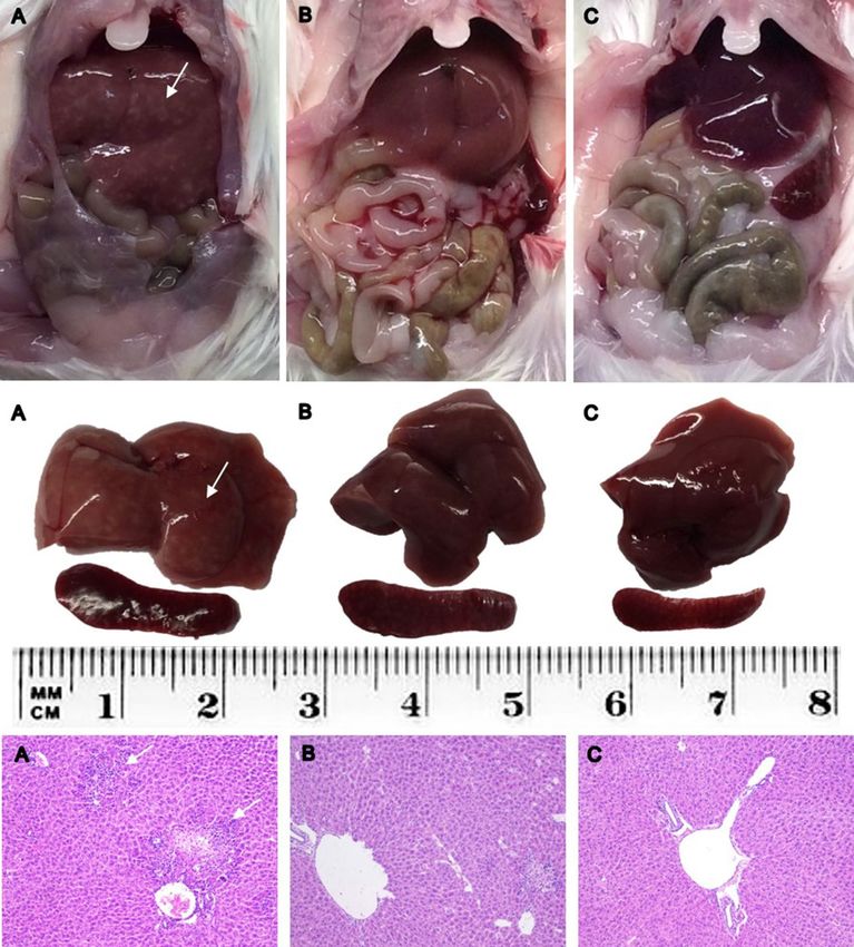
Figure 1. Representative liver and spleen from each group: (A) infected; (B) infected + IVM; (C) control. Upper
panel: abdominal cavity at necropsy; middle panel: dissected liver and spleen; lower panel: HE histological
sections of livers. White arrows indicate white spotted patterns in the liver from infected mice and severe
hepatocellular necrosis and lymphoplasmacytic inflammatory infiltration in the histological images (A). IVM
ivermectin.
infected groups (Fig. 8a), whereas no differences in number of red blood cells (RBCs), hemoglobin (HGC) level,
hematocrit (HCT) level, or number of platelets were found compared with these measures in the control and
preinfection groups (Table 1). Absolute WBC and lymphocyte counts were increased in the control group com-
pared to those at the pre- and postinfection timepoints.
Measurements of the WBC differences revealed that the percentage of neutrophils and monocytes were sig-
nificantly increased in animals in both infected groups. In contrast, the percentage and counts of lymphocytes
were the only WBC parameters that were significantly decreased in animals in the infected groups, regardless
of ivermectin treatment, compared with percentage and counts in the control and preinfection groups (Fig. 8c).
Moreover, animals in the infected group that received ivermectin treatment showed an increase in the number
of neutrophils compared with animals in the infected group (Fig. 8d).
To further characterize the reduction in lymphocytes observed in the animals in the infected groups, B and
T lymphocytes were analyzed by the detection of specific cell surface markers, CD19 (B lymphocytes) or CD8
and CD4 (T lymphocytes), at the endpoint of the experiment. The results showed that both B and T lymphocyte
percentages were reduced in the mice from the virus-infected groups compared to the control group (Fig. 8b),
Scientific Reports | (2021) 11:7132 | https://doi.org/10.1038/s41598-021-86679-0 3
Vol.:(0123456789)www.nature.com/scientificreports/
Figure 2. Body weight at the beginning and the end of the experiment (a) and organ weight and liver
appearance at necropsy five days postinfection (b,c,d, respectively) in MHV-infected (infected group, n = 20),
infected and treated with ivermectin (infected + IVM group, n = 20), and noninfected untreated mice (control
group, n = 16) (mean ± SD). Different letters indicate significant differences (p < 0.05) between pre- and
postinfection timepoints for a and significant differences (p < 0.05) between the indicated groups for (b–d).
Figure 3. Histopathological scores (grades 0 to 3, where 0 is no damage and 3 is the most damaged) of livers
from the MHV-infected (infected group, n = 20), infected and treated with ivermectin (infected + IVM group,
n = 20), and noninfected untreated mice (control group, n = 16). The distribution of animals for each score
showed significant differences between the groups (p < 0.05).
with CD8 + cells being the subpopulation with the greatest reduction (64% and 66% depletion in the infected
and infected + IVM groups, respectively).
Ivermectin reduces the TNFα levels in infected mice. Cytokine levels obtained from the plasma sam-
ples at the endpoint of the experiment (5 days after viral inoculation) were measured in all three groups. From
the panel of 13 inflammatory-related cytokines, only IFNγ and MCP-1 were significantly increased in both
infected groups (p < 0.05) compared with the control group, regardless of ivermectin treatment. On the other
Scientific Reports | (2021) 11:7132 | https://doi.org/10.1038/s41598-021-86679-0 4
Vol:.(1234567890)www.nature.com/scientificreports/
Figure 4. Hepatic viral load measured by qPCR in liver samples from MHV-infected (infected group, n = 20),
infected and treated with ivermectin (infected + IVM group, n = 20), and noninfected untreated mice (control
group, n = 16) (mean ± SD). Different letters indicate significant differences (p < 0.05) among the groups.
Figure 5. Protein levels measured in the blood of mice in the MHV-infected (infected group, n = 20), infected
and treated with ivermectin (infected + IVM group, n = 20), and noninfected untreated mice (control group,
n = 16) before and after infection with MHV A-59. (a) Total proteins; (b) albumin; (c) globulin, (d) albumin/
globulin ratio and (e) total bilirubin (mean ± SD). Different letters indicate significant differences (p < 0.05)
between the pre- and post-infection timepoints; asterisks (*) refer to significant differences (p < 0.05) between
indicated groups.
hand, TNFα values, which were increased in the mice from the infected group, were reduced in the animals that
received ivermectin treatment, with the latter showing no significant difference compared to the control mice
(Fig. 9).
No statistically significant differences were observed for the other analyzed cytokines (IL-1b, IL-1a, IL-23,
IL-12p70, IL-10, or IFNβ), and some of these proteins (IL-6, IL-27, IL-17A and GM-CSF) were below the detec-
tion limit of the assay.
Discussion
This study proposes a mouse experimental model for the in vivo evaluation of pharmacological therapies against
coronavirus diseases. It is well known that preclinical animal models are of utmost importance when develop-
ing new therapies or vaccines that will be applied to humans. In addition, the need to develop animal models
to study SARS-CoV-2 has been recently proposed by many r esearchers23. Our study is based on the previously
tested in vitro reports of the use of ivermectin against several RNA and DNA human and animal viruses12, such
as influenza A, West Nile, Venezuelan equine encephalitis, Zika, chikungunya, Newcastle disease, and porcine
reproductive and respiratory syndrome viruses; HIV-1; dengue fever, yellow fever, tick-borne encephalitis, and
Scientific Reports | (2021) 11:7132 | https://doi.org/10.1038/s41598-021-86679-0 5
Vol.:(0123456789)www.nature.com/scientificreports/
Figure 6. Hepatic enzyme levels in the blood of MHV-infected (infected group, n = 20), infected and treated
with ivermectin (infected + IVM group, n = 20), and noninfected untreated mice (control group, n = 16) before
and after virus infection. (a) Alanine aminotransferase; (b) aspartate aminotransferase and (c) gamma glutamyl
transpeptidase (mean ± SD). Different letters indicate significant differences (p < 0.05) between pre- and
postinfection timepoints; asterisks (*) refer to significant differences (p < 0.05) between the indicated groups.
pseudorabies viruses; porcine circovirus, parvovirus and bovine herpesvirus. However, most of these studies
reported only in vitro results, and information on the effect of ivermectin used in vivo is scarce.
In our model, mice infected with MHV and immediately treated with ivermectin showed a lower hepatic
viral load five days after infection and better general health than infected animals not treated with ivermectin.
A possible explanation for the lower viral load in the infected animals that received ivermectin may be related
to impairment of virus replication in the cells, since this drug has been shown to inhibit nuclear import through
inhibition of IMP α/β1 heterodimer formation24. In the necropsy and histological analysis, the livers of infected
and untreated mice showed the worst appearance, with several animals showing severe hepatocellular necrosis
and lymphoplasmacytic inflammatory infiltration. The treated group showed a lower severity of anomalies,
although the liver and spleen weights were heavier in the mice in both infected groups than in the mice in the
control group. The organ weight increase following infection is indicative of the immune reaction25, something
that cannot be evaluated in an in vitro model. These findings are typical for MHV-infected m ice26, and their
generalized immune reaction are confirmed with the cytokine levels found in our study in both infected groups.
Serum biochemistry of the liver and kidney showed a clear impairment of the metabolic profile, mainly due
to liver damage. Both groups of infected mice showed hypoalbuminemia and hyperglobulinemia, with a decrease
in the A/G ratio compared with the control group. Variations in both proteins reflect hepatic function27, and the
serum concentrations of transaminases such as AST and ALT were significantly higher in the virus-infected mice
that did not receive ivermectin and were associated with other variables indicative of liver damage28. A consider-
able decrease in serum creatinine levels was found in the infected mice, representing major liver damage and
diminished kidney function in the sick animals. Glucose levels were significantly decreased in the infected mice,
probably due to the animals’ loss of appetite related to the impairment of their general health status. Interestingly,
this metabolic profile showed major liver damage in the infected animals, in accordance with the remainder of
the data analyzed during this study. Treatment with ivermectin was effective in reducing the effects of the viral
infection, supporting the use of this model to test novel therapies against coronavirus diseases.
The most relevant hematological findings were an increase in neutrophil and monocyte percentages and a
reduction in WBC and lymphocyte counts (B and T) in both infected groups, regardless of ivermectin treatment.
Scientific Reports | (2021) 11:7132 | https://doi.org/10.1038/s41598-021-86679-0 6
Vol:.(1234567890)www.nature.com/scientificreports/
Figure 7. Blood urea nitrogen (BUN), creatinine (CRE), BUN/CRE ratio and glucose (GLU) levels were
measured before and after infection in MHV-infected (infected group, n = 20), infected and treated with
ivermectin (infected + IVM group, n = 20), and noninfected untreated mice (control group, n = 16). The results
are shown in (a–d) (mean ± SD). Different letters indicate significant differences (p < 0.05) between pre- and
postinfection timepoints; asterisks (*) refer to significant differences (p < 0.05) between the indicated groups.
Infected Infected + IVM Control
Parameter Pre Pos Pre Pos Pre Pos
WBC # ( 109/L) 5.80 (1.07) 3.64 (0.87) 6.35 (1.71) 4.49 (1.25) 5.07 (1.07) 8.56 (2.32)
Neu # (109/L) 0.89 (0.12) 1.59 (0.38) 0.92 (0.33) 2.03 (0.62) 0.742 (0.15) 1.28 (0.25)
Lym # (109/L) 4.77 (1.00) 1.80 (0.49) 5.31 (1.44) 2.15 (0.64) 4.12 (1.02) 6.99 (2.05)
Mon # (109/L) 0.07 (0.03) 0.14 (0.03) 0.07 (0.04) 0.17 (0.05) 0.05 (0.02) 0.13 (0.04)
Eos # (109/L) 0.05 (0.02) 0.05 (0.04) 0.04 (0.03) 0.06 (0.02) 0.05 (0.02) 0.08 (0.03)
Bas # (109/L) 0.01 (0.01) 0.06 (0.02) 0.01 (0.01) 0.08 (0.02) 0.01 (0.01) 0.02 (0.01)
Neu % (%) 15.9 (2.8) 43.6 (4.0) 14.4 (3.3) 45.1 (4.0) 15.2 (3.3) 21.4 (12.7)
Lymp % (%) 81.8 (3.0) 49.2 (4.2) 83.6 (3.5) 47.7 (4.3) 82.2 (3.5) 74.9 (14.2)
Mon % (%) 1.2 (0.5) 3.8 (0.8) 1.1 (0.4) 3.8 (0.7) 1.1 (0.3) 1.5 (0.2)
Eos % (%) 0.8 (0.5) 1.4 (0.4) 0.6 (0.3) 1.3 (0.7) 1.1 (0.4) 1.0 (0.4)
Bas % (%) 0.2 (0.1) 1.8 (0.4) 0.2 (0.0) 1.9 (0.4) 0.3 (0.2) 0.2 (0.1)
RBC (1012/L) 10.0 (0.7) 8.4 (0.6) 10.3 (1.3) 9.0 (0.8) 10.2 (1.1) 8.5 (0.8)
HGC (g/L) 167 (14) 143 (10) 171 (22) 153 (14) 171 (18) 148 (16)
HCT (%) 48.9 (3.7) 42.0 (3.0) 50.3 (6.4) 45.4 (4.2) 49.6 (5.2) 41.7 (4.1)
PLT (109/L) 620 (184) 831 (198) 675 (209) 765 (251) 647 (204) 757 (196)
Table 1. Hematological parameters from peripheral blood samples of the three experimental groups,
measured pre- and postinfection. Data are expressed as the means (SD). Neu neutrophils; Lym lymphocytes;
Mon monocytes; Eos eosinophils; Bas basophils; RBC red blood cells; HGB hemoglobin; HCT hematocrit; PLT
platelets.
Neutrophilia and lymphopenia have been well documented in viral respiratory infectious diseases in mouse
models and humans29–31. The increase in the percentage of neutrophils in the virus-infected groups may be
associated with acute-phase viral infection. On the other hand, the reduction in lymphocytes might be due to
migration/retention of these cells in the liver and/or lymphoid tissue.
The rapid development of lymphopenia has also been observed in COVID-19 patients with adverse out-
comes, whereby CD4 + T-cells were more severely reduced than CD8 + T-cells32,33. The neutrophil count was the
Scientific Reports | (2021) 11:7132 | https://doi.org/10.1038/s41598-021-86679-0 7
Vol.:(0123456789)www.nature.com/scientificreports/
a c preinfection
postinfection
*
preinfection ab, Pwww.nature.com/scientificreports/
ab, Pwww.nature.com/scientificreports/
Merial, Paris, France) diluted in 50 µL of PBS. The mice in the other two groups (infected and control) received
50 µL of PBS s.c.
Five days after infection/treatment, the mice were weighed, and 300 µL of blood was retrieved from the
submandibular vein for plasma cytokine quantification and metabolic and hematological profiles. The mice
were immediately euthanized by cervical dislocation to dissect the liver and spleen for weight recording and
histological and qPCR analysis. At necropsy, the liver appearance was blindly scored (0 to 3) by an independent
and trained technician who considered the main pathologic pattern of the MHV infection39,40. Briefly, gross
hepatic lesions were identified as multifocal to coalescent whitish spots of less than 1 mm diameter and were
defined as hepatic granulomas.
Histological analysis. Immediately after necropsy, the liver and spleen were fixed in 10% neutral buffered
formalin (pH 7.4) for further processing. For evaluation, they were embedded in paraffin, sectioned in 4 µm
sections and stained with hematoxylin–eosin (H&E) according to Kyuwa et al.41. Whole specimens were exam-
ined under a light microscope (BX41, Olympus, Tokyo, Japan) at 10 × in three randomly selected areas or in the
highest incidence areas of each specimen by three different pathologists to establish a histopathological score in
each case. The score was determined on the basis of a previously defined semiquantitative microscopy grading
scale representing typical histopathologic changes caused by MHV (characterized by the presence of hepatocel-
lular necrotic areas and granulomatous inflammatory reaction) according to the following criteria: 0 = normal
(no necrotic areas identified in the whole specimen); 1 ≤ 10 necrotic areas; 2 = 10–20 necrotic areas; and 3 ≥ 20
necrotic areas.
Hepatic viral load. After dissecting and trimming the whole liver, samples (0.5 × 0.5 cm) of the hepatic right
lobe were retrieved for qPCR analysis. The samples were loaded in cryotubes with TRI Reagent (Sigma-Aldrich,
Saint-Louis, MO, US), immediately plunged into liquid nitrogen and stored at − 80 °C until analysis. Total RNA
was isolated according to the manufacturer’s instructions. cDNA was synthesized from 2 µg of total RNA using
M-MLV Reverse Transcriptase (Thermo Fisher, Waltham, MA, US) and random primers (Invitrogen, Carlsbad,
CA, US). Sample analysis was performed with a QuantStudio 3 Real-time PCR system (Thermo Fisher) using
FastStart Universal SYBR Green Master (Rox) (Roche, Basel, CH). The primer sequences were as follows: MHV
forward primer (5′–3′): GGAACTTCTCGTTGGGCATTATACT and MHV reverse primer (5′–3′): ACCACA
AGATTATCATTTTCACAACATA. The reactions were performed according to the following settings: 95 °C for
10 min and 40 cycles of 95 °C for 15 s and 60 °C for 1 min. The quantification of the viral load was performed
with the relative standard curve method using an undiluted positive control of 1 06 arbitrary units (AU).
Blood biochemistry profile. Individual whole blood (100 µL) was analyzed for liver and kidney bio-
chemical profiles using a POINTCARE V2 automatic device (Tianjin MNCHIP Technologies Co., China) at
the beginning (preinfection determination) and at the end of the experiment (postinfection determination).
The analyzed parameters included total proteins (TP), albumin (ALB) level, globulin (GLO) level, ALB/GLO
ratio, total bilirubin (TBIL) level, alanine aminotransferase (ALT) level, aspartate aminotransferase (AST) level,
gamma glutamyl transpeptidase (GGT) level, blood urea nitrogen (BUN) level, creatinine (CRE) level, BUN/
CRE ratio and glucose (GLU) level.
Hematological parameters. For hematologic analysis, aliquots of 20 µL of blood were collected and
stored in 0.5 mL microtubes containing EDTA potassium salts (W anticoagulant, Wiener Lab, Rosario, Argen-
tina) at a ratio of 1:10 (EDTA:blood) during the pre- and postinfection stages. All measurements were performed
within four hours after blood collection. Total WBC count, differential WBC count and percentage, RBC count,
HGC level, HCT level, and PLT count were evaluated using an autohematology analyzer BC-5000Vet (Mindray
Medical International Ltd., Shenzhen, China).
Cytokine quantification and flow cytometry analysis. Bead-based multiplex assays were employed
to quantify cytokines (LEGENDplex mouse Inflammation Panel, BioLegend Inc., San Diego, CA, US) in plasma
samples obtained from the mice pre- and postinfection, according to the manufacturer’s instructions. Briefly,
blood samples were combined with EDTA as an anticoagulant and centrifuged for 10 min at 1000×g, and plasma
was recovered and stored at − 20 °C until use. For the assay, 25 µL of twofold diluted plasma samples, diluted
standards, and blanks were added to the corresponding tubes; 25 µL of premixed beads and detection antibod-
ies were added to all of the tubes. The tubes were incubated for 2 h at room temperature with shaking. Without
washing the samples, 25 µL of streptavidin–phycoerythrin (SA-PE) conjugate was added, and the tubes were
incubated for 30 min. Then, the samples were washed and suspended in 200 µL of wash buffer. The data were
acquired with a BD Accuri C6 flow cytometer (BD Biosciences, CA, US). BD Accuri C6 software was used for
data acquisition. Bead excitation was achieved using 488 and 640 nm lasers, and the emission was detected using
530/30 and 665/20 nm bandpass filters, respectively. For each analyte to be detected, 4,000 beads gated on a for-
ward scatter (FSC) versus side scatter (SSC) were recorded by dot plot. The data were processed with BioLegend
LEGENDplex Data Analysis Software. The results represent the concentration expressed in pg/mL.
B and T lymphocyte analysis by flow cytometry. Lymphocyte surface markers were evaluated in
peripheral blood samples (50 μL) anticoagulated with EDTA. Erythrocytes were removed by suspending cells
in 1 mL of lysis buffer (155 mM N
H4Cl, 12 mM NaHCO3, 0.1 mM EDTA, pH 7.4) for 10 min at room tempera-
ture. After washing in PBS containing 0.2% bovine serum albumin, nucleated cells were incubated on ice for
Scientific Reports | (2021) 11:7132 | https://doi.org/10.1038/s41598-021-86679-0 10
Vol:.(1234567890)www.nature.com/scientificreports/
15 min with an antibody mixture. The following fluorophore-conjugated antibodies were used: anti-CD4-FITC
(#11,004,181, clone GK1.5) and anti-CD8-PE-Cy7 (#25,008,182, clone 53–6.7) from eBioscience (San Diego,
CA, US) and anti-CD19-PerCP-Cy 5.5 (#551,001, clone ID3) from BD Pharmingen (San Diego, CA, US). Flow
cytometry analysis was performed using an Attune Nxt Acoustic Focusing Cytometer (Thermo Fisher) equipped
with a 488 nm laser. Emissions were detected using 530/30, 695/40 and 780/60 nm bandpass filters for FITC,
PerCP-Cy5.5 and PE-Cy7, respectively. FlowJo software, version 10.6.1 (Tree Star, Ashland, Oregon, US), was
used for data analysis. Unstained controls, single-color controls and fluorescence-minus-one controls were used
to establish baseline gate settings for each respective antibody combination.
Lymphocytes were gated based on their FSC and SSC dot plot profiles, and an FSC area vs FSC height dot plot
was used to exclude doublets. B lymphocytes were defined as CD19-PerCP-Cy5.5-positive cells. For T lymphocyte
analysis, a gate was placed on the CD19-negative population, and based on the PE-Cy7 vs FITC dot plot, CD8-
PE-Cy7-positive cells and CD4-FITC-positive cells were defined as CD8 + and CD4 + lymphocytes, respectively.
A minimum of 10,000 events in a single cell region were collected. The results are expressed as percentage of the
specific cell type from the analyzed single-cell population.
Statistical analysis. Statistical analysis was performed using generalized linear mixed models (GLMM,
InfoStat software42), which included the treatments (three groups) and time (pre- and postinfection) as fixed
variables and the animals and replicates as random variables. The data for continuous variables were evaluated
for normality and homogeneity of variance by histograms, q-q plots, and formal statistical tests as part of the
univariate procedure. The type of variance–covariance structure was chosen depending on the magnitude of the
Akaike information criterion (AIC) for models analyzed by heterogeneous compound symmetry, unstructured,
autoregressive, spatial power, and first-order interdependence methods. The model with the lowest AIC was
chosen. The data are presented as the means ± SEM, and the significance level was defined as a p-value of 0.05.
Data availability
All data generated or analyzed during this study are included in this published article.
Received: 30 November 2020; Accepted: 17 March 2021
References
1. Weiss, S. R. & Leibowitz, J. L. Coronavirus pathogenesis. Adv. Virus Res. 81, 85–164. https://doi.org/10.1016/B978-0-12-385885-6.
00009-2 (2011).
2. Korner, R. W., Majjouti, M., Alcazar, M. A. A. & Mahabir, E. Of mice and men: The coronavirus MHV and mouse models as a
translational approach to understand SARS-CoV-2. Viruses 12, 15. https://doi.org/10.3390/v12080880 (2020).
3. Fan, X., Cao, D., Kong, L. & Zhang, X. Cryo-EM analysis of the post-fusion structure of the SARS-CoV spike glycoprotein. Nat.
Commun. 11, 3618. https://doi.org/10.1038/s41467-020-17371-6 (2020).
4. Hu, B., Guo, H., Zhou, P. & Shi, Z. L. Characteristics of SARS-CoV-2 and COVID-19. Nat. Rev. Microbiol. https://doi.org/10.1038/
s41579-020-00459-7 (2020).
5. Timani, K. A. et al. Nuclear/nucleolar localization properties of C-terminal nucleocapsid protein of SARS coronavirus. Virus Res.
114, 23–34. https://doi.org/10.1016/j.virusres.2005.05.007 (2005).
6. Wulan, W. N., Heydet, D., Walker, E. J., Gahan, M. E. & Ghildyal, R. Nucleocytoplasmic transport of nucleocapsid proteins of
enveloped RNA viruses. Front. Microbiol. 6, 553. https://doi.org/10.3389/fmicb.2015.00553 (2015).
7. Crump, A. & Omura, S. Ivermectin, “wonder drug” from Japan: The human use perspective. Proc. Jpn. Acad. B87, 13–28. https://
doi.org/10.2183/pjab.87.13 (2011).
8. Ashraf, S., Chaudhry, U., Raza, A., Ghosh, D. & Zhao, X. In vitro activity of ivermectin against Staphylococcus aureus clinical
isolates. Antimicrob. Resist. Infect. Control 7, 27. https://doi.org/10.1186/s13756-018-0314-4 (2018).
9. Crump, A. Ivermectin: enigmatic multifaceted “wonder” drug continues to surprise and exceed expectations. J. Antibiot. (Tokyo)
70, 495–505. https://doi.org/10.1038/ja.2017.11 (2017).
10. Dominguez-Gomez, G. et al. Ivermectin as an inhibitor of cancer stem-like cells. Mol. Med. Rep. 17, 3397–3403. https://doi.org/
10.3892/mmr.2017.8231 (2018).
11. Wagstaff, K. M., Sivakumaran, H., Heaton, S. M., Harrich, D. & Jans, D. A. Ivermectin is a specific inhibitor of importin α/β-
mediated nuclear import able to inhibit replication of HIV-1 and dengue virus. Biochem. J. 443, 851–856. https://doi.org/10.1042/
BJ20120150 (2012).
12. Heidary, F. & Gharebaghi, R. Ivermectin: A systematic review from antiviral effects to COVID-19 complementary regimen. J.
Antibiot. 73, 593–602. https://doi.org/10.1038/s41429-020-0336-z (2020).
13. Caly, L., Druce, J. D., Catton, M. G., Jans, D. A. & Wagstaff, K. M. The FDA-approved drug ivermectin inhibits the replication of
SARS-CoV-2 in vitro. Antiviral Res. 178, 104787. https://doi.org/10.1016/j.antiviral.2020.104787 (2020).
14. Dias de Melo, G. et al. Anti-COVID-19 efficacy of ivermectin in the golden hamster. bioRvix https://doi.org/10.1101/2020.11.21.
392639 (2020).
15. Chaccour, C. et al. The effect of early treatment with ivermectin on viral load, symptoms and humoral response in patients with
non-severe COVID-19: A pilot, double-blind, placebo-controlled, randomized clinical trial. EClinicalMedicine https://doi.org/10.
1016/j.eclinm.2020.100720 (2021).
16. Vallejos, J. et al. Ivermectin to prevent hospitalizations in patients with COVID-19 (IVERCOR-COVID19): A structured summary
of a study protocol for a randomized controlled trial. Trials 21, 965. https://doi.org/10.1186/s13063-020-04813-1 (2020).
17. Procter, B. C. et al. Clinical outcomes after early ambulatory multidrug therapy for high-risk SARS-CoV-2 (COVID-19) infection.
Rev Cardiovasc Med 21, 611–614. https://doi.org/10.31083/j.rcm.2020.04.260 (2020).
18. Ochoa-Jaramillo, F. (https://clinicaltrials.gov/ct2/show/study/NCT04602507, 2020).
19. Laboratorio Elea Phoenix, S. A. (https://clinicaltrials.gov/ct2/show/NCT04381884, 2020).
20. Benha University. (https://clinicaltrials.gov/ct2/show/NCT04668469, 2020).
21. Gorial, F. I. et al. Effectiveness of ivermectin as add-on therapy in COVID-19 management (Pilot Trial). Medrxiv https://doi.org/
10.1101/2020.07.07.20145979 (2020).
22. Krolewiecki, A. et al. Antiviral effect of high-dose ivermectin in adults with COVID-19: A pilot randomised, controlled, open
label multicentre trial. The Lancet https://doi.org/10.2139/ssrn.3714649 (2020).
Scientific Reports | (2021) 11:7132 | https://doi.org/10.1038/s41598-021-86679-0 11
Vol.:(0123456789)www.nature.com/scientificreports/
23. Johansen, M. D. et al. Animal and translational models of SARS-CoV-2 infection and COVID-19. Mucosal Immunol. 13, 877–891.
https://doi.org/10.1038/s41385-020-00340-z (2020).
24. Kosyna, F. K., Nagel, M., Kluxen, L., Kraushaar, K. & Depping, R. The importin alpha/beta-specific inhibitor Ivermectin affects
HIF-dependent hypoxia response pathways. Biol. Chem. 396, 1357–1367. https://doi.org/10.1515/hsz-2015-0171 (2015).
25. Robinson, M. W., Harmon, C. & O’Farrelly, C. Liver immunology and its role in inflammation and homeostasis. Cell. Mol. Immunol.
13, 267–276. https://doi.org/10.1038/cmi.2016.3 (2016).
26. Barthold, S. W. In Digestive System (eds Jones, T. C. et al.) 134–139 (Springer, Berlin Heidelberg, 1985).
27. Carvalho, J. R. & Verdelho Machado, M. New insights about albumin and liver disease. Ann. Hepatol. 17, 547–560. https://doi.
org/10.5604/01.3001.0012.0916 (2018).
28. Smith, G. S., Walter, G. L. & Walker, R. M. In Haschek and Rousseaux’s Handbook of Toxicologic Pathology (Third Edition) (eds
Haschek, W. M. et al.) 565–594 (Academic Press, Cambridge, 2013).
29. Camp, J. V. & Jonsson, C. B. A role for neutrophils in viral respiratory disease. Front. Immunol. 8, 550. https://doi.org/10.3389/
fimmu.2017.00550 (2017).
30. Feng, Y. et al. Molecular pathology analyses of two fatal human infections of avian influenza A(H7N9) virus. J. Clin. Pathol. 68,
57–63. https://doi.org/10.1136/jclinpath-2014-202441 (2015).
31. Preusse, M., Schughart, K., Wilk, E., Klawonn, F. & Pessler, F. Hematological parameters in the early phase of influenza A virus
infection in differentially susceptible inbred mouse strains. BMC Res. Notes 8, 225. https://doi.org/10.1186/s13104-015-1195-8
(2015).
32. Chen, J. & Subbarao, K. The Immunobiology of SARS*. Annu. Rev. Immunol. 25, 443–472. https://d oi.o
rg/1 0.1 146/a nnure v.i mmun
ol.25.022106.141706 (2007).
33. Guan, W. J. et al. Clinical characteristics of coronavirus disease 2019 in China. N. Engl. J. Med. 382, 1708–1720. https://doi.org/
10.1056/NEJMoa2002032 (2020).
34. Sajid, M. S., Iqbal, Z., Muhammad, G. & Iqbal, M. U. Immunomodulatory effect of various anti-parasitics: A review. Parasitology
132, 301–313. https://doi.org/10.1017/s0031182005009108 (2006).
35. Jans, D. A. & Wagstaff, K. M. Ivermectin as a broad-spectrum host-directed antiviral: The real deal?. Cells 9, 2100. https://doi.org/
10.3390/cells9092100 (2020).
36. Sharun, K. et al. Ivermectin, a new candidate therapeutic against SARS-CoV-2/COVID-19. Ann. Clin. Microbiol. Antimicrob. 19,
23. https://doi.org/10.1186/s12941-020-00368-w (2020).
37. Ci, X. et al. Avermectin exerts anti-inflammatory effect by downregulating the nuclear transcription factor kappa-B and mitogen-
activated protein kinase activation pathway. Fundam. Clin. Pharmacol. 23, 449–455. https://doi.org/10.1111/j.1472-8206.2009.
00684.x (2009).
38. Zhang, X. et al. Ivermectin inhibits LPS-induced production of inflammatory cytokines and improves LPS-induced survival in
mice. Inflamm. Res. 57, 524–529. https://doi.org/10.1007/s00011-008-8007-8 (2008).
39. MacPhee, P. J., Dindzans, V. J., Fung, L. S. & Levy, G. A. Acute and chronic changes in the microcirculation of the liver in inbred
strains of mice following infection with mouse hepatitis virus type 3. Hepatology 5, 649–660. https://doi.org/10.1002/hep.18400
50422 (1985).
40. Perlman, S. Pathogenesis of coronavirus-induced infections. Review of pathological and immunological aspects. Adv. Exp. Med.
Biol. 440, 503–513 (1998).
41. Kyuwa, S. et al. Acute hepatic failure in IFN-gamma-deficient BALB/c mice after murine coronavirus infection. Virus Res. 83,
169–177. https://doi.org/10.1016/s0168-1702(01)00432-4 (2002).
42. InfoStat (http://www.infostat.com.ar 2017).
Acknowledgements
We thank PhDs Tatiana Basika and Sofía Russo for assistance with the qPCR settings. The experiments were
carried out with funds from Institut Pasteur de Montevideo and FOCEM (MERCOSUR Structural Convergence
Fund), COF 03/11. MS, JMV, MH, MB and MC are members of Sistema Nacional de Investigadores (SNI).
Author contributions
A.P.A. and M.C. contributed to the conception and experimental design, data acquisition and analysis, and
writing of the manuscript. R.P., J.L. P, H.D., B.V., K. Y and J.M.V. contributed to data acquisition and analysis
and wrote the corresponding sections of the manuscript. M.S. and M.D.V. contributed to animal model design,
virus preparation and conception discussion. M.H. and M.B.F. contributed to data interpretation and substantive
revision of the article. All authors contributed to revision of the manuscript.
Competing interests
The authors declare no competing interests.
Additional information
Correspondence and requests for materials should be addressed to M.C.
Reprints and permissions information is available at www.nature.com/reprints.
Publisher’s note Springer Nature remains neutral with regard to jurisdictional claims in published maps and
institutional affiliations.
Open Access This article is licensed under a Creative Commons Attribution 4.0 International
License, which permits use, sharing, adaptation, distribution and reproduction in any medium or
format, as long as you give appropriate credit to the original author(s) and the source, provide a link to the
Creative Commons licence, and indicate if changes were made. The images or other third party material in this
article are included in the article’s Creative Commons licence, unless indicated otherwise in a credit line to the
material. If material is not included in the article’s Creative Commons licence and your intended use is not
permitted by statutory regulation or exceeds the permitted use, you will need to obtain permission directly from
the copyright holder. To view a copy of this licence, visit http://creativecommons.org/licenses/by/4.0/.
© The Author(s) 2021
Scientific Reports | (2021) 11:7132 | https://doi.org/10.1038/s41598-021-86679-0 12
Vol:.(1234567890)You can also read