Analysis of intercondylar notch size and shape in patients with cyclops syndrome after anterior cruciate ligament reconstruction
←
→
Page content transcription
If your browser does not render page correctly, please read the page content below
Ficek et al. Journal of Orthopaedic Surgery and Research            (2021) 16:554
https://doi.org/10.1186/s13018-021-02706-w

 RESEARCH ARTICLE                                                                                                                                 Open Access

Analysis of intercondylar notch size and
shape in patients with cyclops syndrome
after anterior cruciate ligament
reconstruction
Krzysztof Ficek1,2* , Jolanta Rajca2, Jerzy Cholewiński2,3,4, Agnieszka Racut2, Paweł Gwiazdoń1,2,5,
Krzysztof Przednowek6 and Grzegorz Hajduk2

  Abstract
  Background: Cyclops lesion is the second most common cause of extension loss after anterior cruciate ligament
  reconstruction. This study focused on the correlation between the anatomy of the intercondylar notch and the
  incidence of cyclops lesion. To determine whether the size and shape of the intercondylar notch are related to
  cyclops lesion formation following anterior cruciate ligament reconstruction according to magnetic resonance
  imaging (MRI) findings.
  Methods: One hundred twenty-five (125) patients were retrospectively evaluated. The notch width index (NWI) and
  notch shape index (NSI) were measured based on coronal and axial MRI sections in patients diagnosed with
  cyclops syndrome (n = 25), diagnosed with complete anterior cruciate ligament (ACL) tears (n = 50), and without
  cyclops lesions or ACL ruptures (n = 50).
  Results: Imaging analysis results showed that the cyclops and ACL groups had lower mean NWI and NSI values
  than the control group. Significant between-group differences were found in NSI (p = 0.0140) based on coronal
  cross-sections and in NWI (p = 0.0026) and NSI (p < 0.0001) based on axial sections.
  Conclusions: The geometry of the intercondylar notch was found to be associated with the risk of cyclops lesion
  formation and ACL rupture.
  Keywords: Cyclops lesion, Anterior cruciate ligament reconstruction, Intercondylar notch

Background                                                                           in 1990, causes extension loss of approximately 5° com-
Cyclops lesion, defined as the local presentation of                                 pared with a healthy lower limb. A cyclops lesion is a fi-
arthrofibrosis, is the second most common cause of ex-                               brous nodule of granulation tissue anterolateral to the
tension loss after anterior cruciate ligament (ACL) re-                              tibial tunnel that has matured in a manner similar to a
construction [1]. In the early postoperative period,                                 healing scar and occasionally develops cartilaginous or
cyclops syndrome, described by Jackson and Schaefer [2]                              bony tissue, and it is usually not associated with any
                                                                                     clinical symptoms of the knee [2, 3]. The incidence of
* Correspondence: krzysztof.ficek@galen.pl                                           cyclops syndrome in patients after ACL reconstruction
1
 Department of Physiotherapy, The Jerzy Kukuczka Academy of Physical
Education, 40-065 Katowice, Poland                                                   ranges from 1.9 to 10.6%, whereas the incidence of cyc-
2
 Deparment of Science, Innovation and Development, Galen-Orthopaedics,               lops lesions that do not cause extension loss ranges from
43-150 Bieruń, Poland
Full list of author information is available at the end of the article

                                      © The Author(s). 2021 Open Access This article is licensed under a Creative Commons Attribution 4.0 International License,
                                      which permits use, sharing, adaptation, distribution and reproduction in any medium or format, as long as you give
                                      appropriate credit to the original author(s) and the source, provide a link to the Creative Commons licence, and indicate if
                                      changes were made. The images or other third party material in this article are included in the article's Creative Commons
                                      licence, unless indicated otherwise in a credit line to the material. If material is not included in the article's Creative Commons
                                      licence and your intended use is not permitted by statutory regulation or exceeds the permitted use, you will need to obtain
                                      permission directly from the copyright holder. To view a copy of this licence, visit http://creativecommons.org/licenses/by/4.0/.
                                      The Creative Commons Public Domain Dedication waiver (http://creativecommons.org/publicdomain/zero/1.0/) applies to the
                                      data made available in this article, unless otherwise stated in a credit line to the data.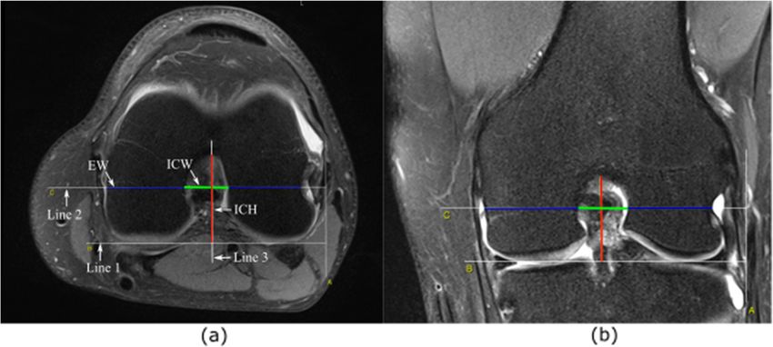
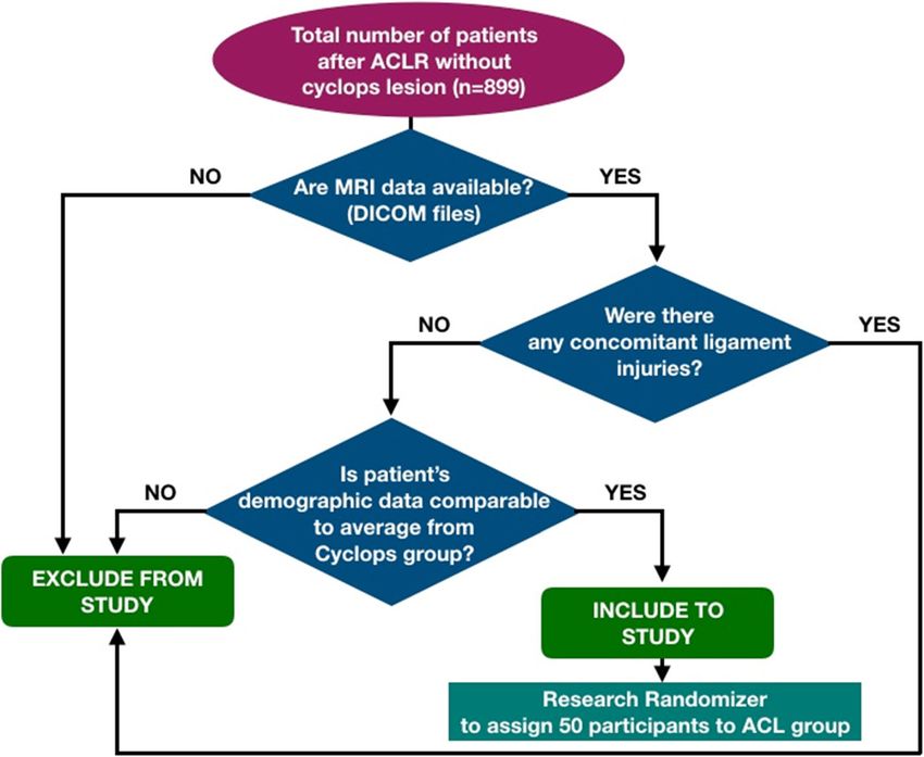
Ficek et al. Journal of Orthopaedic Surgery and Research   (2021) 16:554                                                     Page 2 of 10

2.2 to 46.8% [4–11]. Most of these reports are based on                    an increased risk of ACL injury [31–36]. Based on a re-
single-bundle ACL reconstruction.                                          cent study, it can be predicted that a smaller notch
   Several risk factors have been linked to loss of knee                   might be correlated with ACL impingement [37]. More-
motion, including the mechanism of ACL injury and as-                      over, a cause of notch impingement could potentially
sociated injuries, the timing of surgery, technical factors,               exist for a “mismatch” between the notch size and the
and postoperative/rehabilitation factors [12]. Among the                   ACL size [38]. However, the literature seldom mentions
factors associated with surgery, the timing of surgery re-                 the association between the notch size and the formation
mains particularly controversial. Many authors have re-                    of cyclops lesions. Some authors reported that notch size
ported an association between early surgery and the                        influences cyclops formation [39], and some did not find
development of arthrofibrosis [13–16], while others have                   statistically significant differences in the size of intercon-
found no relationship between the timing of surgery and                    dylar notches in the patient groups with and without
extension loss [17–21]. Patients whose ligaments were                      cyclops lesions [40].
reconstructed within the first week of injury had a statis-                  Therefore, the purpose of this study was to retrospect-
tically significant increase in the incidence of motion                    ively determine whether the size and shape of the inter-
loss compared with those who waited at least 3 weeks                       condylar notch were related to cyclops lesion formation.
[15]. Wasilewski et al. [22] showed that acute ACL re-                     The authors predict that the geometry of the intercondy-
construction (5–10 days after injury) significantly slowed                 lar notch might be a risk for developing cyclops lesions.
down postoperative motion recovery compared to de-
layed surgery.                                                             Methods
   On the other hand, not only the timing of the surgery                   Patients
but also the condition of the knee prior to surgery may                    There were 929 patients who underwent surgery due to
be important. The goals of preoperative rehabilitation                     complete ACL tears between 2011 and 2017, and we
are to restore normal knee range of motion, eliminate                      reviewed the medical records of the patients treated at
swelling, and regain leg control [15]. Preoperative motion                 our clinic for cyclops syndrome. We identified thirty
is an important preoperative predictor of ultimate motion                  (30) patients diagnosed with cyclops lesions after ACL
and may even be the key clinical factor to guide decisions                 reconstruction. All these cases were confirmed by MRI
regarding the timing of ACL reconstruction [23].                           scans and arthroscopic surgery, during which the lesion
   As genetic factors play a proven role in ACL rupture                    was removed. The major criterion for exclusion was the
[24, 25], several genetic mechanisms have been proposed                    lack of digitally available MRI data on the knee joint.
to be related to arthrofibrosis after ACL reconstruction.                  Therefore, the cyclops group included 25 patients.
Platelet-derived growth factor-β (PDGF-β) and trans-                       Among the remaining patients with available MRI data,
forming growth factor-β (TGF-β) may play a central role                    we randomly selected 50 patients to be included in the
in the healing process, but their expression has been                      ACL group, and they had demographic characteristics
associated with unresolved inflammation and fibrotic                       similar to those of the cyclops group. The algorithm for
events [26, 27].                                                           the patients’ inclusion and exclusion is presented in Fig. 1.
   Another risk factor that may be related to the forma-                   The control group (n = 50) included patients without rup-
tion of a cyclops lesion is associated with the anatomy of                 ture of the ACL or other ligaments in the knee who
the femoral intercondylar notch. The geometry of the                       underwent MRI scans of the knee due to suspicion of a
intercondylar notch varies among the population, in-                       meniscus tear and did not have any reports of knee in-
cluding the differences between females and males.                         stability in the medical records. No patient in the selected
Three geometry types of intercondylar notch have been                      cohort underwent notchplasty during ACL reconstruction.
distinguished: (1) A-shaped, (2) U-shaped, and (3) W-                      The demographic data are displayed in Table 1.
shaped [28]. The A-shaped notch is defined as a stenotic
notch which is narrow from the base to the midsection                      Surgical procedure and postoperative management
as well as at the apex. In the U-shaped notch, the mid-                    The transportal femoral tunnel drilling technique for
section does not narrow, allowing for a wider contour to                   endoscopic ACL reconstruction was used in patients in
the notch. The W-shaped notch is similar to type U, but                    the cyclops and ACL groups. The types of grafts used
with two apparent apices [29]. Interestingly, Hirtler et al.               during the procedure are presented in Table 2. In pa-
[30] performed a study in which they described the on-                     tients with additional features of I° medial instability, the
going dynamic morphologic modifications in the inter-                      quadriceps tendon or the patellar tendon was used.
condylar notch at different stages of life, indicating that                   After they were harvested, the semitendinosus and the
the shape and size of the notch and of the femoral con-                    gracilis tendon (ST GR) were doubled and folded over
dyles changed significantly during life. Many authors                      the loop of an EndoButton CL (Smith & Nephew Inc.,
have reported that narrowing of this notch is related to                   Andover, MA, USA), and the distal ends of the grafts
Ficek et al. Journal of Orthopaedic Surgery and Research            (2021) 16:554                                                           Page 3 of 10

  Fig. 1 Patients selection algorithm. The question about demographic data refers to age, sex, and BMI

were sutured. When the quadriceps tendon was harvested,                             the diameter depended on the diameter of the whole graft.
the operator sutured the tendon on both ends with Krakow                            A 5-mm unreamed bone fragment was left to anchor the
sutures. Next, the diameter of each graft was measured                              EndoButton plate. Next, the knee was flexed to 90°, and a
using a cylindrical gauge (sizing system, Acufex, Smith &                           guided pin was placed using a tibial guide. A tibial tunnel
Nephew Inc., Andover, MA, USA). All grafts were preten-                             was created. Femoral fixation was achieved using an Endo-
sioned in full extension, with 20 lbs applied by a tensiome-                        Button system (Smith & Nephew Inc., Andover, MA, USA)
ter (Smith & Nephew Inc., Andover, MA, USA). The                                    or with an interference screw Biosure PEEK system (Smith
surgeon removed only the interposed tissue of the ACL                               & Nephew Inc., Andover, MA, USA), depending on the
remnant. The knee was flexed to 120°, and a guided pin                              graft used. Tibial fixation was performed with a Biosure
was placed in the anatomical femoral footprint using a fem-                         PEEK interference screw. Repeatedly in the last phase of
oral guide. Then, the guided pin was overdrilled using a 4.5                        the surgical procedure, the operator checks for any im-
drill and then a 7–9-mm-diameter cannulated reamer, and                             pingement with the intercondylar notch by flexing and

Table 1 Demographic data
                           Cyclops       ACL           Control           p*           Cyclops-ACL       Cyclops Control   ACL-Control   Cyclops+ACL-
                           (n = 25)      (n = 50)      (n = 50)                                                                         Control
                                                                         Chi-squared test
Sex (male:female)          17:9          37:13         35:15             0.8385       0.5878            0.8594            0.6560        0.8848
Knee (left:right)          18:7          25:25         27:23             0.1716       0.0655            0.1336            0.3889        0.8537
                                                                         K-W          Bonferroni test
Age (years)                32.7 ± 8.8    32.7 ± 10.1   38.0 ± 13.7       0.0072*      0.9999            0.0151*           0.0106*       0.0009*
Body weight                75.7 ± 14.4   80.2 ± 18.6   82.7 ± 19.4       0.3994       0.5378            0.3850            0.9999        0.0241*
Body height                177.6 ± 9.8   176.6 ± 8.4   173.5 ± 18.2      0.7016       0.7741            0.8734            0.9999        0.4368
BMI                        23.8 ± 2.9    25.5 ± 4.8    26.0 ± 4.8        0.1540       0.1969            0.0855            0.9071        0.1140
Graft diameter             0.77 ± 0.10   0.79 ± 0.05   –                 –            0.4266            –                 –             –
K-W Kruskal-Wallis test
*Statistically significant (p < 0.05)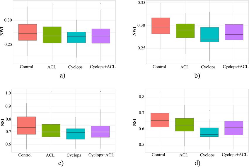
Ficek et al. Journal of Orthopaedic Surgery and Research      (2021) 16:554                                                   Page 4 of 10

Table 2 Types of grafts used in ACL reconstruction in the                     Blumensaat’s line. On the scans, the full contours of the
cyclops and ACL groups                                                        medial and lateral condyles, the notch shape, and a
                                     Cyclops [%]               ACL [%]        groove of the popliteus tendon sulcus of the lateral con-
ST GR                                83                        69             dyle were clearly visible. On these scans, three lines were
Quadriceps tendon                    13                        12             designated (Fig. 2). Line 1 was designated as the line be-
                                                                              tween the lowest points of the cartilage surfaces of the
Patellar ligament                    4                         –
                                                                              medial and lateral condyles. A line parallel to line 1 and
Rectus femoris tendon                –                         2
                                                                              passing through the largest groove of the popliteus ten-
Allogeneic                           –                         16             don sulcus was defined as line 2. With respect to line 2,
                                                                              the intercondylar notch width (ICW) was measured. The
extending the knee. If necessary, the adequate debridement                    distance measured on line 2, determined by the points
of damaged parts of ACL remnant was performed to avoid                        of intersection of the line with the lateral and medial
creating a potential fibrous conflict, simultaneously trying                  walls of the condyles, was marked as the epicondyle
to keep as much remnant tissues as possible, due to the fact                  width (EW). The height of the intercondylar notch was
that remnant preservation enhances early revascularization                    measured on line 3, which was perpendicular to line 1.
of the graft [41].                                                            The intercondylar notch height (ICH) was defined as the
  All patients started rehabilitation protocols 1 week                        distance from the level of the femoral joint surface (line
after surgery. A standardized physical therapy program                        1) to the top of the notch. The ratio of ICW to EW rep-
was prescribed for all patients. Cyclops nodules were di-                     resented the notch width index (NWI), as shown in Eq.
agnosed by the presence of pain, loss of motion (exten-                       (1), and that of ICW to ICH represented the notch shape
sion and/or flexion), and audible clicking during the                         index (NSI), as shown in Eq. (2).
terminal phase of extension and were confirmed by
MRI.                                                                                       ICW ½cm2
                                                                                 NWI ¼                                                ð1Þ
                                                                                            EW½cm2
MRI-based measurement of the femoral intercondylar
notch size and shape
                                                                                          ICW ½cm
Two independent examiners performed each MRI meas-                               NSI ¼                                                ð2Þ
urement, and the average of these measurements was re-                                     ICH½cm
corded and used for further analysis. An open-source
platform, OsiriX MD (v. 7.5, Osiris Foundation, Geneva,
Switzerland), was used for image analysis. The MRI im-                          The femoral intercondylar notch sizes and graft sizes
ages were analyzed in a random order. The MRI mea-                            were compared between the knees with cyclops lesion
surements were performed on the coronal T2 scans and                          (cyclops group) and those without cyclops lesions (ACL
axial T2 scans. The measurement method was based on                           group). In order to determine the effect of the size mis-
the approach proposed by Fuji et al. [39]. We confirmed                       match between the graft and the intercondylar notch on
Blumensaat’s line on a T2-weighted sagittal plane beside                      cyclops lesion formation, the ratio between the graft
the lateral intercondylar wall, and then, we identified the                   diameter and intercondylar notch width (G/ICW) was
coronal and axial planes of the middle point of                               calculated and compared between the two groups.

 Fig. 2 Measurement of the intercondylar notch. On axial cross-section (a). On coronal cross-section (b)
Ficek et al. Journal of Orthopaedic Surgery and Research (2021) 16:554 Page 5 of 10
Statistical analysis
The statistical analysis was performed using the Statis-
tica software (release 10.0, StatSoft, Tulsa, OK, USA)
and GNU R software with an additional package to com-
pare the differences among the tested groups. The nor-
mality (Shapiro-Wilk test) and the homogeneity of
variance (Levene’s test) of all the measured variables
were checked. A nonparametric test (Kruskal-Wallis
test) was used to assess the differences among the cyc-
lops, ACL, and control groups. For multiple compari-
sons, the Bonferroni test was used. The chi-square test
was used to compare the qualitative data (sex and knee)
between the groups. The G/ICW results were statisti-
cally analyzed by the Mann-Whitney U test between the
cyclops and ACL groups. For all tests, differences with p
< 0.05 were regarded as significant. Interobserver reli-
ability between two observers (for parameters ICW,
ICH, and EW, both for coronal and axial cross-sections)
was evaluated using the intraclass correlation coefficient
(ICC). As a general guideline [42], ICCs exceeding 0.75
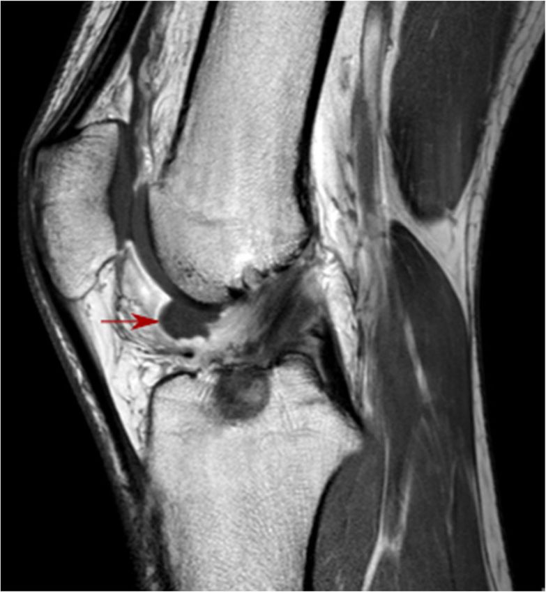
are indicative of good reliability, whereas those below Fig. 3 Cyclops lesion
0.75 indicate poor to moderate reliability.
Ethics The comparison of the NWI and NSI between the
The study was performed according to the Declaration tested groups is shown in Table 3. Compared with the
of Helsinki. The study protocol was approved by the Re- control group, the cyclops and ACL groups demon-
search Ethics Committee of the Jerzy Kukuczka Acad- strated a lower mean NWI and NSI. The results of the
emy of Physical Education (Reference Number: 2/2011). statistical analysis are presented in Fig. 4 and Table 4.
Due to the retrospective nature of the study, the require- Significant differences were found in the coronal cross-
ment for informed consent was waived. sections between the cyclops and control groups in NSI
(p = 0.0086), in the axial cross-sections between the cyc-
Results lops and control groups (p = 0.0016) and between the
The MRI images of the cyclops group, which were ACL and control groups (p = 0.0284) in the NWI, and
reviewed by a musculoskeletal radiologist, showed an ab- between the cyclops and ACL (p = 0.0037), cyclops and
normal signal anteriorly to the ACL graft in the inter- control (p = 0.0001), and ACL and control groups (p =
condylar notch (Fig. 3). The cyclops lesion was detected 0.0339) in the NSI. The analysis between the patients
on the arthroscopy look when the knee was extended. who underwent reconstructive surgery (Cyclops+ACL)
Patients received arthroscopic surgery for the removal of and the control group shows that there are significant
the lesion, and in every patient, the loss of extension in differences in the coronal cross-sections in NSI (p =
the knee joint was improved, followed by rehabilitation 0.0035) and in the axial cross-sections in both parame-
protocol, equal for all patients. However, in six patients ters NWI (p = 0.0007) and NSI (p = 0.0001).
(from the cyclops group (n = 25)), symptomatic cyclops The ratio between the graft diameter and ICW (G/
lesion reappeared in the 6 to 24 months after. In the ICW) was comparable in the cyclops and ACL groups
subsequent control, MR imaging as well as during the (Table 5).
consecutive arthroscopic surgery with lesion removal, Within the cyclops, ACL, and control groups, we did
the variability in increment of the diameter and volume not find significant differences in NWI and NSI between
of the graft which underwent ligamentization was ob- male and female patients.
served. The notchplasty was performed during the sec- For the mean interrater reliability between observers 1
ond arthroscopy with debridement of excess fibrous and 2 for all component parameters (ICW, ICH, EW) of
tissue, which formed from uncontrolled scarring re- NWI and NSI, the ICC of the analyzed features in the
sponse, surrounding the ACL graft. Therefore, the coronal and axial cross-sections was 0.75 and 0.90, re-
second-look arthroscopy comprised invasive widening of spectively. Considering the axial cross-sections, there
the femoral notch to avoid another return of cyclops was no parameter for which ICC was below 0.75. All
lesion. ICCs showed statistical significance (p < 0.05).Ficek et al. Journal of Orthopaedic Surgery and Research (2021) 16:554 Page 6 of 10
Table 3 Comparison of NWI and NSI between the groups
Mean SD Median 95% confidence interval
Cross-section: coronal
NWI Cyclops 0.2629 0.2665 0.2665 0.2541 0.2717
ACL 0.2689 0.0247 0.2672 0.2619 0.2759
Cyclops+ACL 0.2669 0.0236 0.2671 0.2615 0.2723
Control 0.2767 0.0255 0.2723 0.2695 0.2840
NSI Cyclops 0.6871 0.0629 0.6940 0.6611 0.7130
ACL 0.7148 0.0793 0.6982 0.6922 0.7373
Cyclops+ACL 0.7056 0.0750 0.6982 0.6882 0.7228
Control 0.7456 0.0878 0.7326 0.7207 0.7706
Cross-section: axial
NWI Cyclops 0.2790 0.0263 0.2703 0.2681 0.2898
ACL 0.2863 0.0232 0.2381 0.2797 0.2929
Cyclops+ACL 0.2838 0.0243 0.2805 0.2782 0.2894
Control 0.2986 0.0229 0.2970 0.2921 0.3051
NSI Cyclops 0.5800 0.0561 0.5664 0.5569 0.6032
ACL 0.6285 0.0600 0.6252 0.6114 0.6455
Cyclops+ACL 0.6123 0.0627 0.6088 0.5979 0.6268
Control 0.6643 0.0229 0.6534 0.6440 0.6846
According to the guidelines of Portney and Watkins the medial/lateral direction to the notch height in the an-
[42], reliability for many clinical measurements should terior/posterior direction. Knees with lower NSI may not
exceed 0.90 to ensure reasonable validity. Thus, out- permit normal function of the ACL [43]. According to
comes of NWI and NSI based on coronal cross-sections Tillman et al. [43], when the knee is in full extension, the
should be treated with some degree of caution. ACL is pulled tight and will reside in the more anterior
portion of the intercondylar notch. Therefore, a low NSI
Discussion indicates that this particular region of the intercondylar
In our study, we investigated the geometry of the inter- notch will be narrower and thus will provide less space for
condylar notch by measuring the width and shape index the ACL to function correctly.
using MRI on the coronal and axial cross-sections to iden- According to previous studies [39, 40] and to the as-
tify the associations between the geometry and the occur- sumption that the width of the intercondylar notch is
rence of cyclops syndrome. However, considering the important in developing cyclops lesion, we calculated
moderate interrater reliability for coronal cross-sections the ratio between graft diameter and notch width (ICW)
between the two observers, conclusions were based on the to determine the potential size mismatch between the
results for axial cross-sections (mean ICC = 0.90). The graft and intercondylar notch width (G/ICW). The re-
most important finding of this research was that patients sults showed no significant differences between patients
who developed cyclops lesions had significantly lower after ACL reconstruction with and without cyclops le-
NSIs than patients with and without ACL rupture or other sion; hence, not the width alone but the shape may have
ligaments in the knee (ACLs and controls, respectively). an impact on impingement of the graft. The literature
According to our results, the shape of the femoral inter- reports that excessively large grafts may increase the risk
condylar notch, described by NSI, might be a potential of graft notch mismatch, impingement, loss of extension,
risk factor for the development of cyclops lesions. In our and ultimately failure [44]. A great majority of studies
study, we also used a previously known NWI parameter, indicate that intercondylar notch stenosis may be a
which also confirmed our prediction about its influence cause of ACL rupture because of impingement of the
on graft impingement against the notch. Therefore, our ACL. It is suggested that any rotational force while the
hypothesis regarding the association of the geometry of knee joint is near full extension increases the potential
the femoral notch with an increased occurrence of cyclops of ACL tear during knee valgus through shearing or im-
syndrome was confirmed based on the axial cross- pinging mechanisms [45].
sections. A superior measure of notch geometry appears The diagnosis of cyclops syndrome is usually based on
to be NSI, which is a relative measure of notch width in clinical symptoms such as painful extension duringFicek et al. Journal of Orthopaedic Surgery and Research (2021) 16:554 Page 7 of 10
Fig. 4 Boxplots showing the NWI and NSI results based on coronal cross-sections (a, c) and axial cross-sections (b, d). Boxplot legend: line inside
= median, box = 25th and 75th percentiles, whiskers = maximal and minimal value
walking, snapping, and progressive extension loss. Re- hamstring contracture. According to Noailles et al. [49],
habilitation does not bring pain relief and cannot im- factors that were not associated with the occurrence of
prove the extension loss in the developed phase of the cyclops lesions are age [47, 50], level of sports activities
syndrome. In suspicion of cyclops syndrome, MRI is ne- [47], occurrence of bone bruises [47], use of patellar or
cessary. The etiology of cyclops formation is multifactor- hamstring graft [4, 6, 47, 50], preservation of residual ACL
ial and has not yet been clearly explained; however, fibers [10], concomitant injuries (meniscal or antero-
there are several hypotheses regarding it: cartilage and lateral ligament reconstruction) [39, 47, 50], and time
bone residue in the joint following tibial tunnel drilling from surgery to the first rehabilitation session [6, 39].
and preparation for graft passage [2, 11], torn graft fibers Another hypothesis concerns narrow intercondylar
[46], the native ACL stump [47], and repeated graft im- notches. To date, the size of the intercondylar notch has
pingement on the notch [48]. Pinto et al. [47] note the mostly been associated with ACL rupture. Patients with
important role of rehabilitation deficits manifested by narrow intercondylar notches are considered more
Table 4 Results of Kruskal-Wallis analysis
Kruskal-Wallis Cyclops-ACL Cyclops-Control ACL-Control Cyclops+ACL-Control
Coronal section
NWI 0.1239 0.6335 0.0793 0.2475 0.0301*
NSI 0.0140* 0.3847 0.0086* 0.0690 0.0035*
Axial section
NWI 0.0026* 0.2644 0.0016* 0.0284* 0.0007*
NSI 0.0000* 0.0037* 0.0001* 0.0339* 0.0001*
*Statistically significant (p < 0.05)Ficek et al. Journal of Orthopaedic Surgery and Research (2021) 16:554 Page 8 of 10
Table 5 Results of G/ICW based on coronal and axial cross- femoral notch to avoid a return of cyclops lesion. Since
sections long-term consequences of notchplasty are questionable,
Mean ± SD Median p and several studies reported detrimental effects such as
Cross-section: coronal destructive effect on the near cartilage, negative bio-
Cyclops 0.4119 ± 0.0555 0.4020 0.6559 mechanical effects on the graft, and postoperative bleed-
ing caused by notchplasty that may lead to arthrofibrosis
ACL 0.4038 ± 0.0446 0.4092
[54], it was decided that widening of the femoral inter-
Cross-section: axial
condylar notch would not be performed during the first
Cyclops 0.3851 ± 0.0555 0.3920 0.5925 removal of cyclops lesion. Additionally, with regard to
ACL 0.3718 ± 0.0689 0.3734 the unpredictable direction of ligamentization, deeper
evaluation of the notch in preoperative planning for
susceptible to ACL injury. The results of studies [36, 51] ACL surgery based on diagnostic imaging in patients
have shown that NWI and NSI in patients with ACL with narrow notches offers the opportunity to tailor the
rupture were significantly lower than those in the con- size of ACL remnant which is used during the ACL re-
trol group. Many studies have confirmed that the shape construction. The preservation of ACL remnant helps
and width of the femoral intercondylar notch, calculated the biological process of graft healing, enhances early re-
on coronal cross-sections in MRI, are important factors vascularization of the graft, and may improve recovery
that affect ACL rupture [36]. In contrast, Teitz et al. [52] of joint positioning, which is crucial for the rehabilita-
and Schickendantz and Weiker [53] found no significant tion process [41]. However, it increases the volume of
correlation between notch dimensions and risk of ACL the graft. For that reason, detailed analysis of the inter-
injury. In our study, we found significant correlations condylar notch may facilitate the operator’s decision
based on axial and coronal cross-sections, by comparing about reducing the volume of ACL remnant in case of
two groups cyclops and ACL (Cyclops+ACL) to the con- narrow geometry of the notch, which, in the end, in-
trol group. On the other hand, the literature rarely draws creases the likelihood of cyclops lesion formation.
attention to the relation between notch size and cyclops In the literature, we did not find any papers in which
formation. Fujii et al. [39], based on bi-socket ACL re- both width and shape were evaluated in relation to the
construction, concluded that patients with small inter- incidence of cyclops syndrome. To the best of our know-
condylar notches tend to develop cyclops lesions. There ledge, no research has assessed the NSI in MRI in axial
was a significant correlation between notch size and cyc- cross-sections. The majority of research papers related
lops formation. They predicted that the formation of to intercondylar notches present MRI measurements
cyclops lesions may depend on notch impingement in- performed only on coronal sections. In our paper, we
duced by the size mismatch between the intercondylar decided to assess NWI and NSI parameters on the axial
notch and the graft. The authors suggest performing cross-sections to verify and compare the outcomes from
measurements on MRI preoperatively to avoid mismatch two reconstructed sets of images. In fact, the results ob-
of the size of the graft to the intercondylar notch [39]. tained from the axial section showed higher interrater
According to these results, a narrow intercondylar notch reliability between two observers and accordingly higher
might be a potential risk factor for developing cyclops statistical significance.
lesions due to graft impingement against the notch. On This study has some limitations. We did not perform
the other hand, another study [40] showed no differ- microscopic analysis of the extracted nodules and were
ences between cyclops formation and notch size. Both of not able to determine their histologic formation. An-
these studies have limitations. In Fujii et al.’s [39] re- other issue is that part of the control MRI was per-
search, the transtibial technique was applied, which is formed in an external center; therefore, the MRI
not an anatomical method; therefore, it may affect the examination protocols varied. Additionally, in order to
incidence of cyclops syndrome. In Bradley et al.’s [40] draw a solid conclusion, it will be necessary to increase
research, the nonanatomic transtibial technique was also the sample size in the cyclops group (n = 25) because
used, and different measurement methods of the inter- the ACL and control groups are twice as large (n = 50).
condylar notch were applied. In our study, we reported
that the impingement was checked in each patient by Conclusions
flexing and extending the knee by the operator, and In conclusion, our goal was to shed new light on the size
none of the patients from the cyclops and ACL group analysis of the femoral intercondylar notch, which until
had impingement with intercondylar notch—none of now was mostly carried out in a group of patients diag-
these patients underwent notchplasty. However, in six nosed with ACL rupture. Traditional measures of NWI
patients, symptomatic cyclops lesion reappeared; there- in patients with cyclops syndrome address the question
fore, the second arthroscopy comprised widening of the about the influence of narrow notches on the risk ofFicek et al. Journal of Orthopaedic Surgery and Research (2021) 16:554 Page 9 of 10
developing this type of lesion based on axial cross- Received: 15 June 2021 Accepted: 31 August 2021
sections. However, we propose MRI analysis, including
both coronal and axial sections, not only focusing on
NWI parameters but also quantitatively analyzing the References
shape of the femoral intercondylar notch, which is sig- 1. Recht MP, Piraino DW, Cohen MA, Parker RD, Bergfeld JA. Localized anterior
arthrofibrosis (cyclops lesion) after reconstruction of the anterior cruciate
nificantly different in control subjects. In our opinion,
ligament: MR imaging findings. AJR Am J Roentgenol. 1995;165(2):383–5.
the NSI parameter may be of great importance in evalu- https://doi.org/10.2214/ajr.165.2.7618562.
ating notch-graft conflict and thus can have an impact 2. Jackson DW, Schaefer RK. Cyclops syndrome: loss of extension following
on the incidence of appearing cyclops lesion. However, intra-articular anterior cruciate ligament reconstruction. Arthroscopy. 1990;
6(3):171–8. https://doi.org/10.1016/0749-8063(90)90072-L.
additional studies should be conducted using larger sam- 3. Nuccion SL, Hame SL. A symptomatic cyclops lesion 4 years after anterior
ples for the results to be confirmed. cruciate ligament reconstruction. Arthroscopy. 2001;17:E8, 2, 1, 4, DOI:
https://doi.org/10.1053/jars.2001.17997.
4. Ahn JH, Yoo JC, Yang HS, Kim JH, Wang JH. Second-look arthroscopic
Abbreviations findings of 208 patients after ACL reconstruction. Knee Surg Sports
ACL: Anterior cruciate ligament; PDGF-β: Platelet-derived growth factor-β; Traumatol Arthrosc. 2007;15(3):242–8. https://doi.org/10.1007/s00167-006-01
TGF-β: Transforming growth factor-β; ICW: Intercondylar notch width; 77-8.
EW: Epicondyle width; ICH: Intercondylar notch height; NWI: Notch width 5. Sonnery-Cottet B, Lavoie F, Ogassawara R, Kasmaoui H, Scussiato RG, Kidder
index; NSI: Notch shape index JF, et al. Clinical and operative characteristics of cyclops syndrome after
double-bundle anterior cruciate ligament reconstruction. Arthroscopy. 2010;
26(11):1483–8. https://doi.org/10.1016/j.arthro.2010.02.034.
Acknowledgements
6. Wang J, Ao Y. Analysis of different kinds of cyclops lesions with or without
We would like to give special thanks to the staff from Helimed Diagnostic
extension loss. Arthroscopy. 2009;25(6):626–31. https://doi.org/10.1016/j.a
Imaging in Bieruń for their cooperation and support.
rthro.2008.12.006.
7. Dandy DJ, Edwards DJ. Problems in regaining full extension of the knee
Authors’ contributions after anterior cruciate ligament reconstruction: does arthrofibrosis exist?
KF and GH: conception and design of the study. JR and AR: performing the Knee Surg Sports Traumatol Arthrosc. 1994;2(2):76–9. https://doi.org/10.1
experiment. JR, JCh, and PG: analysis and interpretation of the data. KP: 007/BF01476476.
performing the statistical analysis. KF, JR, AR, and GH: drafting of the 8. Cha J, Choi SH, Kwon JW, Lee SH, Ahn JH. Analysis of cyclops lesions after
manuscript. KF, JCh, and PG: critical revision of the article for important different anterior cruciate ligament reconstructions: a comparison of the
intellectual content. The authors read and approved the final manuscript. single-bundle and remnant bundle preservation techniques. Skeletal Radiol.
2012;41(8):997–1002. https://doi.org/10.1007/s00256-011-1347-4.
9. Delcogliano A, Franzese S, Branca A, Magi M, Fabbriciani C. Light and scan
Funding electron microscopic analysis of cyclops syndrome: etiopathogenic
Not applicable. hypothesis and technical solutions. Knee Surg Sports Traumatol Arthrosc.
1996;4(4):194–9. https://doi.org/10.1007/BF01567962.
10. Gohil S, Falconer TM, Breidahl W, Annear PO. Serial MRI and clinical
Availability of data and materials assessment of cyclops lesions. Knee Surg Sports Traumatol Arthrosc. 2014;
The datasets will be available from the corresponding author on reasonable 22(5):1090–6. https://doi.org/10.1007/s00167-013-2480-5.
request. 11. Muellner T, Kdolsky R, Grossschmidt K, Schabus R, Kwasny O, Plenk H Jr.
Cyclops and cyclopoid formation after anterior cruciate ligament
reconstruction: clinical and histomorphological differences. Knee Surg
Declarations Sports Traumatol Arthrosc. 1999;7(5):284–9. https://doi.org/10.1007/s001
670050165.
Ethics approval and consent to participate
12. Millett PJ, Wickiewicz TL, Warren RF. Motion loss after ligament injuries to
The study was performed according to the Declaration of Helsinki. The study
the knee. Part II: prevention and treatment. Am J Sports Med. 2001;29(6):
protocol was approved by the Research Ethics Committee of the Jerzy
822–8. https://doi.org/10.1177/03635465010290062701.
Kukuczka Academy of Physical Education (Reference Number: 2/2011). Due
13. Harner CD, Irrgang JJ, Paul J, Dearwater S, Fu FH. Loss of motion after
to the retrospective nature of the study, the requirement for informed
anterior cruciate ligament reconstruction. Am J Sports Med. 1992;20(5):499–
consent was waived.
506. https://doi.org/10.1177/036354659202000503.
14. Shelbourne KD, Johnson GE. Outpatient surgical management of
Consent for publication arthrofibrosis after anterior cruciate ligament surgery. Am J Sports Med.
Not applicable. 1994;22(2):192–7. https://doi.org/10.1177/036354659402200207.
15. Shelbourne KD, Wilckens JH, Mollabashy A, DeCarlo M. Arthrofibrosis in
acute anterior cruciate ligament reconstruction. The effect of timing of
Competing interests reconstruction and rehabilitation. Am J Sports Med. 1991;19(4):332–6.
The authors declare that they have no competing interests. https://doi.org/10.1177/036354659101900402.
16. Strum GM, Friedman MJ, Fox JM, Ferkel RD, Dorey FH, Del Pizzo W, et al.
Author details Acute anterior cruciate ligament reconstruction. Analysis of complications.
1
Department of Physiotherapy, The Jerzy Kukuczka Academy of Physical Clin Orthop Relat Res. 1990:184-189. doi:https://doi.org/10.1097/00003086-1
Education, 40-065 Katowice, Poland. 2Deparment of Science, Innovation and 99004000-00025, &NA;, 253
Development, Galen-Orthopaedics, 43-150 Bieruń, Poland. 3Department of 17. Bach BR, Jones GT, Sweet FA, Hager CA. Arthroscopy-assisted anterior
Orthopedics and Traumatology, Brothers Hospitallers Hospital, 40-211 cruciate ligament reconstruction using patellar tendon substitution. Two- to
Katowice, Poland. 4Department of Rehabilitation, Faculty of Health Sciences four-year follow-up results. Am J Sports Med. 1994;22(6):758–67. https://doi.
in Katowice, Medical University of Silesia in Katowice, Katowice, Poland. org/10.1177/036354659402200606.
5
Department of Biopharmacy, School of Pharmacy with the Division of 18. Hunter RE, Mastrangelo J, Freeman JR, Purnell ML, Jones RH. The impact of
Laboratory Medicine in Sosnowiec, Medical University of Silesia, 40-055 surgical timing on postoperative motion and stability following anterior
Katowice, Poland. 6College of Medical Sciences, Institute of Physical Culture cruciate ligament reconstruction. Arthroscopy. 1996;12(6):667–74. https://doi.
Studies, University of Rzeszow, 35-959 Rzeszow, Poland. org/10.1016/S0749-8063(96)90168-1.Ficek et al. Journal of Orthopaedic Surgery and Research (2021) 16:554 Page 10 of 10
19. Majors RA, Woodfin B. Achieving full range of motion after anterior cruciate notch dimensions in ACL-injured knees. J Biomech. 2010;43(9):1702–7.
ligament reconstruction. Am J Sports Med. 1996;24(3):350–5. https://doi. https://doi.org/10.1016/j.jbiomech.2010.02.033.
org/10.1177/036354659602400317. 39. Fujii M, Furumatsu T, Miyazawa S, Okada Y, Tanaka T, Ozaki T, et al.
20. Marcacci M, Zaffagnini S, Iacono F, Neri MP, Petitto A. Early versus late Intercondylar notch size influences cyclops formation after anterior cruciate
reconstruction for anterior cruciate ligament rupture. Results after five years ligament reconstruction. Knee Surg Sports Traumatol Arthrosc. 2015;23(4):
of followup. Am J Sports Med. 1995;23(6):690–3. https://doi.org/10.1177/03 1092–9. https://doi.org/10.1007/s00167-014-2891-y.
6354659502300610. 40. Bradley DM, Bergman AG, Dillingham MF. MR imaging of cyclops lesions.
21. Steadman J, Burns T, Peloza J. Surgical treatment of arthrofibrosis of the AJR Am J Roentgenol. 2000;174(3):719–26. https://doi.org/10.2214/ajr.174.3.1
knee. J Orthop Tech. 1993;1:119–27. 740719.
22. Wasilewski SA, Covall DJ, Cohen S. Effect of surgical timing on recovery and 41. Takazawa Y, Ikeda H, Kawasaki T, Ishijima M, Kubota M, Saita Y, et al. ACL
associated injuries after anterior cruciate ligament reconstruction. Am J Sports reconstruction preserving the ACL remnant achieves good clinical
Med. 1993;21(3):338–42. https://doi.org/10.1177/036354659302100302. outcomes and can reduce subsequent graft rupture. Orthop J Sports Med.
23. Cosgarea AJ, Sebastianelli WJ, DeHaven KE. Prevention of arthrofibrosis after 2013;1(4):2325967113505076. https://doi.org/10.1177/2325967113505076.
anterior cruciate ligament reconstruction using the central third patellar 42. Portney LG, Watkins MP. Foundations of clinical research: applications to
tendon autograft. Am J Sports Med. 1995;23(1):87–92. https://doi.org/10.11 practice. Prentice Hall: Upper Saddle River; 2009.
77/036354659502300115. 43. Tillman MD, Smith KR, Bauer JA, Cauraugh JH, Falsetti AB, Pattishall JL.
24. Lulinska-Kuklik E, Rahim M, Domanska-Senderowska D, Ficek K, Differences in three intercondylar notch geometry indices between males
Michalowska-Sawczyn M, Moska W, et al. Interactions between COL5A1 and females: a cadaver study. Knee. 2002;9(1):41–6. https://doi.org/10.1016/
gene and risk of the anterior cruciate ligament rupture. J Hum Kinet. 2018; S0968-0160(01)00135-1.
62(1):65–71. https://doi.org/10.1515/hukin-2017-0177. 44. Sheldon M. The effect of ACL graft size on post-operative knee extension. J
25. O’Connell K, Knight H, Ficek K, Leonska-Duniec A, Maciejewska-Karlowska A, Trauma Treat. 2019;8:447.
Sawczuk M, et al. Interactions between collagen gene variants and risk of 45. Shimokochi Y, Shultz SJ. Mechanisms of noncontact anterior cruciate
anterior cruciate ligament rupture. Eur J Sport Sci. 2015;15(4):341–50. ligament injury. J Athl Train. 2008;43(4):396–408. https://doi.org/10.4085/1
https://doi.org/10.1080/17461391.2014.936324. 062-6050-43.4.396.
26. Wahl SM. Transforming growth factor beta: the good, the bad, and the ugly. J 46. Delince P, Krallis P, Descamps PY, Fabeck L, Hardy D. Different aspects of
Exp Med. 1994;180(5):1587–90. https://doi.org/10.1084/jem.180.5.1587. the cyclops lesion following anterior cruciate ligament reconstruction: a
27. Menon S, Karistinos A, Paulos LE. Stiffness: prevention and treatment. In: multifactorial etiopathogenesis. Arthroscopy. 1998;14(8):869–76. https://doi.
Prodromos CC, editor. The anterior cruciate ligament: reconstruction and org/10.1016/S0749-8063(98)70025-8.
basic science. Philadephia, PA: Elsevier; 2017. p. 550–6. https://doi.org/10.101 47. Pinto FG, Thaunat M, Daggett M, Kajetanek C, Marques T, Guimares T, et al.
6/B978-0-323-38962-4.00137-5. Hamstring contracture after ACL reconstruction is associated with an
28. van Eck CF, Martins CAQ, Vyas SM, Celentano U, van Dijk CN, Fu FH. increased risk of cyclops syndrome. Orthop J Sports Med. 2017;5:
Femoral intercondylar notch shape and dimensions in ACL-injured patients. 2325967116684121.
Knee Surg Sports Traumatol Arthrosc. 2010;18(9):1257–62. https://doi.org/1 48. Marzo JM, Bowen MK, Warren RF, Wickiewicz TL, Altchek DW. Intraarticular
0.1007/s00167-010-1135-z. fibrous nodule as a cause of loss of extension following anterior cruciate
29. Al-Saeed O, Brown M, Athyal R, Brown M, Sheikh M. Association of femoral ligament reconstruction. Arthroscopy. 1992;8(1):10–8. https://doi.org/10.101
intercondylar notch morphology, width index and the risk of anterior 6/0749-8063(92)90129-Y.
cruciate ligament injury. Knee Surg Sports Traumatol Arthrosc. 2013;21(3): 49. Noailles T, Chalopin A, Boissard M, Lopes R, Bouguennec N, Hardy A.
678–82. https://doi.org/10.1007/s00167-012-2038-y. Incidence and risk factors for cyclops syndrome after anterior cruciate
30. Hirtler L, Röhrich S, Kainberger F. The femoral intercondylar notch during ligament reconstruction: a systematic literature review. Orthop Traumatol
life: an anatomic redefinition with patterns predisposing to cruciate Surg Res. 2019;105(7):1401–5. https://doi.org/10.1016/j.otsr.2019.07.007.
ligament impingement. AJR Am J Roentgenol. 2016;207(4):836–45. https:// 50. Sanders TL, Kremers HM, Bryan AJ, Kremers WK, Stuart MJ, Krych AJ.
doi.org/10.2214/AJR.16.16015. Procedural intervention for arthrofibrosis after ACL reconstruction: trends
31. LaPrade RF, Burnett QM. Femoral intercondylar notch stenosis and over two decades. Knee Surg Sports Traumatol Arthrosc. 2017;25(2):532–7.
correlation to anterior cruciate ligament injuries. A prospective study. Am J https://doi.org/10.1007/s00167-015-3799-x.
Sports Med. 1994;22(2):198–202; discussion 3. https://doi.org/10.1177/0363 51. Ouyang X, Wang YH, Wang J, Hong SD, Xin F, Wang L, et al. MRI
54659402200208. measurement on intercondylar notch after anterior cruciate ligament
rupture and its correlation. Exp Ther Med. 2016;11(4):1275–8. https://doi.
32. Shelbourne KD, Davis TJ, Klootwyk TE. The relationship between
org/10.3892/etm.2016.3078.
intercondylar notch width of the femur and the incidence of anterior
52. Teitz CC, Lind BK, Sacks BM. Symmetry of the femoral notch width index.
cruciate ligament tears. A prospective study. Am J Sports Med. 1998;26(3):
Am J Sports Med. 1997;25(5):687–90. https://doi.org/10.1177/036354659702
402–8. https://doi.org/10.1177/03635465980260031001.
500517.
33. Souryal TO, Freeman TR. Intercondylar notch size and anterior cruciate
53. Schickendantz M, Weiker G. The predictive value or radiographs in the
ligament injuries in athletes. A prospective study. Am J Sports Med. 1993;
evaluation of unilateral and bilateral anterior cruciate ligament injuries. Am J
21(4):535–9. https://doi.org/10.1177/036354659302100410.
Sport Med. 1993;21(1):110–3. https://doi.org/10.1177/036354659302100118.
34. Souryal TO, Moore HA, Evans JP. Bilaterality in anterior cruciate ligament
54. Ranuccio F, Familiari F, Tedesco G, La Camera F, Gasparini G. Effects of
injuries: associated intercondylar notch stenosis. Am J Sports Med. 1988;
notchplasty on anterior cruciate ligament reconstruction: a systematic
16(5):449–54. https://doi.org/10.1177/036354658801600504.
review. Joints. 2017;8(03):173–9. https://doi.org/10.1055/s-0037-1605551.
35. Uhorchak JM, Scoville CR, Williams GN, Arciero RA, St Pierre P, Taylor DC.
Risk factors associated with noncontact injury of the anterior cruciate
ligament: a prospective four-year evaluation of 859 West Point cadets. Am J Publisher’s Note
Sports Med. 2003;31(6):831–42. https://doi.org/10.1177/03635465030310061 Springer Nature remains neutral with regard to jurisdictional claims in
801. published maps and institutional affiliations.
36. Zeng C, Gao SG, Wei J, Yang TB, Cheng L, Luo W, et al. The influence of the
intercondylar notch dimensions on injury of the anterior cruciate ligament:
a meta-analysis. Knee Surg Sports Traumatol Arthrosc. 2013;21(4):804–15.
https://doi.org/10.1007/s00167-012-2166-4.
37. Fung DT, Hendrix RW, Koh JL, Zhang LQ. ACL impingement prediction
based on MRI scans of individual knees. Clin Orthop Relat Res. 2007;460:
210–8. https://doi.org/10.1097/BLO.0b013e31804d2339.
38. Simon RA, Everhart JS, Nagaraja HN, Chaudhari AM. A case-control study of
anterior cruciate ligament volume, tibial plateau slopes and intercondylarYou can also read