ATF2-Induced Overexpression of lncRNA LINC00882, as a Novel Therapeutic Target, Accelerates Hepatocellular Carcinoma Progression via Sponging ...
←
→
Page content transcription
If your browser does not render page correctly, please read the page content below
ORIGINAL RESEARCH
                                                                                                                                             published: 27 August 2021
                                                                                                                                        doi: 10.3389/fonc.2021.714264

                                              ATF2-Induced Overexpression of
                                              lncRNA LINC00882, as a Novel
                          Edited by:
                           Fan Feng,          Therapeutic Target, Accelerates
    The 302th Hospital of PLA, China

                        Reviewed by:          Hepatocellular Carcinoma
                             Hua Yang,
                 Hebei University, China
                               Jun Hou,
                                              Progression via Sponging miR-214-
      General Hospital of the Chinese
   People’s Liberation Army Western
                                              3p to Upregulate CENPM
                         Theater, China
                          Xue-Wen Xu,         Hua Ren 1†, Zhi-cheng Wei 2†, Yan-xia Sun 3, Chun-yan Qiu 1, Wen-jue Zhang 1,
    Chongqing Public Health Medical           Wei Zhang 4, Tao Liu 5 and Xu Che 2,4*
                          Center, China
                          Jia-song Shi,       1 Department of Radiation Oncology, National Cancer Center/National Clinical Research Center for Cancer/Cancer Hospital &
    First Affiliated Hospital of Shantou       Shenzhen Hospital, Chinese Academy of Medical Sciences and Peking Union Medical College, Shenzhen, China,
    University Medical College, China         2 Department of Hepatobiliary Surgery, National Cancer Center/National Clinical Research Center for Cancer/Cancer

                          Qianjin Liang,      Hospital & Shenzhen Hospital, Chinese Academy of Medical Sciences and Peking Union Medical College, Shenzhen, China,
      Beijing Normal University, China        3 Department of Etiology and Carcinogenesis, National Cancer Center/Cancer Hospital, Chinese Academy of Medical

                               Wei Ling,      Sciences and Peking Union Medical College, Beijing, China, 4 Department of Pancreatic and Gastric Surgery, National
        Beijing Center for Physical and       Cancer Center/National Clinical Research Center for Cancer/Cancer Hospital, Chinese Academy of Medical Sciences and
   Chemical Analysis (BCPCA), China           Peking Union Medical College, Beijing, China, 5 Department of Oncology Rehabilitation, Shenzhen Luohu People’s Hospital,
                                              Shenzhen, China
                  *Correspondence:
                            Xu Che
                chexu0192@163.com
                                              Background: Long intergenic non-protein coding RNA 882 (LINC00882) are abnormally
     †
      These authors have contributed
                                              expressed in several tumors. Our research aimed to uncover the functions and the
                 equally to this work
                                              potential mechanisms of LINC00882 in hepatocellular carcinoma (HCC) progression.
                 Specialty section:           Methods: RT-qPCR was applied to identify LINC00882 and miR-214-3p levels in HCC
       This article was submitted to
    Molecular and Cellular Oncology,
                                              specimens and cells. Luciferase reporter was applied for the exploration of whether
              a section of the journal        activating transcription factor 2 (ATF2) could bind to the promoter region of LINC00882.
               Frontiers in Oncology
                                              Cell proliferation, invasion, and migration were evaluated. In vivo tumor xenograft models
           Received: 25 May 2021
                                              were constructed to assess tumorigenicity. RT-PCR, Western blot and Luciferase reporter
         Accepted: 09 August 2021
         Published: 27 August 2021            assays were conducted to examine the regulatory relationships among LINC00882, miR-
                            Citation:         214-3p and ATF2.
   Ren H, Wei Z-c, Sun Y-x, Qiu C-y,
Zhang W-j, Zhang W, Liu T and Che X
                                              Results: LINC00882 was markedly upregulated in HCC cells and clinical specimens.
(2021) ATF2-Induced Overexpression            Additionally, ATF2 could bind directly to the LINC00882 promoter region and activate its
   of lncRNA LINC00882, as a Novel
                                              transcription. Loss-of-function studies further demonstrated that LINC00882 knockdown
      Therapeutic Target, Accelerates
Hepatocellular Carcinoma Progression          inhibited proliferation, invasion, and migration of HCC cells. Mechanistically, LINC00882
            via Sponging miR-214-3p           adsorbed miR-214-3p, thus promoting the expressions of CENPM. Rescue assays
               to Upregulate CENPM.
            Front. Oncol. 11:714264.
                                              demonstrated that functions of LINC00882 deficiency in HCC cells were reversed
     doi: 10.3389/fonc.2021.714264            through suppressing miR-214-3p.

Frontiers in Oncology | www.frontiersin.org                                         1                                       August 2021 | Volume 11 | Article 714264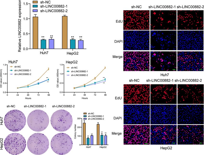
Ren et al.                                                                                                        LINC00882 Accelerates HCC Progression

                                              Conclusion: Our group identified a novel regulatory axis of ATF2/LINC00882/miR-214-
                                              3p/CENPM, which may provide potential therapeutic targets for HCC.
                                              Keywords: lncRNA LINC00882, hepatocellular carcinoma, miR-214-3p, ATF2, metastasis, CENPM

INTRODUCTION                                                                       National Clinical Research Center for Cancer/Cancer Hospital &
                                                                                   Shenzhen Hospital. Chemotherapy or radiotherapy were not
Hepatocellular carcinoma (HCC) is one of the most common                           used for all patients before the operation. All patients gave
malignant cancers (1). It has been reported that there are 783,800                 informed consent and signed a written consent form. This
new cases diagnosed HCC worldwide each year (2). The main risk                     research was ratified by the Ethics Review Committee of
elements for HCC include hepatitis B virus, infection, cirrhosis,                  National Cancer Center/National Clinical Research Center for
and accumulation of aflatoxins in the liver (3). Although more and                  Cancer/Cancer Hospital & Shenzhen Hospital.
more advancements have been achieved to develop life expectancy
of HCC patients, postoperative metastasis and recurrence remain
major disincentives for the improvements of clinical outcomes of                   Cell Lines and Cell Culture
HCC patients (4–6). Therefore, for personalized treatment and                      The normal liver cells (HepaRG) and HCC cell lines
improvement of clinical efficacy, it is an urgent need to develop                   (SMMC-7721, HepG2.2.15, HepG2 and Huh7) were purchased
specific molecular biomarkers.                                                      from ATCC(Manassas, VA, USA). HCC cells were cultured in a
    Long noncoding RNAs (lncRNAs) are RNAs with > 200                              RPMI 1640 medium containing 10% fetal bovine serum (FBS)
nucleotides in length without protein coding capacities (7).                       and HepaRG cells in keratinocyte-SFM (Thermo Fisher, Jijie
They are involved in almost all cellular and pathological                          Technology, Wuhan, China), together with 100 U/mL penicillin
processes, including cellular growth, apoptosis and stem cell                      (Baomanbio, Pudong, Shanghai, China) and 100 mg/mL
maintenance (8, 9). Dysregulation of some lncRNAs has been                         streptomycin(Yita Bio, Pinggu, Haidian, Biejing, China) with
verified in various tumors, such as lung tumor, gastric cancers,                    5% CO2 at 37°C.
bladder cancers, and HCC (10, 11). Growing evidence indicates
that lncRNAs act as tumor supporters or tumor suppressors
by controlling cellular processes, such as differentiation,                        Cell Transfection
proliferation, invasion, and metastasis, highlighting their great                  Short-hairpin RNA oligos directed against LINC00882
potentials in tumor screening, prognosis, and therapies (12, 13).                  (sh-LINC00882-1 and sh-LINC00882-2) were synthesized.
Although the involvements of lncRNAs in HCC initiation and                         Then, oligos were ligated into the shRNA vectors (JunHui Bio,
progression have been studied in several studies such as lncRNA                    Guangzhou, Guandong, China). To increase the expressions of
MIAT and lncRNA-PDPK2P, the potential exploration of                               ATF2 in HCC cells, the expressing plasmids for ATF2 were
lncRNA functions in the HCC contexts was located on their                          PCR-amplified and subcloned into the pcDNA3.1 vector,
infancy (14–16). The novel mechanisms involved in lncRNA                           respectively (JunHui Bio, Guangzhou, Guandong, China). An
effects on HCC progression deserve in-depth explorations for the                   empty pcDNA 3.1 served as a control. Ruibio Biology
development of personalized treatments.                                            (Guangzhou, Guangdong, China) provided miRNA-214-3p
    Long intergenic non-protein coding RNA 882 (LINC00882),                        mimics and miRNA-214-3p inhibitors. ATF2 small interfering
located on 3q13.12, was a new cancer-related lncRNA (17).                          RNA (si-ATF2-1 and si-ATF2-2) and the corresponding control
Several studies have reported its dysregulation in a few cancers,                  RNA (si-NC) were purchased from Biomics Bio (Jiangsu, China)
such as chromophobe renal cell carcinoma and hepatocellular                        Co., Ltd. For cellular transfection, Lipofectamine 3000
carcinoma (18, 19). However, its specific function in HCC has                       (L3000015, Invitrogen, Ruidei Biology, Suzhou, Jiangsu, China)
not been investigated. In this study, we analyzed TCGA datasets,                   was applied based on the operating instructions.
finding LINC00882 was highly expressed in HCC. Then, we
further provided evidences of LINC00882 upregulation in HCC                        Quantitative Real-Time Polymerase
specimens from our patients. Furthermore, our group conducted                      Chain Reaction
functional assays to study the possible influence of LINC00882                      Total RNAs in specimens and several cells were extracted by
dysregulation on tumor behaviors, and also studied the potential                   TRIzol reagent (Invitrogen, Beiyu Bio, Nanjing, China). Then,
molecular mechanisms.                                                              total RNAs were synthesized to cDNA by the use of
                                                                                   PrimeScript RT reagent Kit (TaKaRa, Chengdu, Sichuan,
                                                                                   China) based on the operating instructions. SYBR® Premix
MATERIALS AND METHODS                                                              Ex Taq ™ and StepOne Plus Real-time PCR System were
                                                                                   applied to perform RT-PCR reactions. The primers were
Clinical Specimens                                                                 shown in Table 1. GAPDH or U6 served as an internal
A total of 6 pairs of tumor specimens and the matched non-                         control. The relative expressions were calculated by the use
tumor specimens were collected from National Cancer Center/                        of the DDCt methods.

Frontiers in Oncology | www.frontiersin.org                                    2                                 August 2021 | Volume 11 | Article 714264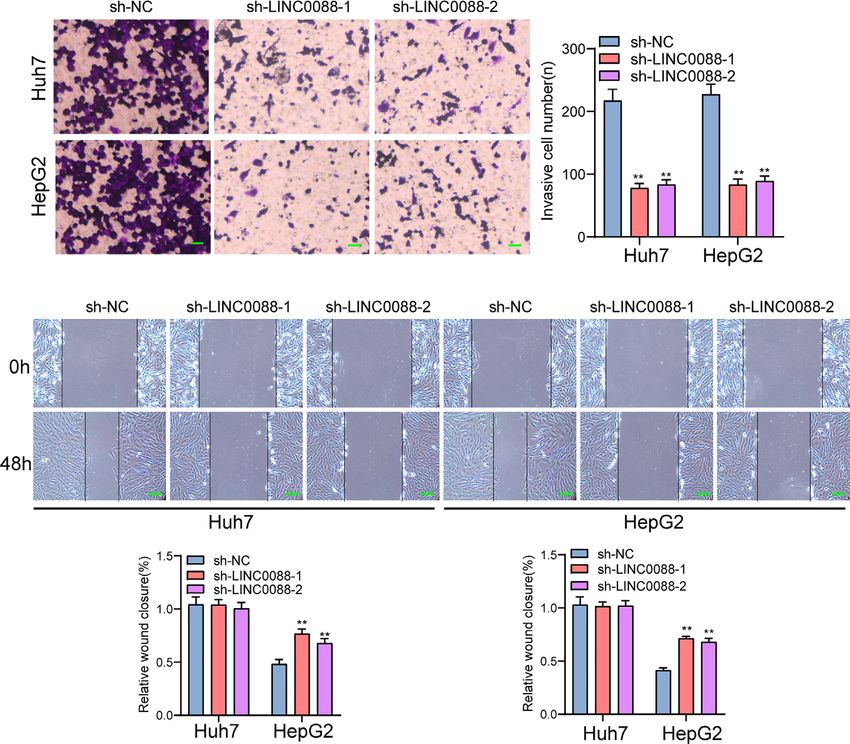
Ren et al.                                                                                                  LINC00882 Accelerates HCC Progression

TABLE 1 | The primers used in this study for RT-PCR.
                                                                            Tranwell Assays
Names                                              Sequences (5’-3’)        The transwell assays were conducted to examine cellular
                                                                            invasion. Briefly, Huh7 and HepG2 cells were seeded on the
LINC00882: Forward                            GCCGATACTTGACCTACGCA
                                                                            top transwell chamber (60 µL of Matrigel at 1:7 dilution;
LINC00882: Reverse                            AGATGGCAGGTGCAATCACA
ATF2: Forward                                 AATTGAGGAGCCTTCTGTTGTAG
                                                                            Shenzhen, China) to the bottom chamber, 500 mL of medium
ATF2: Reverse                                 CATCACTGGTAGTAGACTCTGGG       was added. 90% ethanol was applied to fix the cells after 24 h.
miR-214-3p: Forward                           GCACAGCAGGCACAGACA            Besides, 0.5% crystal violet prior was applied to stain the
miR-214-3p: Reverse                           -CAGAGCAGGGTCAGCGGTA          collected cells. An inverted microscope was used for
CENPM: Forward                                CGACCTGAACAGGGCTACC
                                                                            the observation.
CENPM: Reverse                                ACGCACAGTCATCTTTGAGCA
GAPDH: Forward                                GGAGCGAGATCCCTCCAAAAT
GAPDH: Reverse                                GGCTGTTGTCATACTTCTCATGG
U6: Forward                                   CTCGCTTCGGCAGCACA
                                                                            Dual-Luciferase Reporter Gene Assay
U6: Reverse                                   AACGCTTCACGAATTTGCGT          The wild-type and mutant LINC00882 containing miR-214-3p
                                                                            binding sites on the LINC00882 promoter region were ligated
                                                                            into the pMIR luciferase reporter vectors. Then, using
Subcellular Fractionation                                                   Lipofectamine 2000 (Invitrogen, Xuanwu, Nanjing, China), we
Nuclear and cytoplasmic separation was carried out using the                co-transfected the vectors into cells with LINC00882/NC mimic.
PARIS Kit (Life Technologies, Shenzhen, Guangdong, China)                   To normalize the reporter luciferase activities, the dual luciferase
based on the operating instructions.                                        reporter assays were carried out. In addition, with mutant-type
                                                                            and wild ATF2 binding sites, the LINC00882 promoter were
                                                                            inserted to pGL3 vector (Promega, Haidian, Beijing, China) for
Cell Counting Kit Assays                                                    promoter assays.
Transfected Huh7 and HepG2 cells (4000 cells/well) were
incubated at 37°C in an atmosphere with 5% CO2 for 24, 48,
72 and 96 h, respectively. CCK-8(Dojindo, Beiyu Biology,                    Chromatin Immunoprecipitation
Nanjing, China) was used to incubate the above cells for 4 h.               The ChIP assays were performed following the methods and
Using a microplate reader, the absorbance was observed at                   protocols descripted by the previous publications (20, 21). The
450 nm.                                                                     HCC cells, HepG2, were transfected with vectors and harvested
                                                                            for the ChIP performed by using the ChIP kit (Promega
                                                                            Corporation, USA) according to the protocol by manufacture.
Colony Formation Assays
                                                                            About 2×10 9 amount of HepG2 cells were fixed by 4%
Cells (at a density of 3 × 10³ per well) were seeded in six-well
                                                                            formaldehyde (v/v diluted by PBS) and the final concentration
plates. Subsequently, 4% paraformaldehyde (absin, Pudong,
                                                                            formaldehyde was 1%. Then, the mM dose of glycine solution
Shanghai) was applied to fix formed colonies for 30 min. 0.1%
                                                                            was added and cells were cracked by the lysis-buffer. The nuclear
Crystal violet dye (Merck, Folaisi, Wuxi, China) was used to stain
                                                                            sub-fraction of the cells was separated by centrifugation. Then,
the cells for 15 min. Finally, our group counted and
                                                                            the nuclear sub-fraction lysates were sonicated to generate an
photographed the colonies.
                                                                            average DNA fragments with the size of 0.5–1 kb. At last, the
                                                                            immunoprecipitation was performed with anti-Tubulin (a-, b-
5-Ethynyl-2’-Deoxyuridine Assay                                             and g- Tubulin) (Abcam Corporation, UK). The non-specific
EDU experiments were conducted for exploring cellular                       IgG was used as blank control. The DNA fragments in the
capabilities. Cells (6×10³) were placed in 48-well plates. Under            complex (Tubulin with Centromere related sequence) was
the condition of 5% CO2 and 37°C, 50 mM of EdU diluent was                  analyzed by the quantitative polymerase chain reaction. The
used to cultivate cells for 2 hours. Then, PBS was used rinse the           DNA faction amplified was Human (hg38) chr2 q11.1:
collect cells which was fixated by the use of paraformaldehyde.              92872758- 92873099. The primers are as followed: Forward
Next, Apollo 567 working solution was applied to process cells.             sequence: 5’-tctgcaagaggatatttggatagc-3’; Reverse sequence:
For the counterstain of the nuclei, DAPI was added for 8                    5’-tcaccataggcctgaaagcg-3’.
minutes. Finally, fluorescent microscopy was used to observe
the collected cells.
                                                                            Western Blotting
                                                                            Proteins were extracted from HCC cells, and then quantified.
Scratch Test                                                                Subsequently, 30 mg of proteins was separated via 10%
Cellular scratch experiments were applied for the determination of          SDS-PAGE and transferred on nitrocellulose membranes
the effects of LINC00882 on invasive abilities. Briefly, 1×10⁶ cells         (Pierce, Qingpu, Shanghai, China). After blocked with 3% BSA
were inoculated into a 6-well plate and cultured for 24 h. After a          (YT0230-2, Yita Bio, Pinggu, Beijing, China), the membranes
pipette tip was applied to gently scratch the cells for the formation       were incubated with primary antibodies against CENPM
of the centers of the well, PBS was applied to wash the cells for 3         (ab243820, Abcam, Shenzhen, Guandong), and GAPDH
times. an inverted fluorescence microscope (Nikon, Tokyo, Japan)             (ab8245, Abcam, Shenzhen, Guandong) overnight at 4°C, and
was applied for the observation of scratch distances.                       secondary antibody labeled with horseradish peroxidase

Frontiers in Oncology | www.frontiersin.org                             3                                  August 2021 | Volume 11 | Article 714264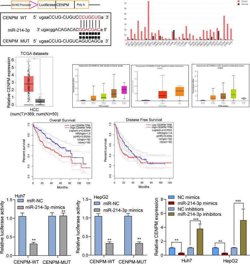
Ren et al.                                                                                                               LINC00882 Accelerates HCC Progression

(Abcam) for 2 h at room temperature. Subsequently, the                             RESULTS
BeyoECL Plus kit (LM0018, Lianmai Technology, Songjiang,
Shanghai, China) was applied to develop the membranes.                             Expressions of LINC00882 Are Increased
GAPDH was used to normalize the expressions of CENPM.                              in Human HCC Tissues
                                                                                   To screen the possible lncRNAs involved in HCC progression,
Tumor Xenograft Analysis                                                           our group searched TCGA datasets and focused on LINC00882
The experimental procedures were approved by Animal Ethics                         which was distinctly overexpressed in HCC samples (n=371)
Committee of National Cancer Center/National Clinical                              compared with normal liver samples (n=50) (Figure 1A). The
Research Center for Cancer/Cancer Hospital & Shenzhen                              results of RT-PCR in samples of our patients also revealed that
Hospital. For in vivo assays, Vital River (Chaoyang, Beijing,                      LINC00882 expressions in HCC tissues (n=6) were distinctly
China) provided 4-weeks old male BALB/c nude mice which                            higher than those in the matched normal liver tissues (n=6)
were randomly divided into two groups: sh-LINC00882-1 group                        (Figure 1B). In addition, we observed that four HCC cells
(n = 6) and sh-NC group (n = 6). LINC00882-1 or sh-NC-                             exhibited a higher level than HepaRG cells (Figure 1C).
transfected Huh7 cells were subcutaneously injected into the                       Survival assays based on TCGA datasets revealed that patients
right flank of nude mice. Digital calipers were applied to measure                  with higher LINC00882 expressions exhibited a poor trend of
tumor volume every 4 days, and the formula (tumor volume = 1/2                     long-term survivals compared with those with lower LINC00882
(length × width2) was used to calculate volumes. 28 days later,                    expressions (Figure 1D). Given that lncRNA functions are
the mice were sacrificed, followed by the examination of the                        dependent on its subcellular localization, we performed
weight of all tumors.                                                              subcellular fractionation, finding that LINC00882 was mainly
                                                                                   localized to the cytoplasm (Figure 1E).
Statistical Analysis
SPSS 19.0 software (SPSS Inc., Chicago, IL, USA) was used to                       ATF2 Activates LINC00882 Transcription
analyze all data which are presented as means ± SD. Student’s                      in HCC Cells
t-test or one-way analysis of variance was applied to compare                      Growing studies suggested that transcription factors such as
the differences between two groups or among multiple                               E2F1, SP1 may exhibit regulatory effects on the expression of
groups, followed by Tukey’s tests. A p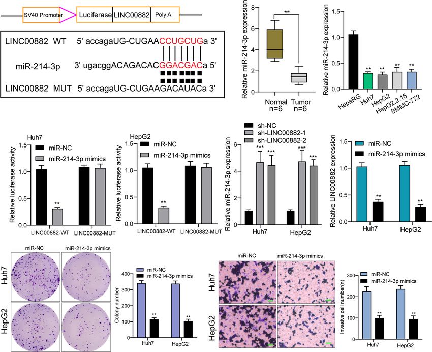
Ren et al.                                                                                                                     LINC00882 Accelerates HCC Progression

             A                                                             B                             C

             D                                  E                                       F

             G                                  H

                                                                                                     I

             J

 FIGURE 2 | ATF2 Activates LINC00882 expression in HCC cells. (A) ATF2 binding site prediction in the LINC00882 promoter region using JASPAR. (B) By analyzing
 TCGA datasets, ATF2 expression was shown in HCC specimens (n=369) and normal liver specimens (n=50). (C, D) RT-PCR for ATF2 expression in HCC specimens
 from our patients and HCC cell lines. (E) Survival assays of 364 patients from TCGA datasets divided into two groups according to LINC00882 expression. (F) QRT-PCR
 for The levels of ATF2 and LINC00882 in HepG2 and Huh7 cells transfected with si-ATF2-1, si-ATF2-2 or si-NC. (G) Overexpression of ATF2 led to the promotion of
 LINC00882 levels in Huh7 and HepG2 cells. (H) ChIP-qPCR analysis of ATF2 occupancy in the LINC00882 promoter in Huh7 and HepG2 cells. (I) Construction of the
 luciferase reporter vector. (J) Dual luciferase reporter assays for the determination of the ATF2 binding site on the LINC00882 promoter region. *p < 0.05, **p < 0.01,
 ***p < 0.001, N.S., no significant.

Frontiers in Oncology | www.frontiersin.org                                         5                                        August 2021 | Volume 11 | Article 714264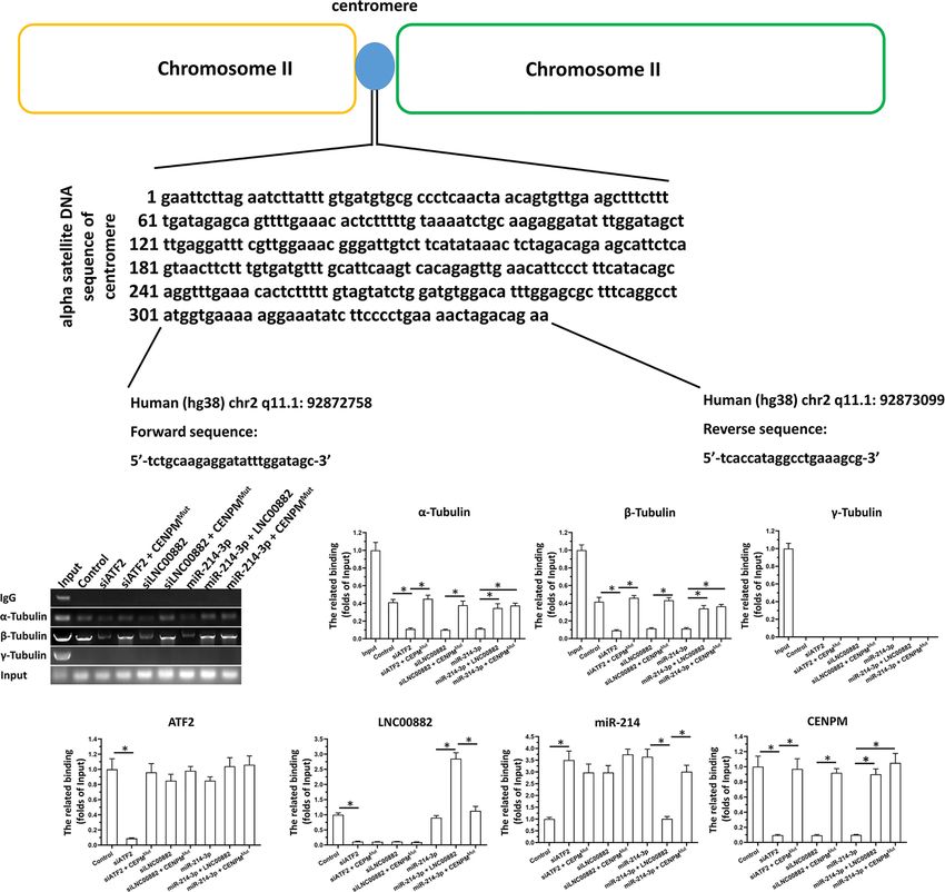
Ren et al.                                                                                                            LINC00882 Accelerates HCC Progression

database (http://jaspar.genereg.net/), finding that ATF2 may                      As examined by RT-PCR, the expressions of LINC00882 were
bound to the LINC00882 promoter region with strong                               significantly inhibited by sh-LINC00882-1 and sh-LINC00882-2
probability (Figure 2A). Based TCGA datasets, ATF2 was                           in both HepG2 and Huh7 cells (Figure 3A). CCK-8 assays
highly expressed in HCC specimens, which was further                             indicated that LINC00882 knockdown distinctly suppressed the
confirmed in our cohort and four cell lines (Figures 2B–D).                       proliferative capabilities of Huh7 and HepG2 cells (Figure 3B).
Survival assays with TCGA datasets revealed patients with higher                 Besides, the inhibitory effects of LINC00882 knockdown on HCC
ATF2 expressions showed a poor trend of overall survival in 40                   cell proliferation were also demonstrated by the colony formation
months (Figure 2E). RT-PCR assays revealed that silence of                       and Edu assays (Figures 3C, D). For further exploration of the
ATF2 resulted in the obvious inhibition of LINC00882                             oncogenic functions of LINC00882, our group conducted in vivo
(Figure 2F), while its overexpression promoted the expression                    experiments. As presented in Figures 4A–C, tumor volume and
of LINC00882 (Figure 2G). By the use of ChIP assays, our group                   weight were reduced by sh-LINC00882-1 compared with
observed distinct ATF2-binding activities on the endogenous                      scramble control. Furthermore, the results of Transwell assays
LINC00882 promoter region around E2 (Figure 2H). Besides,                        and Wound-healing assays showed that the knockdown of
luciferase reporter assays demonstrated that ATF2 did not binds                  LINC00882 weakened the invasion (Figure 5A) and migration
to the other two sites, but only the E2 binding site (Figures 2I, J).            (Figure 5B) ability of Huh7 and HepG2 cells. Therefore,
                                                                                 LINC00882 was an oncogenic lncRNA in HCC.
Silencing of LINC00882 Suppresses the
Proliferation and Metastasis of HCC Cells                                        Reciprocal Modulation Between
To evaluate the roles of LINC00882 in HCC, Huh7 and HepG2                        LINC00882 and miR-214-3p
cells were transfected with shRNA (sh-LINC00882-1,                               We have confirmed that LINC00882 was mostly distributed
sh-LINC00882-1) to silence the expression of LINC00882.                          in the cytoplasm, which suggested that LINC00882

                 A                                                                   D

             B

             C

 FIGURE 3 | Knockdown of LINC00882 suppressed the proliferation of HepG2 and Huh7 cells. (A) The relative expressions of LINC00882 in Huh7 and HepG2
 cells were distinctly reduced by sh-LINC00882-1 or sh-LINC00882-2 compared with the si-NC. (B) CCK-8 assays evaluated cell viability. (C) Clone number of
 Huh7 and HepG2 cell lines after transfected by sh-LINC00882-1, sh-LINC00882-2 or sh-NC. (D) Cell proliferation was determined by EdU assay in Huh7 and
 HepG2 cells. **p < 0.01.

Frontiers in Oncology | www.frontiersin.org                                  6                                      August 2021 | Volume 11 | Article 714264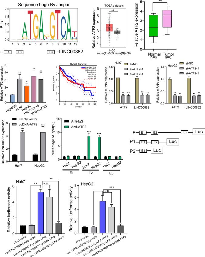
Ren et al.                                                                                                             LINC00882 Accelerates HCC Progression

                 A                                                           B                                         C

 FIGURE 4 | LINC00882 regulates HCC growth in vivo. (A) Tumors derived from mice in two different groups were presented. (B, C) Volume and weight of tumors
 obtained from two groups were measured and shown. **p < 0.01.

                      A

                      B

 FIGURE 5 | Knockdown of LINC00882 suppressed the migration and invasion of HCC cells. (A) Transwell experiments revealed knockdown of LINC00882 inhibited
 Huh7 and HepG2 cell invasion. (B) The wound healing assay showed a significant decrease of Huh7 and HepG2 cells migration after transfected sh-LINC00882-1 or
 sh-LINC00882-2. **p < 0.01.

might exert its biological function by sponging miRNA.                            MUT-LINC00882 was distinctly reduced by miRNA-214-3p
Then, we searched Starbase which can predict the possible                         overexpression in Huh7 and HepG2 cells (Figure 6D). RT-
miRNAs binding sites for LINC00882. The binding sites of                          PCR assays suggested miR-214-3p levels were distinctly
LINC00882 and miR-214-3p were presented in Figure 6A.                             upregulated in Huh7 and HepG2 cells after knockdown of
RT-PCR assays indicated miRNA-214-3p was lowly                                    LINC00882 (Figure 6E), while miR-214-3p overexpression
expressed in HCC specimens and cell lines (Figures 6B, C).                        distinctly suppressed the levels of LINC00882 (Figure 6F).
The luciferase activity of WT- LINC00882 instead of                               Functionally, miR-214-3p upregulation was observed to

Frontiers in Oncology | www.frontiersin.org                                   7                                      August 2021 | Volume 11 | Article 714264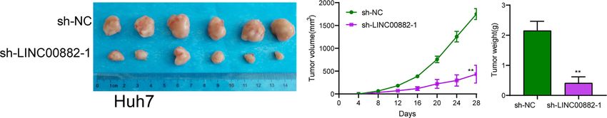
Ren et al.                                                                                                               LINC00882 Accelerates HCC Progression

             A                                                                           B                     C

             D                                                                       E                             F

             G                                                               H

 FIGURE 6 | LINC00882 acts as a ceRNA for miR-214-3p. (A) LINC00882 containing the putative miR-214-3p recognition sites was cloned downstream of the
 luciferase gene. (B, C) RT-PCR for the LINC00882 expression in tumor specimens from our patients and cell lines. (D) Luciferase reporter assay was applied to
 verify the targeted binding effect between LINC00882 and miR-214-3p. (E) RT-PCR for miR-214-3p levels in Huh7 and HepG2 cells after LINC00882 knockdown.
 (F) The levels of LINC00882 levels in Huh7 and HepG2 cells after Overexpression of miR-214-3p. (G) Colony formation assay. (H) The invasive number of Huh7 and
 HepG2 after knockdown of LINC00882. **p < 0.01, ***p < 0.001.

inhibit the proliferation and invasion of HepG2 and Huh7 cells                       outcomes (Figure 7E). The results of luciferase reporter assay s
(Figures 6G, H).                                                                     showed that compared with CENPM 3’UTR-mut and miR-214-
                                                                                     3p mimics, luciferase activity of Huh7 and HepG2 cells
                                                                                     transfected with CENPM 3’UTR-wt and miR-214-3p mimics
LINC00882 Directly Targets CENPM                                                     was distinctly decreased (Figure 7F). Moreover, overexpression
in HCC Cells                                                                         of miRNA-214-3p distinctly suppressed the expressions of
To further study the mechanisms of actions of miR-214-3p in                          CENPM, while its silence displayed an opposite result
HCC, we performed bioinformatics assays to search the targets of                     (Figure 7G). On the other hand, we also performed in vitro
miR-214-3p. CENPM was predicted to harbor putative binding                           assays to explore the function of CENPM in HCC. As shown in
sequences for miR-214-3p (Figure 7A). Then, we focused on                            Supplementary Figure S1A, the results of RT-PCR and western
CENPM due to its important functions during tumor                                    blot showed that the expression of CENPM was distinctly
progression (24, 25). Based on TCGA datasets, we observed                            decreased in Huh7 and HepG2 cells transfected with si-
CENPM displayed a high level in most types of cancers, and                           CENPM compared with si-NC. We further performed
CENPM levels were distinctly upregulated in HCC specimens,                           functional experiments, finding that knockdown of CENPM
especially in those with advanced stages (Figures 7B–D).                             distinctly suppressed the proliferation (Supplementary Figures
Survival assays based on TCGA datasets revealed that high                            S1B, C), invasion (Supplementary Figure S1D) and migration
CENPM expressions were associated with poor clinical                                 (Supplementary Figure S1E) of Huh7 and HepG2 cells.

Frontiers in Oncology | www.frontiersin.org                                      8                                     August 2021 | Volume 11 | Article 714264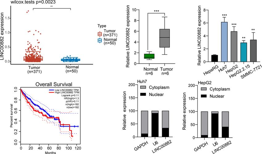
Ren et al. LINC00882 Accelerates HCC Progression
A B
C D
E
F G
FIGURE 7 | CENPM was identified as a direct target of miR-214-3p in HCC cells. (A) Bioinformatics tools reveal the complementary binding sites within miR-214-3p
and CENPM. (B) Pan-cancer analysis of CENPM using TCGA datasets. (C, D) The expression pattern of CENPM in HCC specimens with different stages. (E) Survival
assays of HCC patients according to the expression of CENPM by analyzing TCGA datasets. (F) Luciferase reporter assay validated the molecular binding. (G) RT-PCR
determined the expression of CENPM in Huh7 and HepG2 cells transfected with NC mimics, miR-214-3p mimics, NC inhibitors or miR-214-3p inhibitors. *p < 0.05,
**p < 0.01, ***p < 0.001.
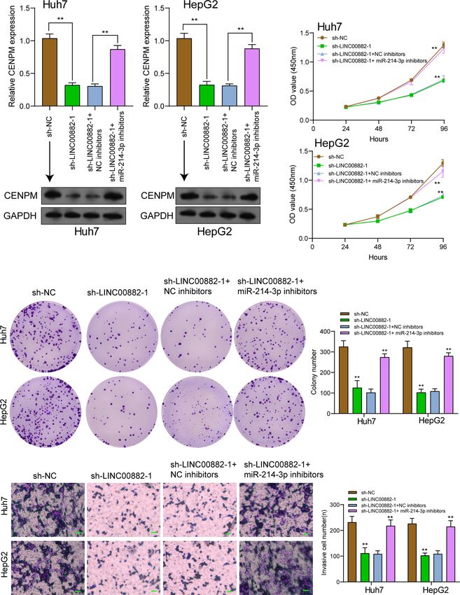
LINC00882 Promoted HCC Progression by sh-LINC00882-1 or sh-NC and miR-214-3p inhibitors or NC
Decreasing CENPM Expression via inhibitors into HCC cells. As presented in Figure 8A, silence of
Sponging miR-214-3p miR-214-3p reversed the distinct suppression of LINC00882
silence on the expressions of CENPM. Besides, functional
To demonstrate that LINC00882 aggravated the tumor behaviors experiments revealed silence of miR-214-3p reversed the
of HCC through the modulation of miR-214-3p/CENPM axis, distinct inhibition of LINC00882 silence on the proliferative
our group conducted rescue experiments via cotransfecting and invasive abilities of HCC cells (Figures 8B–D).
Frontiers in Oncology | www.frontiersin.org 9 August 2021 | Volume 11 | Article 714264Ren et al. LINC00882 Accelerates HCC Progression
A B
C
D
FIGURE 8 | Knockdown of miR-214-3p effectively reversed LINC00882 knockdown-induced inhibition on HCC progression in vitro. (A) RT-PCR and Western blot
examined the expression of CENPM in Huh7 and HepG2 cells transfected with sh-NC, sh-LINC00882-1, sh-LINC00882-1+ NC inhibitor or sh-LINC00882-1+ miR-
214-3p inhibitor. CCK-8 assays (B), colony formation assays (C) and Transwell assays (D) of HepG2 and Huh7 cells after transfection. **p < 0.01.
Frontiers in Oncology | www.frontiersin.org 10 August 2021 | Volume 11 | Article 714264Ren et al. LINC00882 Accelerates HCC Progression
ATF2/LNC000882/miR-214-3p Pathway LNC000882 via siRNA or overexpression of miR-214-3p both
Modulate The Binding of Tubulin With resulted to the knockdown of CENPM and the decreased binding
Centromere Related DNA Sequence by of a-Tubulin or b-Tubulin could to the Centromere related
DNA sequence (Figures 9F–I). Rescued the expression of
Targeting CENPM
CENPM via transfection of CENPM with the mutated
The chromatin immunoprecipitation (ChIP) was performed to miR-214-3p targeting site in 3’UTR (CENPM Mut ) could
detect the binding effect of Tubulin and Centromere related disrupt the effect of ATF2, LNC000882 knockdown or miR-
DNA sequence reflecting the influence of CENPM on the 214-3p overexpression. Moreover, the overexpression of
centromere-microtubule interaction to form the spindle. As LNC000882 also repressed the expression of miR-214-3p and
shown in Figures 9A–E, as expected, a-Tubulin and b- enhanced the expression of CENPM. These results further
Tubulin could binding to the Centromere related DNA confirmed the specificity of ATF2/LNC000882/miR-214-3p
sequence; however, the g-Tubulin could not interacted with the pathway modulate the binding of Tubulin with Centromere
Centromere related DNA sequence. Knockdown of ATF2, related DNA sequence by targeting CENPM.
A
B C D E
F G H I
FIGURE 9 | The effect of ATF2/LNC000882/miR-214-3p/CENPM pathway on the binding of Tubulin to Centromere related DNA sequence. (A–E) The HepG2 cells
were transfected with vectors and harvested for the ChIP assays. The complex of Tubulin with Centromere related DNA sequence was separated by the antibodies
of Tubulins. (F) The expression level of ATF2, (G) LNC000882, (H) miR-214-3p or (I) CENPM in HepG2 cells were examined by the qPCR. The results were shown
as the schematic-diagram images of the Centromere related DNA sequence (A), the images or mean ± SD from qPCR of the ChIP results (B–E) or the mean ± SD
from the qPCR. *p < 0.05.
Frontiers in Oncology | www.frontiersin.org 11 August 2021 | Volume 11 | Article 714264Ren et al. LINC00882 Accelerates HCC Progression
DISCUSSION cytoplasm, which provided a basic possibility for the ceRNA
mechanisms. By the use of bioinformatics assays, miRNA-214-
Several studies reported the functions of lncRNAs in HCC 3p was predicted as a targeting miRNA of LINC00882, followed
progression (26). For instance, lncRNA-PDPK2P was shown to by the demonstration of luciferase reporter and RT-PCR.
exhibit a higher level in HCC and accelerated invasion and Previously, miR-214-3p was found to be lowly expressed in
tumor growth of HCC cells via PDK1/AKT/Caspase 3 pathway HCC and suppressed the proliferative and metastatic abilities
(16). LncRNA MCM3AP-AS1, a possible diagnostic factor for of HCC cells, which was consistent with our findings (42, 43). On
HCC, was reported to promote the growth of HCC through the other hand, we also found that miR-761 may be a potential
modulating miRNA-194-5p (27). The oncogenic or anti- target of LINC00882. However, miR-761 was found to act as a
oncogenic roles of lncRNAs encouraged us to further explore tumor promotor in HCC (44).
the important lncRNAs involved in HCC progression. Here, we Centromere protein M (CENPM), encoding a kinetic protein,
identified a novel lncRNA-related oncogene in HCC. We modulate chromosomal separation during cellular divisions (45).
provided evidences that LINC00882 levels were distinctly The levels of CENPM were observed to appear preferentially in
upregulated in HCC. Using TCGA datasets, we observed immunity cells involving cancer specimens and cancer derived
patients with high LINC00882 showed a poor trend in long- cells (24, 46). In HCC, CENPM silence resulted in the
term survivals. Functionally, we demonstrated knockdown of suppression of the proliferation and metastasis of HCC cells,
LINC00882 distinctly inhibited the invasion, migration and and CENPM was regulated by several miRNAs (47, 48). It has
proliferation of HepG2 and Huh7 cells, suggesting it as a been confirmed that miRNAs can target downstream genes and
tumor promotor in HCC progression. We searched literatures suppress their expression (49–51). Interestingly, using a
and only found LINC00882 was overexpressed in hepatocellular combination of multiple biochemical analyses and mechanistic
carcinoma and chromophobe renal cell carcinoma (18, 19). studies, CENPM was identified as a direct target of miR-214-3p.
However, the studies involved in potential function of We also analyzed TCGA datasets, finding that CENPM was
LINC00882 in tumors have not been found. Our findings may overexpressed in HCC. Knockdown of CENPM suppressed the
provide a new clue for the research of LINC00882 in other types proliferation and invasion of HCC cells. Finally, our group
of tumors. carried out rescue experiments, finding that silence of miR-
Several studies have provided evidences that transcription 214-3p reversed the distinct inhibition of LINC00882
factors can regulated the expressions of lncRNAs just like some knockdown on the expression of CENPM as well as the
protein-coding genes (28, 29). For instance, SP1-induced proliferation and invasion. Therefore, our study unveiled the
upregulation of lncRNA TINCR suppressed the metastasis contribution of the LINC00882/miR-214-3p/CENPM pathway
ability of lung adenocarcinoma cells via regulating miR-107/ in regulating HCC progression.
RAB14 (30). Overexpressed lncRNA HAGLROS induced by For the activity of CENPM itself, we have conducted various
STAT3 promoted the metastasis of gastric cancer (31). In studies. Using siRNA to down-regulate the expression of CENPM
HCC, several HCC-related lncRNAs, such as lncRNA ZFPM2- can significantly inhibit the proliferation of HCC cells. At the same
AS1 (by STAT1) and lncRNA RAET1K (by HIF1A), were also time, for the specific mechanism of CENPM itself, CENPM is a
reported to be regulated by transcription factors (32, 33). In this centromere protein, and its expression level is closely related to the
study, we found ATF2 may bind directly to the LINC00882 normal function of the centromere-spindle. Because cellular
promoter region, followed by a series of experiments confirming immunofluorescence is difficult to quantitatively reflect the
the overexpression of LINC00882 induced by ATF2. ATF2 is a direct interaction between tubulin and centromere, we use
member of the activator protein 1 (AP-1) TF family. Previously, chromatin immunoprecipitation technology to detect the
several studies have reported ATF2 was highly expressed in combination of tubulin and centromere DNA sequence, and
several types of tumors and served as a tumor promotor, finally perform specific effects on CENPM Sexual analysis. Our
including HCC (34, 35). However, whether ATF2 can activate results show that down-regulating the expression of CENPM can
lncRNA transcription was rarely reported. Our finding firstly inhibit the binding of tubulin a and b to the centromere sequence.
reported ATF2 overexpression resulted in the upregulation At the same time, the specificity of the action of ATF2/
of LINC00882. LNC000882/miR-214-3p is determined by setting a reasonable
In recent years, a ceRNA mechanism was developed, which control. Moreover, Tubulin is mainly composed of a- and b-
showed lncRNAs may serve as miRNA sponges, thereby subtypes, while g-subtypes are mainly distributed in the
competitively regulating the targets of miRNAs (36, 37). centrosome. The results of this study also confirmed that in the
More and more evidences have shown that several functional ChIP experiment, only a-Tubulin and b-Tubulin can interact with
lncRNAs could absorb miRNAs to regulate the expressions centromere DNA sequences, while g-Tubulin cannot interact with
of anti-oncogenes or cancer promotors in tumor developments centromere DNA sequences. These methods and results have
(38, 39). However, the ability of several lncRNAs with low advantages and innovations: the combination of centromere and
abundance was not enough in sponging miRNAs. Many tubulin is the basis for the formation of spindles and cell
cellular evidences have demonstrated lncRNAs which exhibit proliferation. The use of chromatin immunoprecipitation
high levels in the cytoplasm are ideal lncRNAs (40, 41). In this technology to detect the combination of tubulin and centromere
study, we observed that LINC00882 was mainly expressed in the can be quantitative and intuitive Reflects the interaction between
Frontiers in Oncology | www.frontiersin.org 12 August 2021 | Volume 11 | Article 714264Ren et al. LINC00882 Accelerates HCC Progression
microtubules and centromeres, which is an ideal method for this Chinese Academy of Medical Sciences and Peking Union
type of research. Medical College. The patients/participants provided their
However, this study has several limitations. Firstly, the sample written informed consent to participate in this study. The
size was relatively small, we will collect more samples for animal study was reviewed and approved by National Cancer
research in the future. Secondly, additional mechanisms of Center/National Clinical Research Center for Cancer/Cancer
LINC00882 such as methylation in regulating HCC Hospital & Shenzhen Hospital, Chinese Academy of Medical
progression require further study. Sciences and Peking Union Medical College.
CONCLUSION AUTHOR CONTRIBUTIONS
Based on results, LINC00882 may be a tumor promotor for
HR, Z-cW, and XC conceived and designed the experiment. HR,
HCC, and ATF2 could activate its transcription. LINC00882
Z-cW and Y-xS performed the experiments and wrote the
exerted oncogenic roles in modulating the proliferation and
manuscript. C-yQ and W-jZ performed the statistical analyses
metastasis of HCC cells. Our findings firstly suggested the
and generated the figures. HR, WZ, and TL collected the public
underlying mechanisms behind this development, that
data. Z-cW and XC collected the patient samples. HR and XC
LINC00882 drove HCC progression by increasing CENPM
revised the manuscript. All authors contributed to the article and
levels through sponging miR-214-3p, providing a novel
approved the submitted version.
therapeutic target for HCC treatments.
DATA AVAILABILITY STATEMENT FUNDING
The raw data supporting the conclusions of this article will be This study was supported by the Sanming Project of Medicine in
made available by the authors, without undue reservation. Shenzhen(No.SZSM201612063 & No.SZSM201911008) and the
Shenzhen Basic Research Program(KJYY20170412153658082).
ETHICS STATEMENT SUPPLEMENTARY MATERIAL
The studies involving human participants were reviewed and The Supplementary Material for this article can be found online
approved by National Cancer Center/National Clinical Research at: https://www.frontiersin.org/articles/10.3389/fonc.2021.
Center for Cancer/Cancer Hospital & Shenzhen Hospital, 714264/full#supplementary-material
10. Huarte M. The Emerging Role of lncRNAs in Cancer. Nat Med (2015) 21
REFERENCES (11):1253–61. doi: 10.1038/nm.3981
1. Siegel RL, Miller KD, Jemal A. Cancer Statistics, 2018. CA: Cancer J Clin 11. Huang Z, Zhou JK, Peng Y, He W, Huang C. The Role of Long Noncoding
(2018) 68(1):7–30. doi: 10.3322/caac.21442 RNAs in Hepatocellular Carcinoma. Mol Cancer (2020) 19(1):77. doi:
2. El Jabbour T, Lagana SM, Lee H. Update on Hepatocellular Carcinoma: 10.1186/s12943-020-01188-4..
Pathologists’ Review. World J Gastroenterol (2019) 25(14):1653–65. doi: 12. Schmitz SU, Grote P, Herrmann BG. Mechanisms of Long Noncoding RNA
10.3748/wjg.v25.i14.1653 Function in Development and Disease. Cell Mol Life Sci: CMLS (2016) 73
3. Barry AE, Baldeosingh R, Lamm R, Patel K, Zhang K, Dominguez DA, et al. (13):2491–509. doi: 10.1007/s00018-016-2174-5
Hepatic Stellate Cells and Hepatocarcinogenesis. Front Cell Dev Biol (2020) 13. Schmitt AM, Chang HY. Long Noncoding RNAs in Cancer Pathways. Cancer
8:709. doi: 10.3389/fcell.2020.00709 Cell (2016) 29(4):452–63. doi: 10.1016/j.ccell.2016.03.010
4. Forner A, Reig M, Bruix J. Hepatocellular Carcinoma. Lancet (London 14. Beermann J, Piccoli MT, Viereck J, Thum T. Non-Coding RNAs in Development
England) (2018) 391(10127):1301–14. doi: 10.1016/S0140-6736(18)30010-2 and Disease: Background, Mechanisms, and Therapeutic Approaches. Physiol Rev
5. Choi SH, Seong J. Strategic Application of Radiotherapy for Hepatocellular (2016) 96(4):1297–325. doi: 10.1152/physrev.00041.2015
Carcinoma. Clin Mol Hepatol (2018) 24(2):114–34. doi: 10.3350/cmh.2017.0073 15. Huang X, Gao Y, Qin J, Lu S. lncRNA MIAT Promotes Proliferation and
6. Li B, Feng F, Jia H, Jiang Q, Cao S, Wei L, et al. Rhamnetin Decelerates the Invasion of HCC Cells via Sponging miR-214. Am J Physiol Gastrointest Liver
Elimination and Enhances the Antitumor Effect of the Molecular-Targeting Physiol (2018) 314(5):G559–65. doi: 10.1152/ajpgi.00242.2017
Agent Sorafenib in Hepatocellular Carcinoma Cells via the miR-148a/PXR 16. Pan W, Li W, Zhao J, Huang Z, Zhao J, Chen S, et al. lncRNA-PDPK2P
Axis. Food Funct (2021) 12(6):2404–17. doi: 10.1039/D0FO02270E Promotes Hepatocellular Carcinoma Progression Through the PDK1/AKT/
7. Kopp F, Mendell JT. Functional Classification and Experimental Dissection of Caspase 3 Pathway. Mol Oncol (2019) 13(10):2246–58. doi: 10.1002/1878-
Long Noncoding RNAs. Cell (2018) 172(3):393–407. doi: 10.1016/ 0261.12553
j.cell.2018.01.011 17. Perry MM, Tsitsiou E, Austin PJ, Lindsay MA, Gibeon DS, Adcock IM, et al.
8. Chen LL. Linking Long Noncoding RNA Localization and Function. Trends Role of Non-Coding RNAs in Maintaining Primary Airway Smooth Muscle
Biochem Sci (2016) 41(9):761–72. doi: 10.1016/j.tibs.2016.07.003 Cells. Respir Res (2014) 15(1):58. doi: 10.1186/1465-9921-15-58
9. Ulitsky I, Bartel DP. lincRNAs: Genomics, Evolution, and Mechanisms. Cell 18. He HT, Xu M, Kuang Y, Han XY, Wang MQ, Yang Q. Biomarker and
(2013) 154(1):26–46. doi: 10.1016/j.cell.2013.06.020 Competing Endogenous RNA Potential of Tumor-Specific Long Noncoding
Frontiers in Oncology | www.frontiersin.org 13 August 2021 | Volume 11 | Article 714264Ren et al. LINC00882 Accelerates HCC Progression
RNA in Chromophobe Renal Cell Carcinoma. OncoTargets Ther (2016) 38. Li JH, Liu S, Zhou H, Qu LH, Yang JH. Starbase V2.0: Decoding miRNA-
9:6399–406. doi: 10.2147/OTT.S116392 ceRNA, miRNA-ncRNA and Protein-RNA Interaction Networks From
19. Zhu L, Huang F, Wan T, Xu H, Zhao Q. Overexpression of Long Noncoding Large-Scale CLIP-Seq Data. Nucleic Acids Res (2014) 42(Database issue):
RNA LINC00882 Is Associated With Poor Prognosis in Hepatocellular D92–7. doi: 10.1093/nar/gkt1248
Carcinoma. OncoTargets Ther (2018) 11:5209–17. doi: 10.2147/OTT.S170825 39. Bai Y, Long J, Liu Z, Lin J, Huang H, Wang D, et al. Comprehensive Analysis
20. Cui J, Yang Y, Zhang C, Hu P, Kan W, Bai X, et al. FBI-1 Functions as a Novel of a ceRNA Network Reveals Potential Prognostic Cytoplasmic lncRNAs
AR Co-Repressor in Prostate Cancer Cells. Cell Mol Life Sci: CMLS (2011) 68 Involved in HCC Progression. J Cell Physiol (2019) 234(10):18837–48. doi:
(6):1091–103. doi: 10.1007/s00018-010-0511-7 10.1002/jcp.28522
21. Kim HJ, Lee J, Chung MY, Hong S, Park JH, Lee SH, et al. Piceatannol 40. Qi X, Zhang DH, Wu N, Xiao JH, Wang X, Ma W. ceRNA in Cancer: Possible
Reduces Resistance to Statins in Hypercholesterolemia by Reducing PCSK9 Functions and Clinical Implications. J Med Genet (2015) 52(10):710–8. doi:
Expression Through P300 Acetyltransferase Inhibition. Pharmacol Res (2020) 10.1136/jmedgenet-2015-103334
161:105205. doi: 10.1016/j.phrs.2020.105205 41. Kong Q, Liang C, Jin Y, Pan Y, Tong D, Kong Q, et al. The lncRNA MIR4435-
22. Wei L, Liu Y, Zhang H, Ma Y, Lu Z, Gu Z, et al. TMPO-AS1, A Novel E2F1- 2HG Is Upregulated in Hepatocellular Carcinoma and Promotes Cancer Cell
Regulated lncRNA, Contributes to the Proliferation of Lung Adenocarcinoma Proliferation by Upregulating miRNA-487a. Cell Mol Biol Lett (2019) 24:26.
Cells via Modulating miR-326/SOX12 Axis. Cancer Manage Res (2020) doi: 10.1186/s11658-019-0148-y
12:12403–14. doi: 10.2147/CMAR.S269269 42. Zhan M, He K, Xiao J, Liu F, Wang H, Xia Z, et al. LncRNA HOXA11-AS
23. Dong H, Wang W, Mo S, Chen R, Zou K, Han J, et al. SP1-Induced lncRNA Promotes Hepatocellular Carcinoma Progression by Repressing miR-214-3p.
AGAP2-AS1 Expression Promotes Chemoresistance of Breast Cancer by J Cell Mol Med (2018) 22(8):3758–67. doi: 10.1111/jcmm.13633
Epigenetic Regulation of Myd88. J Exp Clin Cancer Res (2018) 37(1):202. 43. Li Y, Li Y, Chen Y, Xie Q, Dong N, Gao Y, et al. MicroRNA-214-3p Inhibits
doi: 10.1186/s13046-018-0875-3 Proliferation and Cell Cycle Progression by Targeting MELK in
24. Xiao Y, Najeeb RM, Ma D, Yang K, Zhong Q, Liu Q. Upregulation of CENPM Hepatocellular Carcinoma and Correlates Cancer Prognosis. Cancer Cell Int
Promotes Hepatocarcinogenesis Through Mutiple Mechanisms. J Exp Clin (2017) 17:102. doi: 10.1186/s12935-017-0471-1
Cancer Res (2019) 38(1):458. doi: 10.1186/s13046-019-1444-0 44. Zhou X, Zhang L, Zheng B, Yan Y, Zhang Y, Xie H, et al. MicroRNA-761 Is
25. Zheng C, Zhang T, Li D, Huang C, Tang H, Ni XF, et al. Upregulation of Upregulated in Hepatocellular Carcinoma and Regulates Tumorigenesis by
CENPM Facilitates Tumor Metastasis via the mTOR/P70s6k Signaling Targeting Mitofusin-2. Cancer Sci (2016) 107(4):424–32. doi: 10.1111/
Pathway in Pancreatic Cancer. Oncol Rep (2020) 44(3):1003–12. doi: cas.12904
10.3892/or.2020.7673 45. Foltz DR, Jansen LE, Black BE, Bailey AO, Yates JR3rd, Cleveland DW. The
26. Lai Y, Feng B, Abudoureyimu M, Zhi Y, Zhou H, Wang T, et al. Non-Coding Human CENP-A Centromeric Nucleosome-Associated Complex. Nat Cell
RNAs: Emerging Regulators of Sorafenib Resistance in Hepatocellular Biol (2006) 8(5):458–69. doi: 10.1038/ncb1397
Carcinoma. Front Oncol (2019) 9:1156. doi: 10.3389/fonc.2019.01156 46. Yu Z, Wang R, Chen F, Wang J, Huang X. Five Novel Oncogenic Signatures
27. Wang Y, Yang L, Chen T, Liu X, Guo Y, Zhu Q, et al. A Novel lncRNA Could Be Utilized as AFP-Related Diagnostic Biomarkers for Hepatocellular
MCM3AP-AS1 Promotes the Growth of Hepatocellular Carcinoma by Carcinoma Based on Next-Generation Sequencing. Dig Dis Sci (2018) 63
Targeting miR-194-5p/FOXA1 Axis. Mol Cancer (2019) 18(1):28. doi: (4):945–57. doi: 10.1007/s10620-018-4961-3
10.1186/s12943-019-0957-7 47. Wu ZH, Yang DL. High CENPM mRNA Expression and Its Prognostic
28. Holmes ZE, Hamilton DJ, Hwang T, Parsonnet NV, Rinn JL, Wuttke DS, et al. Significance in Hepatocellular Carcinoma: A Study Based on
The Sox2 Transcription Factor Binds RNA. Nat Commun (2020) 11(1):1805. Data Mining. Cancer Cell Int (2020) 20:406. doi: 10.1186/s12935-020-
doi: 10.1038/s41467-020-15571-8 01499-y
29. Liu M, Zhong J, Zeng Z, Huang K, Ye Z, Deng S, et al. Hypoxia-Induced 48. Zou Y, Sun Z, Sun S. LncRNA HCG18 Contributes to the Progression of
Feedback of HIF-1a and lncRNA-CF129 Contributes to Pancreatic Cancer Hepatocellular Carcinoma via miR-214-3p/CENPM Axis. J Biochem (2020)
Progression Through Stabilization of P53 Protein. Theranostics (2019) 9 168(5):535–46. doi: 10.1093/jb/mvaa073
(16):4795–810. doi: 10.7150/thno.30988 49. Wang C, Ding S, Sun B, Shen L, Xiao L, Han Z, et al. Hsa-miR-4271
30. Gao YW, Ma F, Xie YC, Ding MG, Luo LH, Jiang S, et al. Sp1-Induced Downregulates the Expression of Constitutive Androstane Receptor and
Upregulation of the Long Noncoding RNA TINCR Inhibits Cell Migration Enhances In Vivo the Sensitivity of Non-Small Cell Lung Cancer to
and Invasion by Regulating miR-107/miR-1286 in Lung Adenocarcinoma. Gefitinib. Pharmacol Res (2020) 161:105110. doi: 10.1016/j.phrs.2020.105110
Am J Trans Res (2019) 11(8):4761–75. 50. Chen B, Liao Z, Qi Y, Zhang H, Su C, Liang H, et al. miR-631 Inhibits
31. Chen JF, Wu P, Xia R, Yang J, Huo XY, Gu DY, et al. STAT3-Induced lncRNA Intrahepatic Metastasis of Hepatocellular Carcinoma by Targeting PTPRE.
HAGLROS Overexpression Contributes to the Malignant Progression of Front Oncol (2020) 10:565266. doi: 10.3389/fonc.2020.565266
Gastric Cancer Cells via mTOR Signal-Mediated Inhibition of Autophagy. 51. Ji J, Rong Y, Luo CL, Li S, Jiang X, Weng H, et al. Up-Regulation of hsa-miR-
Mol Cancer (2018) 17(1):6. doi: 10.1186/s12943-017-0756-y 210 Promotes Venous Metastasis and Predicts Poor Prognosis in
32. Zhang XW, Li QH, Xu ZD, Dou JJ. STAT1-Induced Regulation of lncRNA Hepatocellular Carcinoma. Front Oncol (2018) 8:569. doi: 10.3389/
ZFPM2-AS1 Predicts Poor Prognosis and Contributes to Hepatocellular fonc.2018.00569
Carcinoma Progression via the miR-653/GOLM1 Axis. Cell Death Dis
(2021) 12(1):31. doi: 10.1038/s41419-020-03300-4 Conflict of Interest: The authors declare that the research was conducted in the
33. Zhou Y, Huang Y, Hu K, Zhang Z, Yang J, Wang Z. HIF1A Activates the absence of any commercial or financial relationships that could be construed as a
Transcription of lncRNA RAET1K to Modulate Hypoxia-Induced Glycolysis potential conflict of interest.
in Hepatocellular Carcinoma Cells via miR-100-5p. Cell Death Dis (2020) 11
(3):176. doi: 10.1038/s41419-020-2366-7 Publisher’s Note: All claims expressed in this article are solely those of the authors
34. Lv G, Hu Z, Tie Y, Du J, Fu H, Gao X, et al. MicroRNA-451 Regulates and do not necessarily represent those of their affiliated organizations, or those of
Activating Transcription Factor 2 Expression and Inhibits Liver Cancer Cell the publisher, the editors and the reviewers. Any product that may be evaluated in
Migration. Oncol Rep (2014) 32(3):1021–8. doi: 10.3892/or.2014.3296 this article, or claim that may be made by its manufacturer, is not guaranteed or
35. Inoue S, Mizushima T, Ide H, Jiang G, Goto T, Nagata Y, et al. ATF2 Promotes endorsed by the publisher.
Urothelial Cancer Outgrowth via Cooperation With Androgen Receptor
Signaling. Endocr Connect (2018) 7(12):1397–408. doi: 10.1530/EC-18-0364 Copyright © 2021 Ren, Wei, Sun, Qiu, Zhang, Zhang, Liu and Che. This is an open-
36. Tay Y, Rinn J, Pandolfi PP. The Multilayered Complexity of ceRNA Crosstalk access article distributed under the terms of the Creative Commons Attribution
and Competition. Nature (2014) 505(7483):344–52. doi: 10.1038/nature12986 License (CC BY). The use, distribution or reproduction in other forums is permitted,
37. Yang H, Ren L, Wang Y, Bi X, Li X, Wen M, et al. FBI-1 Enhanced the provided the original author(s) and the copyright owner(s) are credited and that the
Resistance of Triple-Negative Breast Cancer Cells to Chemotherapeutic original publication in this journal is cited, in accordance with accepted academic
Agents via the miR-30c/PXR Axis. Cell Death Dis (2020) 11(10):851. doi: practice. No use, distribution or reproduction is permitted which does not comply with
10.1038/s41419-020-03053-0 these terms.
Frontiers in Oncology | www.frontiersin.org 14 August 2021 | Volume 11 | Article 714264You can also read


