Identifying the Effects of Reactive Oxygen Species on Mitochondrial Dynamics and Cytoskeleton Stability in Dictyostelium discoideum - MDPI
←
→
Page content transcription
If your browser does not render page correctly, please read the page content below
cells
Article
Identifying the Effects of Reactive Oxygen Species on
Mitochondrial Dynamics and Cytoskeleton Stability in
Dictyostelium discoideum
Evan Downs, Amber D. Bottrell and Kari Naylor *

                                          Department of Biology, University of Central Arkansas, Conway, AR 72035, USA; edowns3@cub.uca.edu (E.D.);
                                          abottrell1@cub.uca.edu (A.D.B.)
                                          * Correspondence: kknaylor@uca.edu; Tel.: +1-501-450-5826

                                          Abstract: Defects in mitochondrial dynamics, fission, fusion, and motility have been implicated in
                                          the pathogenesis of multiple neurodegenerative diseases, including Parkinson’s disease, Alzheimer’s
                                          disease, Huntington’s disease, and Charcot–Marie–Tooth disease. Another key feature of neurode-
                                          generation is the increase in reactive oxygen species (ROS). Previous work has shown that the
                                          cytoskeleton, in particular the microtubules, and ROS generated by rotenone significantly regulate
                                          mitochondrial dynamics in Dictyostelium discoideum. The goal of this project is to study the effects of
                                          ROS on mitochondrial dynamics within our model organism D. discoideum to further understand
                                          the underlying issues that are the root of neurodegenerative diseases such as Alzheimer’s disease
                                          and Parkinson’s disease. We chose three likely ROS inducers, cumene hydroperoxide, hydroxy-

                                   lamine hydrochloride, and Antimycin A. Our work demonstrates that alteration of the microtubule
                                          cytoskeleton is not required to alter dynamics in response to ROS and there is no easy way to predict
Citation: Downs, E.; Bottrell, A.D.;
Naylor, K. Identifying the Effects of
                                          how mitochondrial dynamics will be altered based on which ROS generator is used. This research
Reactive Oxygen Species on                contributes to the better understanding of the cellular mechanisms that induce the pathogenesis of
Mitochondrial Dynamics and                incurable neurodegenerative diseases with the hope that it will translate into developing new and
Cytoskeleton Stability in                 more effective treatments for patients afflicted by them.
Dictyostelium discoideum. Cells 2021,
10, 2147. https://doi.org/10.3390/        Keywords: mitochondrial dynamics; fission; fusion; motility; ROS; microtubules
cells10082147

Academic Editors: Paul Fisher and
Sarah Annesley                            1. Introduction
                                               Parkinson’s disease affects over four million people worldwide and in the United
Received: 9 July 2021
Accepted: 16 August 2021
                                          States approximately 60,000 new cases are diagnosed every year, making it one of the
Published: 20 August 2021
                                          most common neurodegenerative diseases worldwide [1]. Neurons are reliant upon
                                          mitochondrial dynamics, fission, fusion, and motility, as well as mitochondrial localization
Publisher’s Note: MDPI stays neutral
                                          within the cell to properly function [2,3]. Defects in these equilibriums are associated with
with regard to jurisdictional claims in
                                          several neurodegenerative diseases [4–7]. Another key feature of neurodegeneration is the
published maps and institutional affil-   increase in reactive oxygen species (ROS) [8]. The goal of this project was to study the effects
iations.                                  of ROS on mitochondrial dynamics within our model organism Dictyostelium discoideum
                                          to further understand these underlying issues that are associated with neurodegenerative
                                          diseases. D. discoideum is a well-established model system for studying mitochondrial
                                          dynamics [9,10] and mitochondrial disease including the cellular aspects of Parkinson’s
                                          disease in the presence of rotenone [11–14].
Copyright: © 2021 by the authors.
Licensee MDPI, Basel, Switzerland.
                                               Mitochondria are mobile organelles that move throughout the cell, continuously divid-
This article is an open access article
                                          ing and joining through processes known as fission and fusion [5,15]. Fusion is the process
distributed under the terms and           by which stressed or damaged mitochondria join together as a form of stress mitigation.
conditions of the Creative Commons        Mitochondria that have been damaged by environmental factors, developed mutations
Attribution (CC BY) license (https://     within their genome, or that are experiencing oxidative stress can merge with healthy
creativecommons.org/licenses/by/          mitochondria within a cell as a form of compensation in which the newly formed mitochon-
4.0/).                                    dria shares components of the two previously independent organelles [16]. This process

Cells 2021, 10, 2147. https://doi.org/10.3390/cells10082147                                                  https://www.mdpi.com/journal/cells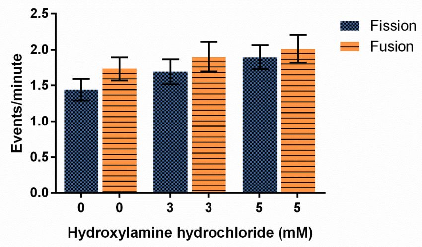
Cells 2021, 10, 2147                                                                                           2 of 14

                       serves to maximize the oxidative capacity and to regulate the performance of individual
                       mitochondria. Fission is another quality control process that occurs in equilibrium with
                       fusion in which mitochondria split in order to create new mitochondria as well as to remove
                       damaged fragments that are inhibiting the function of the organelle [16]. Microtubules
                       are critical structures for these processes as well as being important in mitochondrial
                       motility [10].
                             Mitochondrial motility is the ability of mitochondria to move within a cell and is often
                       facilitated by cytoskeleton components including intermediate filaments, actin filaments,
                       motor proteins, and in particular microtubules [6]. This movement is crucial to cellular
                       processes, such as cell cycle progression and apoptosis that require the proper localization
                       of mitochondria. Motility demands are especially high in elongated and polarized cells,
                       including animal neurons, due to the length of their structure and specialized function [6].
                             Disruptions in mitochondrial fission, fusion, and motility have been implicated in
                       the pathogenesis of multiple neurodegenerative diseases, including Parkinson’s disease,
                       Alzheimer’s disease, Huntington’s disease, and Charcot–Marie–Tooth disease [5–7]. Al-
                       though the production of ATP is the primary function of mitochondria, they are also
                       involved extensively in a myriad of other cellular functions. Mitochondria associated with
                       the endoplasmic reticulum are involved in calcium homeostasis which in turn can initiate
                       apoptosis or improve oxidative metabolism [5]. Other mitochondria located within the
                       pulmonary arterial system function as oxygen sensors by altering the production of ROS in
                       response to fluctuating cellular oxygen levels which facilitates the redox regulation of ion
                       channels, enzymes, and transcription factors. Defects in these complex pathways result in
                       decreased metabolic efficiency and increased oxidative stress which are the hallmarks of
                       diseased neurological cells [5].
                             The cytoskeleton, in particular the microtubules, play a key role in mitochondrial
                       dynamics in D. discoideum. The disruption of microtubules has been shown to result in a
                       drastic decrease in mitochondrial motility as well as fission and fusion events [10]. Recent
                       work by our lab has also shown that rotenone (an inducer of Parkinson’s disease) increases
                       mitochondrial velocity, inhibits fusion, increases ROS, and disrupts microtubule struc-
                       tures [11]. ROS are produced as byproducts of cellular metabolism and include molecules
                       derived from molecular oxygen that include hydrogen peroxide (H2 O2 ), superoxide anions
                       (O2 − ), and hydroxyl radicals (− OH). These molecules play a role in apoptosis, immuno-
                       logical functions, and autophagy. If the production and elimination of ROS falls out of
                       equilibrium, ROS accumulation and neurodegeneration can occur [7,17]. ROS are typically
                       produced via complex I and complex III of the electron transport chain and if not dealt with,
                       can lead to mitochondrial dysfunction, resulting in the neurological symptoms common
                       within neurodegenerative diseases [18,19].
                             For this project, three likely ROS inducers were chosen and their effects on ROS
                       production, mitochondrial dynamics and cytoskeleton stability in D. discoideum analyzed.
                       Here, we chose to look specifically at the relationship between ROS and mitochondrial
                       dynamics to determine whether mitochondrial dynamics are identically altered across
                       different types of ROS inducers and whether the cytoskeleton is always involved in these
                       changes. By using a simple model to study mitochondrial dynamics that clearly are
                       associated with neurodegeneration and ROS imbalance [5,20,21], we can contribute to
                       a greater understanding of the mechanisms behind the degradation of neurons. Finally,
                       little work has been performed in D. discoideum studying the effects of ROS, and the
                       ROS inducers presented here are ones that induce detectable ROS by dihydroethidium
                       (DHE). Cumene hydroperoxide is an organic molecule and known oxidizing agent that
                       affects complex III of the electron transport chain, thus decreasing ATP concentrations
                       and mitochondrial membrane potential [22,23]. Hydroxylamine hydrochloride (HA) is
                       an organic crystalline solid used in organic syntheses and photographic development. It
                       was selected for this study due to its known ability to inhibit catalase thereby impeding
                       hydrogen peroxide decomposition within the cell, which in turn increases the likelihood
                       of oxidative damage to cellular components [24,25]. Antimycin A (AMA) is a known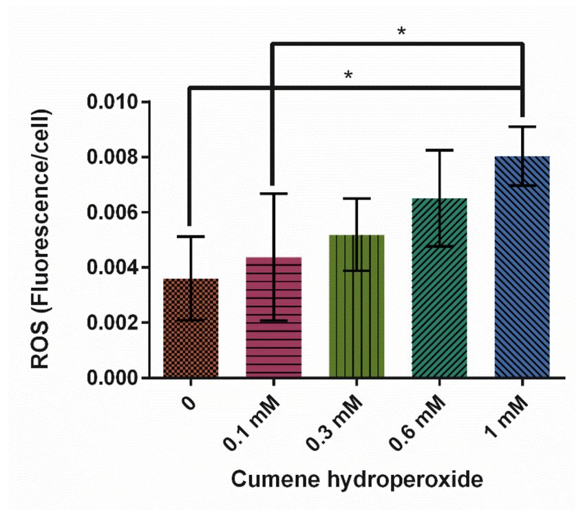
Cells 2021, 10, 2147                                                                                         3 of 14

                       inducer of ROS in human pulmonary fibroblasts and functions by inhibiting succinate and
                       NADH oxidases as well as by binding to complex III of the electron transport chain [26,27].
                       The inhibition of complex III results in disruption of the proton gradient across the inner
                       mitochondrial membrane, leading to an overproduction of ROS within the cell [28,29].
                            Mitochondria possess highly efficient antioxidant systems which work to neutralize
                       excess ROS production and prevent accumulation that can result in increased oxidative
                       stress. When these systems are disrupted by environmental toxins, mutations, or cellular
                       aging, the resulting oxidative stress leads to an alteration in mitochondrial redox home-
                       ostasis [30]. Long-lived non-mitotic cells such as neurons are particularly susceptible to
                       irreparable damage as a result of ROS accumulation. This accumulation has been shown to
                       disrupt redox homeostasis within these cells, which has been linked to the pathogenesis of
                       neurodegenerative diseases such as Alzheimer’s disease and Parkinson’s disease [30,31].
                       In this work, we build on the results of Chernivec et al., 2018 and analyze the relationship
                       between ROS, mitochondrial dynamics and microtubule structure [11].

                       2. Materials and Methods
                       2.1. Strain Growth
                            The AX4 (wild-type) strain of Dictyostelium discoideum was obtained from Bill Loomis
                       via Dicty Stock Center (www.dictybase.com (accessed on 12 August 2021)). AX4 cells were
                       cultured in liquid HL5 media [32,33] containing streptomycin (300 µg/mL) and ampicillin
                       (150 µg/mL) at 22 ◦ C with shaking at 125 rpm.

                       2.2. ROS Induction
                            Log-phase cells in HL5 were exposed to varying concentrations of cumene hydroper-
                       oxide (0–1.5 mM) for 30 min, hydroxylamine hydrochloride (HA) (0–5 mM for) for 1 h, or
                       Antimycin A (AMA) (0–50 µM) for 1 h with shaking at 22 ◦ C, then cells underwent ROS
                       measurement, fixation for immunofluorescence, or confocal imaging. The vehicle control
                       for cumene and AMA was ethanol, and the vehicle for HA was water.

                       2.3. ROS Assay
                             Log-phase cells were pelleted and resuspended in SS6.4 Buffer Solution to
                       6 × 10⁶ cells/mL for cumene assays or 2.68 × 10⁶ to 2.75 × 10⁶ cells/mL for HA and
                       AMA assays. Fluorescence values were normalized to cell number for each assay. A
                       volume of 50 µL of treated cells was added to appropriate wells of a black 96-well plate
                       in triplicate. A volume of 50 µL of 60 µM dihydroethidium (DHE) (Invitrogen, Thermo
                       Fisher Scientific, Grand Island, NY, USA) was added to each well and fluorescence was
                       measured using BioTek’s Synergy H1. Cells were excited at 522 nm and emission was
                       measured at 605 nm for 10 min, with a reading taking place every 30 s. Cumene readings
                       over the 10 min were averaged, then a mean for the triplicate readings was calculated. For
                       HA and AMA, only the first triplicate readings were averaged. Assays were carried out a
                       minimum of 5 times. Statistical analysis was performed using GraphPad Prism version
                       6.07 for Windows (GraphPad Software, La Jolla, CA, USA, www.graphpad.com (accessed
                       on 12 August 2021)). One-way ANOVA with Tukey’s post hoc was used.

                       2.4. Live-Cell Imaging
                             Log-phase cells were washed one time with Lo-Flo media (Formedium, Hunstanton,
                       Norfolk, UK), resuspending to original volume in Lo-Flo. Mitotracker CMXRos (Invitrogen,
                       Thermo Fisher Scientific, Grand Island, NY, USA) at a final concentration of 0.1 µM was
                       added and cells were incubated for 4 h at 22 ◦ C with shaking at 125 rpm. Cells were
                       washed 3 times with Lo-Flo and placed in Nunc Lab-TekII 4-well chambered cover glass
                       for imaging. Cells were treated with 0, 0.3 or 0.6 mM cumene for 30 min prior to imaging,
                       0, 3, or 5 mM HA for 60 min prior to imaging, or 0, 30, or 50 µM AMA 60 min prior to
                       imaging. Cells were imaged with Nikon A1R confocal on a Ti2-E inverted microscope
                       (Nikon, Melville, NY, USA). Live-cell imaging settings included resonance scanning of a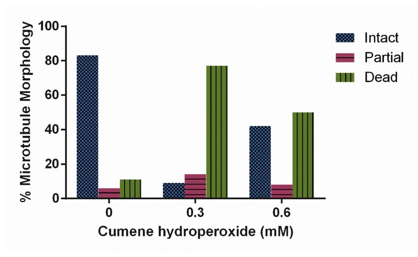
Cells 2021, 10, 2147                                                                                         4 of 14

                       single section with a pinhole of 45.98, and movies were obtained for 2 min with 0.065 s
                       timepoints. Cells were imaged on a minimum of 3–4 separate occasions with approximately
                       5 time-lapse images obtained each time.

                       2.5. Quantification of Fission and Fusion
                             Image analysis of fission and fusion was performed using NIS Elements AR 5.20.02
                       (Nikon, Melville, NY USA) similarly to past work [9–11,34]. Fission was determined by a
                       single organelle splitting into two organelles. Fusion was quantified by two mitochondria
                       nearing each other for multiple frames and then fusing into one organelle. The rates of
                       fission and fusion were determined by calculating the average number of events/min./cell.
                       Images were denoised and ≥30 cells were analyzed. The Kruskal–Wallis test with Dunn’s
                       post hoc, at an alpha level of 0.05 statistical analysis, was performed using GraphPad Prism
                       version 6.07 for Windows (GraphPad Software, La Jolla, CA, USA, www.graphpad.com
                       (accessed on 12 August 2021)).

                       2.6. Quantification of Motility
                            Using NIS Elements AR 5.20.02 (Nikon, Melville, NY USA) live-cell images were
                       denoised and then kymographs by line were created, similarly to past work [10,11,34]. A
                       curvilinear line was drawn across three vertical areas of a cell, ensuring a minimum of
                       3 mitochondria were captured. On the kymograph, a simple line was drawn across
                       every visible mitochondria until direction changed and then a new line was drawn
                       (50–460 lines/measurements per kymograph). The slope of each line is the velocity of
                       movement. A total of 30 cells across 3–4 imaging experiments were analyzed, and slopes
                       were averaged for each cell, ignoring zero slopes. The velocity in µm/s for each cell
                       was then averaged across the 30 cells. To calculate % motility, the number of velocities
                       above 0 µm/s was divided by the total number of velocities measured. Thus, % motil-
                       ity was not calculated per mitochondria but essentially the number of times organelles
                       were not moving. To determine differences in velocity, GraphPad Prism version 6.07
                       for Windows (GraphPad Software, La Jolla, CA, USA, www.graphpad.com (accessed on
                       12 August 2021)), the Kruskal–Wallis test with Dunn’s post hoc test or one-way ANOVA
                       with Tukey’s post hoc test was performed depending on whether the data were parametric.
                       An alpha level of 0.05 was used.

                       2.7. Quantification of Microtubule Stability
                             Immunofluorescence imaging was utilized to determine the morphology of micro-
                       tubules. Log-phase cells were treated with 0, 3, or 5 mM of HA, or 0, 30, or 50 µM for AMA
                       for 30 min shaking at 22 ◦ C (log-phase cells were treated with cumene only on coverslips,
                       see below). Then, 15 mL of cells were washed twice with Lo-Flo and resuspended to
                       4–8 × 10⁶ cells/mL. Further, 0.5 mL of washed cells were added directly to a coverslip in
                       a 6-well plate and incubated for 30 min at room temperature with appropriate treatment
                       (0, 0.3, or 0.6 mM for cumene; 0, 3, or 5 mM for HA, or 0, 30, or 50 µM for AMA). Cells
                       were washed two times with 10 mM MES-NaOH, then fixed with 3% paraformaldehyde
                       pH 6.0 for 30 min. The cells were quenched with 100 mM glycine in 1 × PBS for 5 min
                       then permeabilized with 0.02% Triton X-100 for 5 min. After three quick 1 × PBS washes,
                       cells were incubated in 0.045% fish gelatin and 0.5% BSA in 1 × PBS (PBG) for 1 h at room
                       temperature. Mouse anti-tubulin (12G10, Developmental Studies Hybridoma Bank, Iowa
                       City, IA, USA) diluted 1:150 in PBG solution was added to the coverslips and incubated
                       at 4 ◦ C 12–18 h. Three 5 min washes with 1 × PBS were performed and the secondary
                       antibody was added, AlexaFluor 488 goat alpha mouse IgG, (Life Tech, A11001, Thermo
                       Fisher Scientific Grand Island, NY, USA) 1:250 in PBG solution for 1 h at 4 ◦ C. The cover-
                       slips were kept in the dark from this point forward. Three final 5 min washes in 1 × PBS
                       were performed then coverslips were mounted to glass slides with SlowFade Diamond
                       (Invitrogen, Thermo Fisher Scientific Grand Island, NY, USA) and stored in the dark at
                       4 ◦ C. Immunofluorescence imaging was performed on a Nikon A1R confocal on a Ti2-E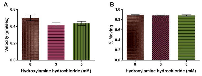
Cells 2021, 10, 2147                                                                                                 5 of 14

                       inverted microscope (Nikon, Melville, NY, USA), settings included Galvano scanning with
                       a pinhole of 26.82. A total of 61 z-sections, 0.1 um thick, were obtained. Image analysis
                       was performed using NIS Elements AR 5.20.02 (Nikon, Melville, NY USA). Microtubules
                       were categorized into intact, partial, spot, or dead based on microtubule morphology for
                       at least 50 cells per treatment by two independent researchers (Figure 1). Intact refers
                       to a complete microtubule complex extending throughout the cell, partial means less a
                       complete complex and shorter microtubule filaments that do not reach periphery of the
                       cell, spot indicates only the center of the microtubule organizing center is present with
                       no radiating tubules, and dead represents cells where microtubules were depolymerized
                       due to cellular death prior to fixation. Statistical analysis was performed using Chi Square
                       analysis in GraphPad Prism version 6.07 for Windows (GraphPad Software, La Jolla, CA,
                       USA, www.graphpad.com (accessed on 12 August 2021)).

                       Figure 1. Microtubule Morphologies. (A) Fully intact microtubule structures with numerous tubules
                       reaching toward the plasma membrane are characterized as intact. (B) A cytoskeleton that demon-
                       strates an astral shape but does not extend to the plasma membrane is designated as partial. (C) Spot
                       morphologies are where only the microtubule organizing center is present and (D) dead cells are
                       when there is no detectable fluorescence (as shown here) or the plasma membrane is obviously no
                       longer intact (data not shown). Scale bar is 5 µM.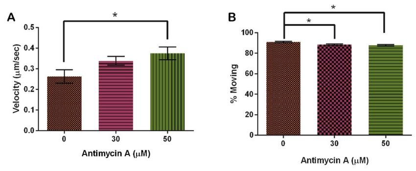
Cells 2021, 10, 2147                                                                                               6 of 14

                       3. Results
                       3.1. Cumene Hydroperoxide Decreases Mitochondrial Velocity and Disrupts the Cytoskeleton
                            Cumene hydroperoxide has been used in previous studies as a positive ROS control
                       and to induce oxidative stress in Dictyostelium discoideum [11,35,36]. In this study, cumene
                       was shown to significantly increase ROS production (Figure 2, p = 0.0116). A total of
                       1 mM cumene induced the highest levels of ROS and was on average 123% higher than the
                       0 mM control.

                       Figure 2. Effect of cumene hydroperoxide on average reactive oxygen species (ROS) levels in D.
                       discoideum ± SE (N = 5). Cumene significantly increased ROS levels as indicated by * (p = 0.0116).

                             Immunofluorescence analysis was used to determine microtubule morphology within
                       cells treated with 0, 0.3 and 0.6 mM concentrations of cumene hydroperoxide. Increasing
                       cumene concentration significantly decreased the stability of microtubules in D. discoideum
                       (Figure 3, p < 0.0001). A total of 88% of the untreated cell’s microtubule structures were
                       intact in contrast to only 21% of 0.3 mM and 53% of 0.6 mM cumene-treated cells (Figure 3).
                       Cumene increased AX4 mortality at all concentrations tested—0 mM yielded 10% dead
                       cells, 0.3 mM 28%, and 0.6 mM 39%.

                       Figure 3. Effect of cumene hydroperoxide on microtubule morphology in D. discoideum. Cumene
                       significantly decreased microtubule stability (n > 50 cells, p < 0.0001).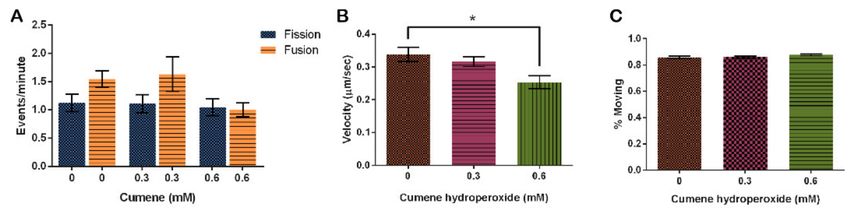
Cells 2021, 10, 2147                                                                                                               7 of 14

                                        Cumene hydroperoxide did not have a significant effect on the rate of mitochondrial
                                  fission and fusion in this study. However, fusion rates did trend higher at the control and
                                  0.3 mM concentrations (Figure 4A, p = 0.0896). Fission rates remained relatively stable
                                  throughout all concentrations tested, while fusion at the 0.6 mM concentration decreased
                                  to match fission (Figure 4A).

      Figure 4. Effect of cumene hydroperoxide on mitochondrial dynamics. (A) Fusion trends faster in vehicle control and
      0.3 mM cumene, but cumene does not significantly alter fission and fusion (n = 30 cells, p = 0.0896). (B) Cumene significantly
      decreases mitochondrial velocity but (C) has no effect on the number of mitochondria moving (n = 30 cells, p= 0.0062 and
      0.1669, respectively). Columns are averages, error bars represent the standard error and * indicates significance.

                                       Finally, we analyzed the effect of cumene on mitochondrial motility. Mitochondria on
                                  average move 0.34 ± 0.02 µm/s when treated with the vehicle control, EtOH. This speed
                                  decreased with the addition of cumene (Figure 4B, p = 0.0062), decreasing by 6.3% with
                                  0.3 mM cumene and 36% with 0.6 mM cumene from the control (Figure 4B). Though the
                                  rate significantly slows down, the percentage of mitochondria actually moving remained
                                  the same (Figure 4C, p = 0.1669).
                                       In summary, cumene increased ROS in D. dictyostelium, destabilized the microtubules,
                                  has no significant effect on mitochondrial fission, fusion, and percent motility, but did
                                  significantly decrease the velocity at which the mitochondria move.

                                  3.2. Hydroxylamine Hydrochloride Does Not Significantly Alter Dynamics
                                       To determine the effect of hydroxylamine hydrochloride in our model system on
                                  ROS levels, we again used DHE to detect ROS. Our results indicate that ROS increased
                                  at 3 mM HA by 23% and 4 mM HA by 44% though these increases were not significant
                                  (p = 0.1320). At 5 mM, ROS decreased similarly to the untreated control (Figure 5A).
                                  Analysis of microtubule structure in cells treated with HA indicated that HA had no effect
                                  on microtubules (Figure 5B, p = 0.9495).

                                  Figure 5. Effect of hydroxylamine hydrochloride on in vivo reactive oxygen species production
                                  and microtubule stability in D. discoideum. (A) HA increases ROS at 3 and 4 mM but this is not
                                  significant (N = 5, p = 0.3120). (B) HA has no effect on microtubule morphology in D. discoideum
                                  (n > 50, p = 0.9495). Columns are averages and error bars represent the standard error.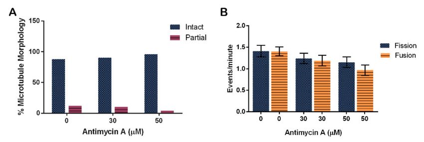
Cells 2021, 10, 2147 8 of 14
Hydroxylamine hydrochloride also had no significant effect on fission and fusion fre-
quencies at the concentrations tested, though fission and fusion events did trend upwards
as HA concentrations increased with the highest rates of both fission and fusion occurring
at the 5 mM concentration (Figure 6, p = 0.2450).
Figure 6. Effect of hydroxylamine hydrochloride on mitochondrial fission and fusion frequency in D.
discoideum. Increasing HA does not significantly alter average ± SE fission and fusion events, though
these rates trend faster with more HA (n = 34 cells for each treatment, p = 0.2450).
Finally, we show that HA had no effect on the amount of mitochondria moving at
any one time (Figure 7B, p = 0.1519). The 3 mM HA concentration decreased the rate of
mitochondria movement by 22% from the vehicle control and 5 mM HA concentration
decreased by 30% from the control, but it is not significant (Figure 7A, p = 0.2670).
Figure 7. Effect of hydroxylamine hydrochloride on mitochondrial motility. (A) Increasing HA does
not significantly alter velocity though it decreases slightly with increasing concentrations of HA.
(B) Increasing HA does not alter the percentage of mitochondria moving (n = 30, p = 0.2670 and
0.1519, respectively). Columns are averages and error bars represent the standard error.
The conclusion of HA treatments on D. discoideum cells is that HA did not signifi-
cantly alter any measured effects in this study. Trends indicate that HA increased ROS,
increased fission and fusion, decreased mitochondrial velocity, and showed no changes to
microtubule structure or percentage of mitochondria moving.
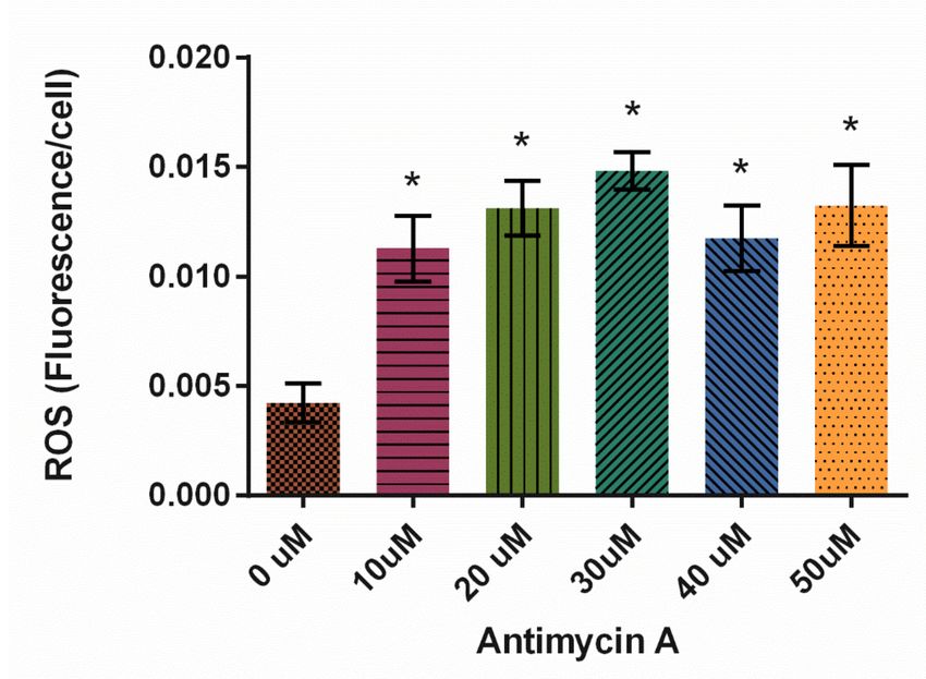
3.3. Antimycin a Decreases Fission and Fusion but Increases Mitochondrial Velocity
In this study, Antimycin A significantly increased ROS production at all concentrations
tested, with 30 µM resulting in the highest levels of ROS (Figure 8, p < 0.0001).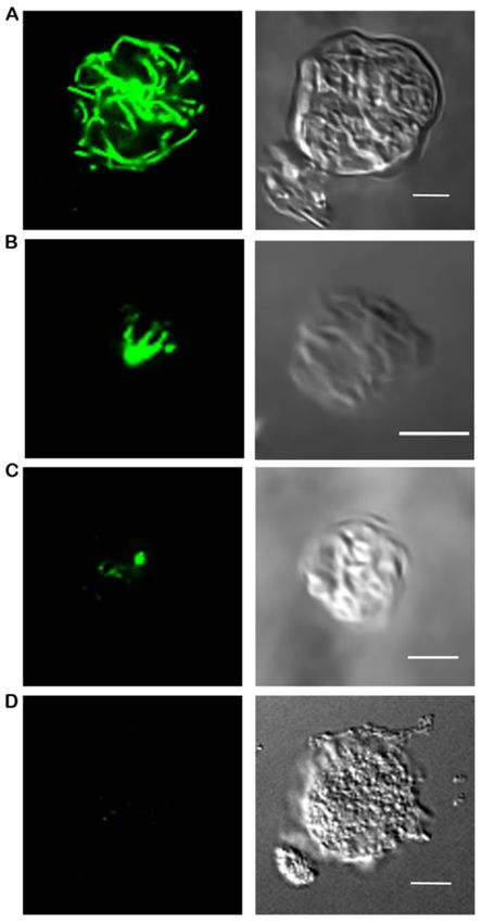
Cells 2021, 10, 2147                                                                                                             9 of 14

                                  Figure 8. Effect of Antimycin A on average reactive oxygen species (ROS) levels in D. discoideum ± SE
                                  (N = 6). AMA significantly increased ROS levels compared to control, as indicated by * (p < 0.0001).

                                        Analysis of microtubule structure in the presence of AMA using immunofluorescence
                                  showed that AMA had no effect on tubulin structures (Figure 9A, p = 0.2846). The trend
                                  suggests that perhaps AMA stabilizes microtubule structures. Quantification determined
                                  that 88% of control cells, 90% of 30 µM AMA-treated cells, and 96% of 50 µM AMA-treated
                                  cells have intact microtubule structure.

      Figure 9. Effect of Antimycin A on microtubule morphology and mitochondrial fission and fusion in D. discoideum.
      (A) AMA did not significantly affect microtubule stability (n > 50 cells, p = 0.2846), nor (B) fission and fusion rates
      (n = 46 cells, p = 0.0516), though these rates trend faster with less AMA. Columns are averages and error bars represent the
      standard error.

                                        Fission and fusion events per minute were quantified in cells at different concen-
                                  trations of AMA. There were on average 1.4 ± 0.2 fission and 1.4 ± 0.1 fusion events in
                                  cells treated with the vehicle control EtOH. Fission rates decreased by 17% for the 30 µM
                                  treatment and by 26% for the 50 µM treatment when compared to control rates. Fusion rates
                                  also decreased in the 30 and 50 µM treatments by 21% and 44%, respectively (Figure 9B,
                                  p = 0.0516).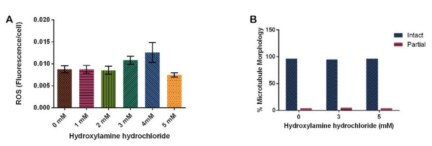
Cells 2021, 10, 2147                                                                                                              10 of 14

                                       Analysis of motility in the presence of AMA indicated that velocity increased with
                                  rising AMA concentration. In the vehicle control, mitochondria moved 0.26 ± 0.03 µm/s, in-
                                  creasing by 24% with 30 µM and 29% with 60 µM AMA from the control group (p = 0.0436)
                                  (Figure 10A). Interestingly, increasing AMA decreased the amount of movement of mito-
                                  chondria even though velocity increased (p = 0.0041) (Figure 10B).

      Figure 10. Effect of Antimycin A on mitochondrial motility. (A) AMA significantly increases mitochondrial velocity and
      (B) has a significant effect on the number of mitochondria moving (n = 30 cells, p = 0.0436 and 0.0041, respectively). Columns
      are averages, error bars represent the standard error and * indicates significance.

                                       Finally, AMA has no effect on microtubule organization similar to HA. However,
                                  it decreased fission and fusion rates while simultaneously increasing organelle velocity.
                                  AMA was significantly increased ROS levels at every concentration tested suggesting it’s
                                  potential to efficiently induce ROS production in future D. discoideum studies.

                                  4. Discussion
                                        In our study, all three treatments, cumene, HA, and AMA, induced ROS but only
                                  cumene and AMA did so significantly (Figures 2, 5A and 8). Cumene is the only chemical
                                  tested that significantly affected microtubule morphologies (Figures 3, 5B and 9A). No
                                  ROS inducer significantly altered mitochondrial fission and fusion but all three did alter
                                  these dynamics slightly. Cumene decreased fusion from the control but did not alter fission
                                  rates, HA increased both fission and fusion, while AMA decreased the number of fission
                                  and fusion events (Figures 4A, 6 and 9B). These chemicals alter mitochondrial motility in
                                  different manners as well. Cumene and HA decreased the speed at which the mitochondria
                                  move while AMA increased mitochondrial velocity and decreased the percent of organelles
                                  moving (Figure 4B,C, Figures 7 and 10). This work indicates that the presence of ROS alone
                                  does not alter mitochondrial dynamics and therefore either the type of ROS, the additional
                                  effects caused by the ROS inducing chemicals, or the method of ROS creation appears to
                                  play a role in altering mitochondrial dynamics.
                                        Cumene is known to inhibit mitochondrial respiration and alters the function of com-
                                  plex III of the electron transport chain, resulting in superoxide formation [22,23]. Cumene
                                  does not appear to alter complex I directly but in conjunction with inhibition of complex
                                  III, complex I activity is altered [22]. Cumene likely also increases ROS through mecha-
                                  nisms not requiring oxygen [22]. Hydroxylamine hydrochloride causes ROS generation
                                  by inhibiting catalase enzymes, resulting in an increase in H2 O2 [25,37,38]. Antimycin
                                  A also inhibits complex III, specifically through the ubiquinol oxidation center as well
                                  as inhibiting the mitochondrial multiple conductance channel, resulting in superoxide
                                  formation [19,26,27,29].
Cells 2021, 10, 2147 11 of 14
Looking at the results from this study, we can see that inhibition of complex III by
cumene or AMA does not result in a similar effect on mitochondrial dynamics. Cumene
decreased fusion and decreased mitochondrial velocity while AMA slightly decreased both
fission and fusion and significantly increased velocity. While HA increases ROS through the
inhibition of catalase rather than the electron transport chain, it does increase mitochondrial
fission and fusion, the opposite of cumene and AMA, but there is no obvious pattern in
terms of mitochondrial velocity Thus, we suggest that ROS generation via inhibition of
the electron transport chain decreases fission and fusion but does not predict the effect on
mitochondrial velocity, supported by Chernivec et al., as described below [11].
What about the effect on the cytoskeletal structure? It is well known that rotenone
induces ROS and alters microtubule organization [39,40]. Rotenone inhibits complex I
activity of the electron transport chain creating superoxides and H2 O2 [18,41]. Recently we
published the results of rotenone on D. discoideum dynamics [11]. We showed that rotenone
disrupts the D. discoideum tubulin cytoskeleton, which cannot be returned to normal by the
addition of reducing agents such as vitamin C [11]. Rotenone decreases fusion, which can
be reversed by the addition of vitamin C, and it increases mitochondrial velocity with no
effect on the number of mitochondria moving [11]. Here, we compare these results with the
current study to identify the relationship between mitochondrial dynamics, microtubules
and ROS.
ROS inducers that disrupt the D. discoideum microtubule cytoskeleton are rotenone
and cumene, while AMA slightly stabilizes. Cumene, AMA and rotenone alter fission
and fusion similarly, while cumene decreases velocity when rotenone and AMA increase
it. This suggests that cytoskeletal disruption, which is known to alter mitochondrial
dynamics [10] is not the only factor at play leading to these effects. We conclude that
ROS alters mitochondrial dynamics, and these alterations do not require a change in
microtubule organization.
So why the various alterations to mitochondrial dynamics? Why do they not cause
the exact same effect across all of the inducers? ROS inducers do not just generate ROS.
For example, rotenone has clearly been shown to bind to tubulin [40,42]. Cumene de-
creases cytosolic calcium levels and disrupts membranes [23]. In general, ROS function
as signaling molecules sometimes directly inhibiting complex I of the electron transport
chain [43–45], inducing transcription factors such as Nf-κB and HIF-1, as well as increasing
antioxidant activity and regulating many other proteins. In terms of mitochondria, ROS
have been shown to induce additional fragmentation of mammalian cells with Mfn2 mu-
tations significantly altering mitochondrial quality control [46], it is also clear that intact
mitochondrial structure is essential to regulate ROS imbalances [20]. Here, we suggest that
while it appears that ROS generated from the electron transport chain generally decrease
fission and fusion, the alteration of dynamics is dependent upon which mitochondrial
dynamic is affected first by the ROS generator. For example, if fusion is increased, then the
larger organelles might move slower, thus we see velocity is decreased as seen for HA or
vice versa with rotenone and AMA. Or if velocity increases, it is logical to assume that the
mitochondria are moving past each other too fast for fission and fusion to properly take
place such as with AMA. Mammalian studies indicate that alterations of calcium levels
will alter mitochondrial dynamics and an increase in fusion will increase ATP synthesis, a
possible mechanism to overcome a decrease in ATP due to excessive ROS [27]. Additionally,
it is known that fission and fusion are used to repair and maintain mitochondrial quality,
thus changes to fission and fusion occur in response to ROS damage. Finally, it is important
to note that D. discoideum cells have a high resistance to oxidative stress [25], and therefore
all the aforementioned reasons may be occurring simultaneously, resulting in the various
responses by mitochondrial dynamics in D. discoideum.
5. Conclusions
This study has shed light on the ROS-producing capabilities of a variety of chemicals
on D. discoideum. Finding reliable ROS-stimulating compounds within model organisms isCells 2021, 10, 2147 12 of 14
critical to furthering research on neurodegenerative diseases related to cellular oxidative
stress, such as seeking to mitigate the effects of ROS accumulation on a cellular level.
Additionally, we show the effect of these various ROS generators on mitochondrial
dynamics. Our work demonstrates that alteration of the microtubule cytoskeleton is not
required to alter dynamics in response to ROS and it appears that ROS generated from
the electron transport chain decrease fission and fusion. By understanding that different
ROS alter mitochondrial dynamics in different ways, we know that more research is
needed to better understand the effects of ROS on the cellular mechanisms that induce
the pathogenesis of incurable neurodegenerative diseases. Further research in this field
is needed with the hope that it will someday translate into developing new and more
effective treatments for patients afflicted by neurodegenerative diseases.
Author Contributions: Conceptualization, E.D.; methodology, K.N.; formal analysis, K.N.; inves-
tigation, E.D. and A.D.B.; resources, K.N.; data curation, E.D., A.D.B. and K.N.; writing—original
draft preparation, E.D.; writing—review and editing, E.D., A.D.B. and K.N.; visualization, K.N.;
supervision, K.N.; project administration, K.N.; funding acquisition, K.N. All authors have read and
agreed to the published version of the manuscript.
Funding: This research was funded by the University of Central Arkansas and the Biology Depart-
ment. This publication was made possible by the Arkansas INBRE program, supported by a grant
from the National Institute of General Medical Sciences, (NIGMS), P20GM103429 from the National
Institutes of Health.
Data Availability Statement: Data are contained within the article.
Acknowledgments: The authors would like to thank UCA’s College of Natural Sciences and Mathe-
matics for the purchase of access to the Nikon AR1 confocal microscope.
Conflicts of Interest: The funders had no role in the design of the study; in the collection, analyses,
or interpretation of data; in the writing of the manuscript, or in the decision to publish the results.
References
1. Marras, C.; Beck, J.C.; Bower, J.H.; Roberts, E.; Ritz, B.; Ross, G.W.; Abbott, R.D.; Savica, R.; Van Den Eeden, S.K.; Willis, A.W.;
et al. Prevalence of Parkinson’s disease across North America. Npj Parkinsons Dis. 2018, 4, 21. [CrossRef]
2. MacAskill, A.F.; Kittler, J.T. Control of mitochondrial transport and localization in neurons. Trends Cell Biol. 2010, 20, 102–112.
[CrossRef]
3. Itoh, K.; Nakamura, K.; Iijima, M.; Sesaki, H. Mitochondrial dynamics in neurodegeneration. Trends Cell Biol. 2013, 23, 64–71.
[CrossRef] [PubMed]
4. Chen, H.; Chan, D. Mitochondrial dynamics—Fusion, fission, movement, and mitophagy—In neurodegenerative diseases. Hum.
Mol. Genet. 2009, 18, R169–R176. [CrossRef] [PubMed]
5. Archer, S.L. Mitochondrial dynamics—Mitochondrial fission and fusion in human diseases. N. Engl. J. Med. 2013, 369, 2236–2251.
[CrossRef]
6. Wu, M.; Kalyanasundaram, A.; Zhu, J. Structural and biomechanical basis of mitochondrial movement in eukaryotic cells. Int. J.
Nanomed. 2013, 8, 4033–4042. [CrossRef]
7. Nunnari, J.; Suomalainen, A. Mitochondria: In Sickness and in Health. Cell 2012, 148, 1145–1159. [CrossRef]
8. Singh, A.; Kukreti, R.; Saso, L.; Kukreti, S. Oxidative Stress: A Key Modulator in Neurodegenerative Diseases. Molecules 2019, 24,
1583. [CrossRef]
9. Schimmel, B.; Berbusse, G.; Naylor, K. Mitochondrial fission and fusion in Dictyostelium discoideum: A search for proteins involved
in membrane dynamics. BMC Res. Notes 2012, 5, 505. [CrossRef]
10. Woods, L.C.; Berbusse, G.W.; Naylor, K. Microtubules Are Essential for Mitochondrial Dynamics—Fission, Fusion, and Motility—
In Dictyostelium discoideum. Front. Cell Dev. Biol. 2016, 4, 19. [CrossRef]
11. Chernivec, E.; Cooper, J.; Naylor, K. Exploring the Effect of Rotenone—A Known Inducer of Parkinson’s Disease—On Mitochon-
drial Dynamics in Dictyostelium discoideum. Cells 2018, 7, 201. [CrossRef]
12. Annesley, S.; Fisher, P. Dictyostelium discoideum—A model for many reasons. Mol. Cell. Biochem. 2009, 329, 73–91. [CrossRef]
[PubMed]
13. Francione, L.M.; Annesley, S.J.; Carilla-Latorre, S.; Escalante, R.; Fisher, P.R. The Dictyostelium model for mitochondrial disease.
Semin. Cell Dev. Biol. 2011, 22, 120–130. [CrossRef]
14. Pearce, X.G.; Annesley, S.J.; Fisher, P.R. The Dictyostelium model for mitochondrial biology and disease. Int. J. Dev. Biol. 2019, 63,
497–508. [CrossRef]Cells 2021, 10, 2147 13 of 14
15. Nunnari, J.; Marshall, W.; Straight, A.; Murray, A.; Sedat, J.; Walter, P. Mitochondrial transmission during mating in S. cerevisiae
is determined by mitochondrial fusion and fission and the intramitochondrial segregation of mtDNA. Mol. Biol. Cell 1997, 8,
1233–1242. [CrossRef]
16. Youle, R.J.; van der Bliek, A.M. Mitochondrial Fission, Fusion, and Stress. Science 2012, 337, 1062–1065. [CrossRef]
17. Weng, M.; Xie, X.; Liu, C.; Lim, K.L.; Zhang, C.W.; Li, L. The Sources of Reactive Oxygen Species and Its Possible Role in the
Pathogenesis of Parkinson’s Disease. Parkinson’s Dis. 2018, 2018, 9163040. [CrossRef]
18. Liu, Y.; Fiskum, G.; Schubert, D. Generation of reactive oxygen species by the mitochondrial electron transport chain. J. Neurochem.
2002, 80, 780–787. [CrossRef]
19. Bleier, L.; Dröse, S. Superoxide generation by complex III: From mechanistic rationales to functional consequences. Biochim.
Biophys. Acta 2013, 1827, 1320–1331. [CrossRef]
20. Lin, T.-K.; Lin, K.-J.; Lin, K.-L.; Liou, C.-W.; Chen, S.-D.; Chuang, Y.-C.; Wang, P.-W.; Chuang, J.-H.; Wang, T.-J. When Friendship
Turns Sour: Effective Communication Between Mitochondria and Intracellular Organelles in Parkinson’s Disease. Front. Cell Dev.
Biol. 2020, 8, 607392. [CrossRef]
21. Berbusse, G. Investigating the Importance of Mitochondrial Dynamics: Fission, Fusion, Trafficking and Fragmentation in the
Single-Celled Organism Dictyostelium discoideum and CD-1 Mice In Vitro. Master’s Thesis, University of Central Arkansas,
Conway, AR, USA, 2014.
22. Sanz, A.; Caro, P.; Gómez, J.; Barja, G. Testing the vicious cycle theory of mitochondrial ROS production: Effects of H2O2 and
cumene hydroperoxide treatment on heart mitochondria. J. Bioenerg. Biomembr. 2006, 38, 121–127. [CrossRef]
23. Vimard, F.; Saucet, M.; Nicole, O.; Feuilloley, M.; Duval, D. Toxicity induced by cumene hydroperoxide in PC12 cells: Protective
role of thiol donors. J. Biochem. Mol. Toxicol. 2011, 25, 205–215. [CrossRef]
24. Kono, Y.; Fridovich, I. Inhibition and reactivation of Mn-catalase. Implications for valence changes at the active site manganese. J.
Biol. Chem. 1983, 258, 13646–13648. [CrossRef]
25. Rajawat, J.; Vohra, I.; Mir, H.A.; Gohel, D.; Begum, R. Effect of oxidative stress and involvement of poly(ADP-ribose) polymerase
(PARP) in Dictyostelium discoideum development. FEBS J. 2007, 274, 5611–5618. [CrossRef]
26. Park, W.H.; You, B.R. Antimycin A induces death of the human pulmonary fibroblast cells via ROS increase and GSH depletion.
Int. J. Oncol. 2016, 48, 813–820. [CrossRef] [PubMed]
27. Kim, J.; Cheong, J.H. Role of Mitochondria-Cytoskeleton Interactions in the Regulation of Mitochondrial Structure and Function
in Cancer Stem Cells. Cells 2020, 9, 1691. [CrossRef] [PubMed]
28. Balaban, R.S.; Nemoto, S.; Finkel, T. Mitochondria, oxidants, and aging. Cell 2005, 120, 483–495. [CrossRef]
29. Campo, M.L.; Kinnally, K.W.; Tedeschi, H. The effect of antimycin A on mouse liver inner mitochondrial membrane channel
activity. J. Biol. Chem. 1992, 267, 8123–8127. [CrossRef]
30. Angelova, P.R.; Abramov, A.Y. Role of mitochondrial ROS in the brain: From physiology to neurodegeneration. FEBS Lett. 2018,
592, 692–702. [CrossRef]
31. Gandhi, S.; Abramov, A.Y. Mechanism of Oxidative Stress in Neurodegeneration. Oxidative Med. Cell. Longev. 2012, 2012, 428010.
[CrossRef]
32. Ashworth, J.M.; Watts, D.J. Metabolism of the cellular slime mould Dictyostelium discoideum grown in axenic culture. Biochem. J.
1970, 119, 175–182. [CrossRef]
33. Available online: http://dictybase.org/techniques/media/media.html (accessed on 29 July 2021).
34. Vogel, E.; Pittman, P.B.; Naylor, K. The FtsZ Homolog, FszB, Inhibits Mitochondrial Dynamics in Dictyostelium discoideum. Cells
2019, 9, 64. [CrossRef]
35. Katoch, B.; Begum, R. Biochemical basis of the high resistance to oxidative stress in Dictyostelium discoideum. J. Biosci. 2003, 28,
581–588. [CrossRef]
36. Kim, H.-L.; Choi, Y.K.; Kim, D.H.; Park, S.O.; Han, J.; Park, Y.S. Tetrahydropteridine deficiency impairs mitochondrial function in
Dictyostelium discoideum Ax2. FEBS Lett. 2007, 581, 5430–5434. [CrossRef]
37. Kono, Y.; Fridovich, I. Isolation and characterization of the pseudocatalase of Lactobacillus plantarum. J. Biol. Chem. 1983, 258,
6015–6019. [CrossRef]
38. Lee, J.N.; Dutta, R.K.; Maharjan, Y.; Liu, Z.Q.; Lim, J.Y.; Kim, S.J.; Cho, D.H.; So, H.S.; Choe, S.K.; Park, R. Catalase inhibition
induces pexophagy through ROS accumulation. Biochem. Biophys. Res. Commun. 2018, 501, 696–702. [CrossRef]
39. Alam, M.; Schmidt, W.J. Rotenone destroys dopaminergic neurons and induces parkinsonian symptoms in rats. Behav. Brain Res.
2002, 136, 317–324. [CrossRef]
40. Brinkley, B.R.; Barham, S.S.; Barranco, S.C.; Fuller, G.M. Rotenone inhibition of spindle microtubule assembly in mammalian cells.
Exp. Cell Res. 1974, 85, 41–46. [CrossRef]
41. Sherer, T.B.; Betarbet, R.; Testa, C.M.; Seo, B.B.; Richardson, J.R.; Kim, J.H.; Miller, G.W.; Yagi, T.; Matsuno-Yagi, A.; Greenamyre,
J.T. Mechanism of Toxicity in Rotenone Models of Parkinson’s Disease. J. Neurosci. 2003, 23, 10756–10764. [CrossRef]
42. Feng, J. Microtubule: A Common Target for Parkin and Parkinson’s Disease Toxins. Neurosci. 2006, 12, 469–476. [CrossRef]
43. Chernyak, B.V.; Bernardi, P. The mitochondrial permeability transition pore is modulated by oxidative agents through both
pyridine nucleotides and glutathione at two separate sites. Eur. J. Biochem. 1996, 238, 623–630. [CrossRef]
44. Zamzami, N.; Hirsch, T.; Dallaporta, B.; Petit, P.X.; Kroemer, G. Mitochondrial implication in accidental and programmed cell
death: Apoptosis and necrosis. J. Bioenerg. Biomembr. 1997, 29, 185–193. [CrossRef]Cells 2021, 10, 2147 14 of 14
45. Abou-Sleiman, P.M.; Muqit, M.M.; Wood, N.W. Expanding insights of mitochondrial dysfunction in Parkinson’s disease. Nat. Rev.
Neurosci. 2006, 7, 207–219. [CrossRef]
46. Wolf, C.; Zimmermann, R.; Thaher, O.; Bueno, D.; Wüllner, V.; Schäfer, M.K.E.; Albrecht, P.; Methner, A. The Charcot-Marie Tooth
Disease Mutation R94Q in MFN2 Decreases ATP Production but Increases Mitochondrial Respiration under Conditions of Mild
Oxidative Stress. Cells 2019, 8, 1289. [CrossRef]You can also read