USP18 deficiency in cervical carcinoma is crucial for the malignant behavior of tumor cells in an ERK signal dependent manner
←
→
Page content transcription
If your browser does not render page correctly, please read the page content below
ONCOLOGY LETTERS 21: 421, 2021

                      USP18‑deficiency in cervical carcinoma is
                   crucial for the malignant behavior of tumor cells
                         in an ERK signal‑dependent manner
          AONAN PAN1, YUE LI2, JIAN GUAN3, PENGXIA ZHANG4, CHUNBIN ZHANG4, YUPENG HAN5,
           TAO ZHANG2, YAO CHENG6, LUO SUN6, SHIZHEN LU4, JINRU WENG3, QIAOSHENG REN3,
                         SHENGJIE FAN7, WEIQUN WANG8 and JINGTAO WANG9

 1
  Department of Clinical Medicine, The Affiliated Second Hospital, Harbin Medical University, Harbin, Heilongjiang 150081;
    2
      Departments of Immunology and Etiology, Basic Medical College, Jiamusi University, Jiamusi, Heilongjiang 154007;
       3
        Department of Maxillofacial Surgery, Stomatological College, Jiamusi University, Jiamusi, Heilongjiang 154002;
      4
       Department of Biochemistry and Cell and Molecular Biology, Basic Medical College, Jiamusi University, Jiamusi,
         Heilongjiang 154007; Departments of 5Gastroenterology and 6Clinical Laboratory, The First Affiliated Hospital,
 Jiamusi University, Jiamusi, Heilongjiang 154002; 7Department of Rehabilitation Medicine, Rehabilitation Medical College,
             Jiamusi University, Jiamusi, Heilongjiang 154007; Departments of 8Physiology and 9Human Anatomy,
                     Basic Medical College, Jiamusi University, Jiamusi, Heilongjiang 154007, P.R. China

                                      Received November 14, 2020; Accepted February 25, 2021

                                                        DOI: 10.3892/ol.2021.12682

Abstract. Ubiquitin‑specific peptidase (USP)18 belongs to                 proliferation, colony formation, migration and aggressive-
the USP family, and is involved in cleaving and removing                  ness of HeLa cells. Mechanistic analysis demonstrated that
ubiquitin or ubiquitin‑like molecules from their target                   USP18‑knockdown increased the levels of Bcl‑2, STAT3
molecules. Recently, increasing evidence has suggested                    and phosphorylated‑ERK in HeLa cells. Notably, USP18
that USP18 is constitutively expressed in different types of              silencing‑induced malignant phenotypes were interrupted
human tumors, and ectopic expression or downregulation of                 following exogenous administration of the ERK1/2 inhibitor
USP18 expression may contribute to tumorigenesis. However,                PD98059. Overall, the results of the present study suggested
the role of USP18 in uterine cervical cancer (UCC) remains                that USP18 may be a potent inhibitor involved in UCC
unclear. Thus, the present study aimed to investigate USP18               tumor‑associated biological behaviors, which are associated
expression in a human tissue microarray constructed using                 with the ERK signaling pathway.
UCC and non‑cancer cervical tissues, and to determine the
potential role and molecular mechanism by which USP18                     Introduction
is implicated in the tumor biology of human UCC HeLa
cells. Microarray analysis demonstrated that USP18 protein                Uterine cervical carcinoma (UCC) is one of the most
expression was downregulated in tumor tissues compared                    common types of malignant cancer in women, representing
with in normal tissues. In addition, in vitro analysis                    the fourth most frequent malignancy worldwide (1). In 2018,
revealed that USP18‑knockdown markedly promoted the                       ~570,000 new cases of UCC were identified, and up to
                                                                          311,000 associated deaths were recorded worldwide (2).
                                                                          Epidemiological data have demonstrated that the incidence
                                                                          and mortality rates of UCC vary across different regions, with
Correspondence to: Professor Weiqun Wang, Department of                   more cases in Sub‑Saharan Africa and South‑Eastern Asia,
Physiology, Basic Medical College, Jiamusi University, 258 Xuefu          and less cases in North America, Australia, New Zealand and
Street, Jiamusi, Heilongjiang 154007, P.R. China                          Western Asia (2). Although it has been reported that UCC inci-
E‑mail: wangweiqun1974@163.com                                            dence and mortality rates have decreased in several regions of
                                                                          the world over the last few decades (3), UCC remains a serious
Professor Jingtao Wang, Department of Human Anatomy, Basic
Medical College, Jiamusi University, 258 Xuefu Street, Jiamusi,           health issue in China, with an estimated 106,430 new cases
Heilongjiang 154007, P.R. China                                           and 47,739 mortalities in 2018 (3).
E‑mail: wjingtao@jmsu.edu.cn                                                  Ubiquitin‑specific peptidase (USP)18, also known as
                                                                          ubiquitin‑specific protease 43 (UBP43), is a member of the
Key words: uterine cervical carcinoma, ubiquitin‑specific peptidase 18,   USP family and is involved in deubiquitinating activity,
malignant behavior, ERK                                                   thereby resulting in stabilization of substrates (4). It is well
                                                                          known that USPs are the largest sub‑family of deubiquitinase
                                                                          enzymes, and exert biological roles through their cysteine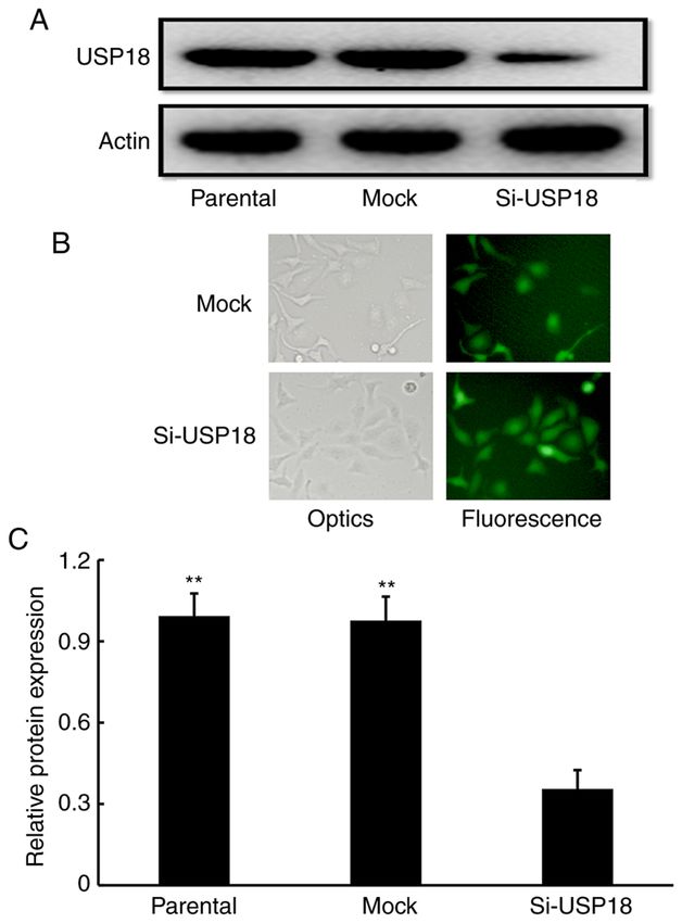
2 PAN et al: USP18 CONTRIBUTES TO THE MALIGNANT BEHAVIOR OF CERVICAL CANCER CELLS
endopeptidase activity (5). The USP family consists of vector‑carrying lentiviruses, according to the manufacturer's
>100 members, which predominantly differ in amino acid protocol, cells were subsequently treated with 2 ng/ml puro-
sequence and protein size, but are characterized by several mycin (Gibco; Thermo Fisher Scientific, Inc.) and transfection
highly homologous sequences around the essential domains efficiency was observed under a fluorescence microscope
important for their catalytic activity (6,7). USP18 was origi- (magnification, x200). Western blot analysis was subsequently
nally identified from acute myelogenous leukemia 1‑ RUNX1 performed to detect USP18 protein expression.
partner transcriptional co‑repressor 1 knock‑in mice and was
characterized by Liu et al (8). Previous studies have demon- Immunohistochemistry (IHC). A commercial cervix tissue
strated that USP18 expression is present in multiple types of microarray containing 15 squamous cell carcinoma tissues,
tissues, including liver, lung, spleen, thymus, bone marrow and five adenosquamous carcinoma tissues, 20 adenocarcinoma
adipose tissues (8,9), and is also expressed in different types tissues, 20 cervical intraepithelial lesions tissues, 14 cervicitis
of cells, such as macrophages, lymphocytes and hematopoi- tissues, four unpaired para‑cancerous tissues and two cervical
etic cells (10,11). The function of USP18 has predominantly canal tissues (cat. no. F801301; Bioaitech Co., Ltd.) was used
been associated with the regulation of cell proliferation, cell to detect USP18 expression. The characteristics of the tissues
differentiation, stress, inflammatory reaction and immune are listed in Table I.
response (9‑11). Additionally, it has been suggested that USP18 The microarray was processed routinely via deparaffiniza-
serves a vital role in regulating T‑cell activation and T helper tion, rehydration, endogenous peroxidase quenching and antigen
17 cell differentiation through its ability to remove the trans- retrieval, and subsequently blocked with 20% normal goat serum
forming growth factor β ‑activated kinase 1‑TAK1‑binding (Wuhan Boster Biological Technology, Ltd.) for 30 min at 37˚C
protein 1 complex (12). to remove the background from staining. The microarray was
Several studies have suggested that USP18 may be involved incubated with a mouse antibody against human USP18 (1:50;
in tumor biology (10,13). USP18 is overexpressed in several cat. no. sc‑374064; Santa Cruz Biotechnology, Inc.) overnight
types of human cancer including glioblastoma, hepatocellular at 4˚C. Following the primary antibody incubation, the micro-
carcinoma, bladder cancer and breast cancer, and its high array was incubated for 35 min at 37˚C using the SABC kit
expression is associated with a poor prognosis in patients with (cat. no. SA1021; Wuhan Boster Biological Technology, Ltd.).
glioblastoma and bladder cancer (14‑17). Furthermore, some DAB and hematoxylin were used for visualization and nuclear
studies have demonstrated that USP18 is important for the counterstaining at room temperature for 30 and 5 sec, respec-
malignant behaviors of tumor cells, including cellular prolif- tively. The USP18 protein expression profiles were estimated
eration, migration, apoptosis and epithelial‑to‑mesenchymal based on the staining intensity and the percentage of positive
transition (EMT) (14,17‑19). Conversely, it has been demon- cells using a fluorescence microscope (magnification, x200). The
strated that USP18 may suppress tumorigenesis due to its staining intensity was ranked as follows: 1, weak; 2, moderate;
involvement in the antitumor immune response (20). 3, intensive; and 4, super intensive. The percentage of positive
Although the biological functions and clinical presenta- cells was scored as follows: 1, 75%. The total score for each sample was the sum of the two
of human cancer including glioblastoma, breast cancer and parameters, as previously described (21).
melanoma (14,17,19,20), its underlying molecular mechanisms
in UCC remain unclear. Thus, the present study aimed to Cell proliferation assay. The Cell Counting Kit‑8
investigate USP18 expression in a cervix tissue microarray, (CCK‑8; Wuhan Boster Biological Technology, Ltd.)
and determine its potential role and molecular mechanism in assay was performed to assess the proliferation of HeLa
UCC malignant phenotypes. cells following USP18‑knockdown. Briefly, mock‑ and
USP18‑siRNA‑transfected HeLa cells were seeded into
Materials and methods 96‑well plates at a density of 1.5x10³ cells/well in a final volume
of 100 µl RPMI‑1640 medium supplemented with or without
Cell line, cell culture and cell transfection. Human UCC 80 µΜ of the ERK1/2 blocker PD98059 (Sigma‑Aldrich;
HeLa cells were purchased from the American Type Culture Merck KGaA). Following incubation at 37˚C for 24, 48 or 72 h,
Collection and maintained in RPMI‑1640 medium supple- 10 µl CCK‑8 reagent was added to each well and incubated
mented with 10% fetal bovine serum, 100 U/ml penicillin and at 37˚C for 1 h. Cell proliferation was subsequently analyzed
100 mg/ml streptomycin (all Gibco; Thermo Fisher Scientific, at a wavelength of 450 nm, using a microplate reader (Omega
Inc.) at 37˚C with 5% CO2 in a humidified incubator. Bio‑Tek, Inc.). The inhibitory role of PD98059 on the prolif-
To generate USP18‑deficient HeLa cells, an RNA interfer- eration of HeLa cells was estimated as follows: Inhibition rate
ence silencing strategy was used to design and construct a (%)=(treatment with 0 µM PD98059‑treatment with 80 µM
lentivirus vector carrying small interfering (si)RNA sequences PD98059)/treatment with 0 µM PD98059 x100.
targeting USP18 by GeneCopoeia, Inc. The lentiviruses
containing USP18‑siRNA vector and scrambled negative Clonogenic ability assay. Mock‑ and USP18‑siRNA‑transfected
control vector (mock) were synthesized by GeneCopoeia, HeLa cells were seeded into 24‑well plates at a density of
Inc (cat. nos. HSH117922‑LVRU6GP‑c and CSHCTR001‑ 5x10² cells/well in 500 µl RPMI‑1640 medium supplemented
3‑LVRU6GP, respectively). The sequences targeting USP18 with 10% fetal bovine serum. Cells were cultured at 37˚C and
were 5'‑CCAACAT TAATTC CATATG AA‑3', and the the medium was replaced every 2‑3 days. Following incubation
scrambled sequences of 5'‑ACGCGTATTCGTT TACTGT‑3' for 10 days, the medium was removed and cells were stained
were used as negative control. Following infection with the with 0.01% crystal violet for 15 min at room temperature.ONCOLOGY LETTERS 21: 421, 2021 3
A minimum of 3 mm diameter or more was considered as a Table I. Patient characteristics of tissues in cervix tissue
colony. Images of cell colonies were captured using an imaging microarray.
system (Tanon Science & Technology Co., Ltd.) and counted
under a fluorescence microscope (magnification, x50). Cancer tissues, Non‑cancer tissues,
Characteristics n=40 n=40
Cell migration assay. The migratory ability of HeLa cells
was assessed using 24‑well Transwell chambers with poly- Age, years
carbonate filter of 8‑µm pore size. A total of 2x10⁴ mock‑ and Range 26‑63 32‑66
USP18‑siRNA‑transfected HeLa cells were plated in the upper Median 48.68 48.20
chambers of Transwell plates in 100 µl serum‑free RPMI‑1640 Stage, n
medium. A total of 600 µl RPMI‑1640 medium containing 10% I 29
FBS (Gibco; Thermo Fisher Scientific, Inc.) supplemented with II 9
or without 80 µΜ of the ERK1/2 blocker PD98059 was plated III 2
in the lower chambers. Following incubation for 24 h at 37˚C,
cells in the upper chambers were removed using a cotton swab, Grade, n
while the migratory cells were fixed and stained using a solu- 1 10
tion of crystal violet in ethanol for 15 min at room temperature. 2 15
Stained cells were counted using a fluorescence microscope 3 10
(magnification, x200). The inhibitory role of PD98059 on the Lymph node
migration of HeLa cells was estimated as follows: Inhibition metastasis, n
rate (%)=(treatment with 0 µM PD98059‑treatment with 80 µM Yes 4
PD98059)/treatment with 0 µM PD98059 x100. No 34
Wound healing assay. Mock‑ and USP18‑siRNA‑transfected
HeLa cells were seeded into 6‑well plates at a density of
2x10⁶ cells/well in 1 ml RPMI‑1640 medium supplemented normalized to total ERK expression using ImageJ software
with 10% fetal bovine serum. After incubation until confluent, (version 1.45 s; National Institutes of Health).
the culture medium was replaced with serum‑free medium,
and a cell‑free wound zone was created by scraping the mono- Statistical analysis. Statistical analysis was performed
layer with a sterile pipette tip. The images of the wounds were using SPSS 22.0 software (IBM Corp.). All experiments
captured and the numbers of migrating cells were counted were performed in triplicate and data are presented as the
at 0 and 24 h after wounding using a fluorescence microscope mean ± SD. Comparisons between two groups were analyzed
by eye (magnification, x100). using unpaired Student's t‑test (if the variance was homo-
geneous) or Cochran and Cox separate variance estimation
Western blotting. Untransfected parental, mock‑ and t‑test (if the variance was not homogeneous). Comparisons
USP18‑siRNA‑transfected HeLa cells were harvested and lysed among multiple groups were analyzed using one‑way ANOVA
using a commercial RIPA buffer kit (cat. no. P0013C; Beyotime followed by Student‑Newman‑Keuls post‑hoc test. P4 PAN et al: USP18 CONTRIBUTES TO THE MALIGNANT BEHAVIOR OF CERVICAL CANCER CELLS
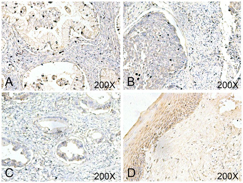
Figure 1. Downregulated USP18 expression in tumor tissues of a cervix tissue microarray. UCC tissues with grade (A) 1 and (B) 2 exhibited moderate
staining, and grade (C) 3 exhibited weak staining of USP18. (D) Non‑cancer tissues exhibited super intensive staining of USP18. Magnification, x200.
USP18, ubiquitin‑specific peptidase 18.
Table II. Ubiquitin‑specific peptidase 18 expression in cervix
tissue microarray (mean ± S).
Characteristics USP18 level t P‑value
Tissues
Non‑cancer 5.25±1.532 2.1750 0.0327
Cancer 4.50±1.553
Stage
Ⅰ 4.62±1.568 0.7945 0.4318
II‑III 4.18±1.537
Grade
1 5.00±1.155 1.0299 0.3105
2‑3 4.40±1.683
Age, years
≤50 4.35±1.522 0.8511 0.4001
≥50 4.79±1.626
Lymph node
metastasis
No 4.62±1.615 1.0452 0.3029
Yes 3.75±0.957
cells compared with untransfected parental and mock‑transfected
HeLa cells (Fig. 2A and C), which indicated that USP18‑deficient
HeLa cells were successfully established.
Figure 2. USP18 expression in HeLa cells. (A) Representative images of
USP18 protein expression from western blot assay. (B) Fluorescence images
Downregulation of USP18 promotes the proliferation, colony of mock‑ and USP18‑siRNA‑transfected HeLa cells. Magnification, x200.
formation, migration and aggressiveness of HeLa cells. The (C) Statistical analysis of USP18 protein expression. **PONCOLOGY LETTERS 21: 421, 2021 5 Figure 3. Roles of USP18‑deficiency in HeLa cell malignant behaviors in vitro. (A) Proliferation analysis from mock‑ and USP18‑siRNA‑transfected HeLa cells using Cell Counting Kit‑8 reagent. (B) Representative images of clonogenic analysis from mock‑ and USP18‑siRNA‑transfected HeLa cells. (C) Statistical anal- ysis of colony formation ability. (D) Representative images of Transwell analysis from mock‑ and USP18‑siRNA‑transfected HeLa cells. Magnification, x200. (E) Statistical analysis of cell migratory capacity. (F) Representative images of wound healing assay from mock‑ and USP18‑siRNA‑transfected HeLa cells. Magnification, x100. (G) Statistical analysis of cell aggressiveness capacity. **P
6                     PAN et al: USP18 CONTRIBUTES TO THE MALIGNANT BEHAVIOR OF CERVICAL CANCER CELLS

Figure 4. Effects of USP18‑deficiency on regulating ERK‑associated gene expression levels in HeLa cells. (A) Representative western blotting images of
Bcl‑2 and STAT3 from mock‑ and USP18‑siRNA‑transfected HeLa cells. (B) Quantification of Bcl‑2 and STAT3 protein expression. (C) Representative
western blotting images of ERK and p‑ERK form mock‑ and USP18‑siRNA‑transfected HeLa cells. (D) Quantification of p‑ERK levels. **P
ONCOLOGY LETTERS 21: 421, 2021                                                        7

treatment with PD98059, the inhibition rates exerted by               to specific immune responses (20). In human leiomyosarcoma,
PD98059 on the proliferation (Fig. 5B) and migration (Fig. 5E)        downregulation of USP18 is associated with a poor clinical
of siRNA‑transfected HeLa cells were significantly higher             outcome, and USP18‑deficient mice exhibited an enhanced
compared with those in mock‑transfected HeLa cells. Overall,          ability to develop these sarcomas (26).
the present results suggested that USP18 silencing‑induced                Although a recent study demonstrated that USP18 is a critical
malignant responses in HeLa cells depend on the activation of         regulator for the tumorigenicity of cervical cancer CaSki and
the ERK signaling pathway.                                            SiHa cells (27), the results of the present study demonstrated
                                                                      that USP18 expression was downregulated in UCC compared
Discussion                                                            with in normal tissues. USP18 expression was knocked down
                                                                      in HeLa cells, and the malignant behaviors in cells, including
USP18 is a major isopeptidase, which was initially identified         proliferation, colony formation and migration, were enhanced.
based on its role to efficiently deconjugate interferon‑stimu-        Mechanistically, p‑ERK expression was significantly upregu-
lated gene 15 (ISG15), a two‑domain ubiquitin‑like protein,           lated following USP18‑knockdown in HeLa cells. Previous
from ISGylation (9). In addition to ISG15, USP18 is highly            studies have reported that several signaling pathways, such as the
induced by type I and III interferons, and it has been proposed       PTEN/AKT (28), AKT/Skp2 (16) and JAK/STAT (29) signaling
that USP18 is a vital blocker of the type I interferons signaling     pathways, are implicated in USP18 associated‑biological roles. To
pathway (22). Furthermore, a study demonstrated that type III         the best of our knowledge, the present study was the first to illus-
interferons may induce USP18 production (23). In the pres-            trate an involvement of ERK signaling in the function of USP18
ence of USP18, type III interferons acquire higher properties         in tumorigenesis. ERKs are a family of protein‑serine/threonine
to weaken type I interferons‑mediated actions by repressing           kinases, which serve vital roles in the control of diverse cell
JAK‑STAT signaling (23).                                              functions, such as cell differentiation, proliferation and survival,
     Increasing evidence has suggested that USP18 is implicated       by phosphorylating several substrates including transcription
in a variety of physiological and pathological processes in           factors, protein kinases and phosphatases (30). Increasing
different tissues and cells, including cell development, viral        evidence has suggested that amplification or activation of ERK
infection, viral replication and antibacterial response (10,24,25).   signaling frequently occurs in several malignant tumors, such as
However, a vast expansion in the understanding of USP18               gastric adenocarcinoma and lung cancer (31,32), which results in
expression and its association with tumor biology has occurred.       increased cell proliferation, promotion of cell cycle progression
It has been reported that USP18 is frequently overexpressed           and repressed apoptosis of tumor cells (33). Given that the ERK
in different types of cancer, including breast cancer, bladder        signaling pathway participates in several aspects of tumorigen-
cancer and hepatocellular carcinoma, and its overexpression           esis by regulating the expression of its downstream signaling
is positively associated with several pathological tumor char-        molecules, such as NF‑κB (34), Bcl‑2 (35) and STAT3 (36), the
acteristics (15‑17). For example, a recent study indicated that       present study assessed whether the expression levels of these
USP18 methylation is predominantly downregulated, whereas             genes were affected following USP18‑knockdown. The results
its expression is upregulated in breast cancer, which is positively   demonstrated that Bcl‑2 and STAT3 expression was upregu-
associated with increasing TNM stage, worse disease‑free              lated, whereas NF‑κB expression remained unchanged (data not
survival rate and HER2+ patients, but negatively associated           shown) following USP18‑knockdown in HeLa cells. Bcl‑2 is a
with apoptosis (17). Accordingly, it has been suggested that          novel gene encoding a unique apoptosis inhibitor that efficiently
USP18 may be used as a predictive marker for poor prognosis           suppresses apoptosis induced by the p53 tumor suppressor
in muscle invasive bladder cancer, since high USP18 expres-           protein (37). STAT3 is an important proto‑oncogene essential
sion is a significant risk factor for cancer‑specific death, and      for modulating the transition from the G1 to S phase of the cell
decreased USP18 expression is markedly associated with                cycle (38), and most cancer cases arise due to proliferating
longer cancer‑specific survival (16). USP18 has also been the         cells losing control of cell cycle regulation, in which loss of the
focus of investigations evaluating its functions in tumorigenesis.    G1/S‑phase transition checkpoint is a major cause of cancer (39).
USP18 silencing in a mouse model for breast cancer exhibited              In conclusion, the results of the presents study demonstrated
a significant decrease in tumor growth, and USP18‑deficiency          that USP18 expression was downregulated in UCC tissues,
in breast cancer MCF‑7 cells in vitro triggered an increase in        and USP18‑knockdown facilitated tumor cell proliferation and
the induction of apoptosis (18,19). In addition, downregulation       migration by affecting the expression levels of genes associated
of USP18 expression in glioblastoma cells may protect against         with the ERK signaling pathway. Overall, the current results
tumor cell invasion and migration by repressing EMT (14),             provide a novel mechanism for USP18‑deficiency, which may
an essential event for cancer metastasis, by which tumor cells        serve a crucial role in UCC progression in an ERK‑dependent
obtain increased motility and invasiveness. Gain‑of‑function          manner.
assays in vitro have demonstrated that overexpression of USP18
has an important role in regulating tumor progression due to          Acknowledgements
its contribution in enhancing breast cancer tumor cell prolifera-
tion, colony formation and cell cycle progression (17). However,      Not applicable.
studies have also revealed that USP18 may exert an opposing
role in the control of cancer development. For example, ectopic       Funding
USP18 expression in B16 melanoma cancer cells may suppress
tumorigenesis, restraining cancer cell‑mediated inhibition of         The present study was supported by the National Natural
T‑cell proliferation and activation, thus facilitating cancer cells   Science Foundation of China (grant no. 81272854), Key
8 PAN et al: USP18 CONTRIBUTES TO THE MALIGNANT BEHAVIOR OF CERVICAL CANCER CELLS
Projects of Natural Science Foundation of Heilongjiang 3. Arbyn M, Weiderpass E, Bruni L, de Sanjosé S, Saraiya M,
Ferlay J and Bray F: Estimates of incidence and mortality of
Province (grant no. ZD2019H008), Excellent Innovation Team cervical cancer in 2018: A worldwide analysis. Lancet Glob
Construction Project of Basic Scientific Research Business Health 8: e191‑e203, 2020.
Fee of Provincial Colleges and Universities in Heilongjiang 4. Oikonomaki M, Bady P and Hegi ME: Ubiquitin specific
peptidase 15 (USP15) suppresses glioblastoma cell growth via
Province (grant no. 2019‑KYYWF‑1334), Double First‑class stabilization of HECTD1 E3 ligase attenuating WNT pathway
Discipline Construction Project in Heilongjiang Province activity. Oncotarget 8: 110490‑110502, 2017.
(grant name. northern medicine and functional food), Young 5. Baker RT, Wang XW, Woollatt E, White JA and Sutherland GR:
Identification, functional characterization, and chromosomal
innovative talents training project of regular undergraduate localization of USP15, a novel human ubiquitin‑specific protease
colleges and universities in Heilongjiang Province (grant related to the UNP oncoprotein, and a systematic nomenclature for
no. UNPYSCT‑2020054), Youth Academic Backbone Support human ubiquitin‑specific proteases. Genomics 59: 264‑274, 1999.
Program for Institution of Common Higher Education in 6. Wilkinson KD: Regulation of ubiquitin‑dependent processes by
deubiquitinating enzymes. FASEBJ 11: 1245‑1256, 1997.
Heilongjiang Province (grant no. 1252G059), Personnel 7. Chung CH and Baek SH: Deubiquitinating enzymes: Their diver-
Training Project of Basic Scientific Research Business sity and emerging roles. Biochem Biophys Res Commun 266:
Expenses of Department of Education in Heilongjiang Province 633‑640, 1999.
8. Liu LQ, Ilaria R Jr, Kingsley PD, Iwama A, van Etten RA, Palis J
(grant no. 2019‑KYYWF‑1338), Science and Innovation Team and Zhang DE: A novel ubiquitin‑specific protease, UBP43,
Foundation of Jiamusi University (grant no. cxtd‑2016‑03) cloned from leukemia fusion protein AML1‑ETO‑expressing
and Biology Team Project of Jiamusi University (grant mice, functions in hematopoietic cell differentiation. Mol Cell
Biol 19: 3029‑3038, 1999.
no. jdxktd‑2019003). 9. Malakhov MP, Malakhova OA, Kim KI, Ritchie KJ and
Zhang DE: UBP43 (USP18) specifically removes ISG 15 from
Availability of data and materials conjugated proteins. J Biol Chem 277: 9976‑9981, 2002.
10. Honke N, Shaabani N, Zhang DE, Hardt C and Lang KS: Multiple
functions of USP18. Cell Death Dis 7: e2444, 2016.
The datasets used and/or analyzed during the current study are 11. Friedrich SK, Schmitz R, Bergerhausen M, Lang J, Cham LB,
available from the corresponding author on reasonable request. Duhan V, Häussinger D, Hardt C, Addo M, Prinz M, et al: Usp18
expression in CD169 + macrophages is important for strong
immune response after vaccination with VSV‑EBOV. Vaccines
Authors' contributions (Basel) 8: 142, 2020.
12. Liu X, Li H, Zhong B, Blonska M, Gorjestani S, Yan M, Tian Q,
Zhang DE, Lin X and Dong C: USP18 inhibits NF‑κ B and NFAT
AP and YL participated in cell experiments and drafted the activation during Th17 differentiation by deubiquitinating the
manuscript. JG and PZ participated in statistical analyses. TAK1‑TAB1 complex. J Exp Med 210: 1575‑1590, 2013.
YH, LS and JRW performed immunohistochemistry assay. 13. Dziamałek‑Macioszczyk P, Haraźna J and Stompór T: Versatility
of USP18 in physiology and pathophysiology. Acta Biochim
CZ, YC, and QR participated in cell transfection and cell Pol 66: 389‑392, 2019.
experiments. SL, SF and TZ performed western blot analyses. 14. Cai X, Feng S, Zhang J, Qiu W, Qian M and Wang Y: USP18
AP, YL and JTW confirm the authenticity of all the raw data. deubiquitinates and stabilizes Twist1 to promote epithe-
lial‑mesenchymal transition in glioblastoma cells. Am J Cancer
WW and JTW designed the study and performed the revision Res 10: 1156‑1169, 2020.
of the manuscript. All authors read and approved the final 15. Tong HV, Hoan NX, Binh MT, Quyen DT, Meyer CG, Hang DT,
manuscript. Hang DT, Son HA, Van Luong H, Thuan ND, et al: Upregulation
of Enzymes involved in ISGylation and Ubiquitination in patients
with hepatocellular carcinoma. Int J Med Sci 17: 347‑353, 2020.
Ethics approval and consent to participate 16. Kim YH, Kim WT, Jeong P, Ha YS, Kang HW, Yun SJ, Moon SK,
Choi YH, Kim IY and Kim WJ: Novel combination markers
for predicting survival in patients with muscle invasive bladder
The medical ethics committee at Jiamusi University (Jiamusi, cancer: USP18 and DGCR2. J Korean Med Sci 29: 351‑356, 2014.
China) approved all procedures performed in the present 17. Tan Y, Zhou G, Wang X, Chen W and Gao H: USP18 promotes
study involving animals and human participants, which were breast cancer growth by upregulating EGFR and activating the
AKT/Skp2 pathway. Int J Oncol 53: 371‑383, 2018.
in accordance with ethical standards, and all patients provided 18. Burkart C, Arimoto K, Tang T, Cong X, Xiao N, Liu YC,
written informed consent prior to participation in this study. Kotenko SV, Ellies LG and Zhang DE: Usp18 deficient mammary
epithelial cells create an antitumour environment driven by
hypersensitivity to IFN‑λ and elevated secretion of Cxcl10.
Patient consent for publication EMBO Mol Med 5: 1035‑1050, 2013.
19. Potu H, Sgorbissa A and Brancolini C: Identification of USP18
Not applicable. as an important regulator of the susceptibility to IFN‑alpha and
drug‑induced apoptosis. Cancer Res 70: 655‑665, 2010.
20. Hong B, Li H, Lu Y, Zhang M, Zheng Y, Qian J and Yi Q: USP18
Competing interests is crucial for IFN‑ γ‑mediated inhibition of B16 melanoma
tumorigenesis and antitumor immunity. Mol Cancer 13: 132,
2014.
The authors declare that they have no competing interests. 21. Wu S, Shang H, Cui L, Zhang Z, Zhang Y, Li Y, Wu J, Li RK and
Xie J: Targeted blockade of interleukin‑8 abrogates its promotion
of cervical cancer growth and metastasis. Mol Cell Biochem 375:
References 69‑79, 2013.
22. François‑Newton V, Magno de Freitas Almeida G, Payelle-
Brogard B, Monneron D, Pichard‑Garcia L, Piehler J, Pellegrini S
1. Van Hede D, Langers I, Delvenne P and Jacobs N: Origin and and Uzé G: USP18‑Based negative feedback control is induced
immunoescape of uterine cervical cancer. Presse Med 43: by type I and type III interferons and specifically inactivates
e413‑e421, 2014. interferon a response. PLoS One 6: e22200, 2011.
2. Bray F, Ferlay J, Colombet M, Soerjomataram I, Siegel RL, 23. Fan W, Xie S, Zhao X, Li N, Chang C, Li L, Yu G, Chi X, Pan Y,
Torre A and Jemal A: Global cancer statistics 2018: GLOBOCAN Niu J, et al: IFN‑λ4 desensitizes the response to IFN‑α treatment
estimates of incidence and mortality worldwide for 36 cancers in in chronic hepatitis C through long‑term induction of USP18.
185 countries. CA Cancer J Clin 68: 394‑424, 2018. J Gen Virol 97: 2210‑2220, 2016.ONCOLOGY LETTERS 21: 421, 2021 9
24. Dagenais‑Lussier X, Loucif H, Cadorel H, Blumberger J, 33. Guo YJ, Pan WW, Liu SB, Shen ZF, Xu Y and Hu LL:
Isnard S, Bego MG, Cohen ÉA, Routy JP and van Grevenynghe J; ERK/MAPK signalling pathway and tumorigenesis. Exp Ther
Montreal Primary Infection Study Group: USP18 is a significant Med 193: 1997‑2007, 2020.
driver of memory CD4 T‑cell reduced viability caused by type I 34. Lin CW, Shen SC, Chien CC, Yang LY, Shia LT and
IFN signaling during primary HIV‑1 infection. PLoS Pathog 15: Chen YC: 12‑O‑tetradecanoylphorbol‑13‑acetate‑induced
e1008060, 2019. invasion/migration of glioblastoma cells through activating
25. Kang JA and Jeon YJ: Emerging roles of USP18: From biology to PKCalpha/ERK/NF‑kappaB‑dependent MMP‑9 expression.
pathophysiology. Int J Mol Sci 21: 6825, 2020. J Cell Physiol 225: 472‑481, 2010.
26. Chinyengetere F, Sekula DJ, Lu Y, Giustini AJ, Sanglikar A, 35. Yang T, Xu F, Sheng Y, Zhang W and Chen Y: A targeted
Kawakami M, Ma T, Burkett SS, Eisenberg BL, Wells WA, et al: proteom ics approach to the quantitative analysis of
Mice null for the deubiquitinase USP18 spontaneously develop ERK/Bcl‑2‑mediated anti‑apoptosis and multi‑drug resistance in
leiomyosarcomas. BMC Cancer 15: 886, 2015. breast cancer. Anal Bioanal Chem 408: 7491‑7503, 2016.
27. Diao W, Guo Q, Zhu C, Song Y, Feng H, Cao Y, Du M and 36. Mu X, Shi W, Xu Y, Xu C, Zhao T, Geng B, Yang J, Pan J, Hu S,
Chen H: USP18 promotes cell proliferation and suppressed Zhang C, et al: Tumor‑derived lactate induces M2 macrophage
apoptosis in cervical cancer cells via activating AKT signaling polarization via the activation of the ERK/STAT3 signaling
pathway. BMC Cancer 20: 741, 2020. pathway in breast cancer. Cell Cycle 17: 428‑438, 2018.
28. Mustachio LM, Kawakami M, Lu Y, Rodriguez‑Canales J, 37. Miyake H, Hanada N, Nakamura H, Kagawa S, Fujiwara T,
Mino B, Behrens C, Wistuba I, Bota‑Rabassedas N, Yu J, Hara I, Eto H, Gohji K, Arakawa S, Kamidono S and Saya H:
Lee JJ, et al: The ISG15‑specific protease USP18 regulates Overexpression of Bcl‑2 in bladder cancer cells inhibits apoptosis
stability of PTEN. Oncotarget 8: 3‑14, 2017. induced by cisplatin and adenoviral‑mediated p53 gene transfer.
29. Gu T, Lu L, An C, Zhang Y, Wu X, Xu Q and Chen G: Negative Oncogene 16: 933‑943, 1998.
regulation of the RLR‑mediated IFN signaling pathway by 38. Luo J, Yan R, He X and He J: Constitutive activation of STAT3
duck ubiquitin‑specific protease 18 (USP18). J Cell Physiol 234: and cyclin D1 overexpression contribute to proliferation, migra-
3995‑4004, 2019. tion and invasion in gastric cancer cells. Am J Transl Res 9:
30. Yoon S and Seger R: The extracellular signal‑regulated kinase: 5671‑5677, 2107.
Multiple substrates regulate diverse cellular functions. Growth 39. Ragkousi K and Gibson MC: Epithelial integrity and cell divi-
Factors 24: 21‑44, 2006. sion: Concerted cell cycle control. Cell Cycle 17: 399‑400, 2018.
31. Bang YJ, Kwon JH, Kang SH, Kim JW and Yang YC: Increased
MAPK activity and MKP‑1 overexpression in human gastric This work is licensed under a Creative Commons
adenocarcinoma. Biochem Biophys Res Commun 250: 43‑47, Attribution-NonCommercial-NoDerivatives 4.0
1998. International (CC BY-NC-ND 4.0) License.
32. Tang Q, Wu J, Zheng F, Hann SS and Chen Y: Emodin increases
expression of insulin‑like growth factor binding protein 1 through
activation of MEK/ERK/AMPKα and interaction of PPAR γ and
Sp1 in lung cancer. Cell Physiol Biochem 41: 339‑357, 2017.You can also read