Intracellular alpha fetoprotein interferes with all trans retinoic acid induced ATG7 expression and autophagy in hepatocellular carcinoma cells ...
←
→
Page content transcription
If your browser does not render page correctly, please read the page content below

www.nature.com/scientificreports
OPEN Intracellular alpha‑fetoprotein
interferes with all‑trans retinoic
acid induced ATG7 expression
and autophagy in hepatocellular
carcinoma cells
Shanshan Wang1, Rilu Feng2, Ying Shi1, Dexi Chen1, Honglei Weng2, Huiguo Ding3 &
Chenguang Zhang4*
Retinoic acid and retinoid acid receptor (RA-RAR) signaling exhibits suppressive functions in the
progression of hepatocellular carcinoma (HCC) through multiple mechanisms. However, whether
RA-RAR signaling induces autophagy that contributes its anti-tumor activity in HCC remains elusive.
In the current study, the effects of RA-RAR pathway on autophagy were investigated in two HCC
cell lines: alpha-fetoprotein (AFP) positive PLC/PRF/5 and AFP negative HLE cells. Cell autophagy
was analyzed with western blot for detection of LC3 conversion and p62/SQSTM1 degradation
while autophagy flux was assayed using the mRFP-GFP-LC3 reporter. Cell apoptosis and viability
were analyzed by caspase-3 activity, TdT-mediated dUTP nick end labeling (TUNEL) assay, and Cell
Counting Kit (CCK)-8, respectively. Chromatin immunoprecipitation (ChIP) was employed to detect
the binding of RAR onto the promoter of autophagy-relevant 7 (ATG7), and co-immunoprecipitation
(CoIP) was used to analyze the interaction of AFP and RAR. The results showed that ATRA dosage and
time-dependently induced high levels of cell autophagy in both the PLC/PRF/5 and HLE cells, which
was accompanied with up-regulation of ATG7. ChIP assay showed that RAR was able to bind to its
responsive elements on ATG7 promoter. Impairment of ATG7 induction or blockade of autophagy
with chloroquine aggravated ATRA induced apoptosis of HCC cells. Furthermore, intracellular AFP
was able to complex with RAR in PLC/PRF/5 cells. Knockdown of AFP in PLC/PRF/5 cells augmented
the up-regulation of ATG7 by ATRA while overexpression of AFP in HLE cells attenuated ATRA
induced ATG7 expression and autophagy. Thus, ATRA induced ATG7 and autophagy participated in
its cytotoxicity on HCC cells and AFP interfere with the induction of ATG7 and autophagy through
forming complex with RAR.
Alpha-fetoprotein (AFP) is the most widely used biomarker for clinical diagnosis of HCC, the fifth most com-
mon malignancy and the third leading cause of cancer related death worldwide1. Dynamics of serum AFP
closely related to the development of the tumor2. Besides its clinical utilization as a biomarker, growing evidence
has revealed that intracellular AFP functioned as an important signaling molecule, through interaction with
a list of proteins, to promote the progression of HCC3–5. A series work by us showed that AFP could perturb
RA-RAR signaling through interaction with RARα, resulting in transcriptional dysregulation of RAR targets,
like survivin3, Fn144, GADD455, GADD1536 etc. in HCC cells. ATRA or its chemical derivatives have long been
tested as candidates for treatment of HCC as single reagent or in combination with other clinically used d rugs7,8,
the results was, however, far to be satisfactory, which could be partially attributed to the perturbation of AFP
and the resultant dysregulation of RAR target genes. Given the profound effect of ATRA on HCC cells and the
1
Beijing Institute of Hepatology, Beijing You’ An Hospital, Capital Medical University, Beijing 100069,
China. 2Department of Medicine II, Medical Faculty Mannheim, Heidelberg University, 68167 Mannheim,
Germany. 3Department of Gastroenterology and Hepatology, Beijing You’An Hospital, Capital Medical University,
Beijing 100069, China. 4Department of Biochemistry and Molecular Biology, School of Basic Medical Sciences,
Capital Medical University, Beijing 100069, China. *email: chzhang@ccmu.edu.cn
Scientific Reports | (2021) 11:2146 | https://doi.org/10.1038/s41598-021-81678-7 1
Vol.:(0123456789)
www.nature.com/scientificreports/
broad distribution of retinoic acid response elements (RAREs) in human genome, whether other target genes of
RA-RAR signaling and related biological process is regulated by AFP in HCC cells is tempting to investigation.
Macroautophagy (referred to as autophagy hereafter) is a conserved degradation system for damaged, mis-
folded, or senescent cellular components, like organelles or certain proteins to maintain cellular h omeostasis9.
About 40 autophagy related genes (ATGs) have been identified to date and participate in the whole process of
autophagy that was mainly composed of initiation and elongation of the phagophore, autophagosome forma-
tion, autophagosome fusion with lysosomes and final degradation of the intracapsular products, in a highly
ordered manner10. Important signaling molecules like AMPK, mTOR, PI3K/Akt etc. showed potent regulation
on autophagy11. For example, mTORC1 inhibited autophagosome formation elicited by ULK1 (ATG1) while acti-
vated AMPK was able to inhibit mTORC1and directly phosphorylates ULK1, leading to autophagy initiation12.
RA-RAR signaling has also been implicated in the modulation of autophagy through multiple mechanisms in
different cell types. In acute promyelocytic leukemia (APL) cells, ATRA was able to induce autophagy through
inhibition of mTOR pathway, which contributed to the degradation of the fusion oncoprotein PML/RARα,
resulting in cell differentiation and the remission of the t umor13,14. In breast cancer cells, ATRA was reported to
induce autophagy dependent on RARα, and ablation of autophagy promoted ATRA induced apoptosis of the
cancer cells15. Fang et al. also suggested induction of autophagy and expression of a panel of ATGs by ATRA
in Hepa1-6 mouse hepatoma c ells16, however, the generality of autophagy induction by ATRA in HCC and the
underlying mechanism remains to be further addressed.
Conventional chemotherapeutic drugs for HCC like doxorubicin, oxaliplatin, cisplatin have all been reported
to induce autophagy in vitro and in vivo that seemed be protective for the cells under treatment, for inhibition
of autophagy was able to enhance the anti-tumor activity of these d rugs17. Multiple ATGs and related signaling
pathways were shown to regulate sensitivity of HCC cells to chemo- or targeted reagents, which might hold
potential therapeutic potentials18. We recently provided intriguing evidence that AFP played a suppressive role
in the maintenance of the basal level of autophagy in HCC cells through interaction with PTEN, which led to
inhibition of its phosphatase activity and subsequent over-activation of PI3K/Akt/mTOR, and finally promoted
cell survival19. As AFP also interacted with RARα and perturbed RA-RAR signaling as well as the anti-tumor
effect of ATRA in HCC cells, whether this perturbation also participates in regulation of cell autophagy is of
great interest to be investigated.
In the present study, we found that ATRA robustly induced autophagy and transcriptional up-regulation of
ATG7 in human HCC cells, which played protective roles for ATRA treated cells. Furthermore, AFP interacted
with RARα and attenuated its regulation on ATG7 expression and autophagy. Our results were supposed to be
helpful for developing novel therapeutics for HCC composed of ATRA and autophagy inhibition reagents, where
the level of AFP needs to be taken into consideration.
Results
ATRA induced autophagy in PLC/PRF/5 and HLE cells. To investigate if ATRA was able to induce
autophagy in HCC cells, HCC cells were treated with 40 μM ATRA, and Ethyl Alcohol (Alc), the solvent of
ATRA, was used as negative control. As shown in Fig. 1A,A’, ATRA treatment significantly promoted cell
autophagy in a time dependent manner, in both PLC/PRF/5 and HLE cells, as demonstrated by up-regulation
of LC3-II and decrement of p62/SQSTM1 at the protein level. Chloroquine, an inhibitor of autophagy, blocked
ATRA induced LC3 conversion and p62/SQSTM1 degradation (supplementary Figure 3A and B), supporting
the induction of autophagic flux by ATRA in HCC. To further evaluate the autophagic flux in ATRA treated
HCC cells, we employed the mRFP-GFP-LC3 adenovirus vectors. PLC/PRF/5 and HLE cells transfected with
mRFP-GFP-LC3 adenovirus were added with 40 μM ATRA and cultured for 24 h. Numbers of GFP and mRFP
dots per cell were both significantly increased under ATRA treatment (Fig. 1B,B’, and quantification in supple-
mentary Figure 1C). Immunofluorescence analyses further confirmed that ATRA significantly reduced the level
of p62/SQSTM1 in both PLC/PRF/5 and HLE cells (Fig. 1C,C’). All these results indicated that ATRA induced
autophagy in HCC cells. The activation of RA-RAR signaling in HCC cells was verified with nuclear accumula-
tion of RAR as demonstrated with western blot analyses for nuclear proteins (supplementary Figure 1A) and
cellular immunofluorescence (supplementary Figure 1B).
ATRA‑RAR signaling regulated transcription of ATG7. To further reveal the potential molecu-
lar mechanism underlying ATRA induced autophagy in HCC cells. Expressions of ATG5, Beclin1 and ATG7
were evaluated with RT-qPCR in PLC/PRF/5 and HepG2 cells. According to preliminary experimental results,
expression of ATG7, an E1-like activating enzyme for autophagosome formation20, was most significantly up-
regulated under ATRA treatment in both HCC cell lines, while Beclin1 and ATG5 were not (supplementary
Figure 2A, B, C). ATG5 even manifest a decrement at the mRNA level, but no similar expression trend was seen
at the protein level which needs further investigation (supplementary Figure 2D and E). We thus focused on
potential regulation of ATG7 by ATRA-RAR in the following studies. Western blotting and qRT-PCR showed
that ATRA induced robust increment of ATG7 in both PLC/PRF/5 (Fig. 2A,B) and HLE (Fig. 2A’,B’) cells, in a
dose-dependent manner, reaching maximum at 40 μM. Similar results were observed at the mRNA level with
the qRT-qPCR assay (Fig, 2C,C’). The alteration of ATG7 at the mRNA level prompted us to investigate if ATG7
was transcriptionally regulated by RAR. Two adjacent binding sequence for RAR was discovered at the proximal
promoter of ATG7 (Fig. 2D). To validate if RAR was able to bind to the region, ChIP assays were performed. As
shown in Fig. 2E,2E’, RAR was able to bind to the 5′-flanking regions containing its responsive elements at the
ATG7 promoter in both PLC/PRF/5 and HLE cells, indicating a direct transcriptional regulation of ATRA-RAR
signaling on ATG7 via RAR.
Scientific Reports | (2021) 11:2146 | https://doi.org/10.1038/s41598-021-81678-7 2
Vol:.(1234567890)
www.nature.com/scientificreports/

                                  Figure 1.  ATRA promoted autophagy in PLC/PRF/5 and HLE cells. A. Western blotting analyses of the
                                  level of p62/SQSTM1 and LC3 in PLC/PRF/5 (A) and HLE (A’) cells upon ATRA treatment at indicated time
                                  points (Top). GAPDH was used as loading control. Densitometry of the blots were quantified, and the ratio
                                  of p62/SQSTM1 to GAPDH, or LC3II to LC3I were calculated (Bottom). **P < 0.01, one-way ANOVA. ns: no
                                  significance. B. PLC/PRF/5 (B) and HLE (B’) cells were transduced with tandem mRFP-GFP-LC3 adenovirus
                                  and were then subjected to ATRA for 24 h. Representative images of fluorescent LC3 puncta are shown. (C)
                                  and (C’). The expression of p62/SQSTM1 was detected with immunofluorescence and was observed under the
                                  fluorescence microscopy. Nuclei were stained with DAPI (blue). p62/SQSTM1 were labeled with TRITC (red).
                                  All the shown images are representative of three independent experiments. Ethyl Alcohol (Alc) was used as a
                                  solvent and as a control.

                                  ATG7 played a protective role in HCC during ATRA treatment. We next investigated whether the
                                  induction of ATG7 expression and autophagy were functional in ATRA treated HCC, or merely indicators for
                                  the activity of ATRA. CCK-8 and caspase-3 activity assays were carried out. The CCK-8 results shown that
                                  cell viability was further decreased with knockdown of ATG7 in response to ATRA in both PLC/PRF/5 and
                                  HLE cells (Fig. 3A, A´), which was accompanied with increment of caspase-3 activity (Fig. 3B,B´). Meanwhile,
                                  knockdown of ATG7 mildly reduced basic level of autophagy in HCC cells, as evidenced with P62/SQSTM1
                                  accumulation and reduced LC3 conversion (Fig. 3C,C’, lane 3 versus lane 1). Impairment ATG7 expression also
                                  attenuated ATRA induced LC3II conversion. However, degradation of p62/SQSTM1 upon ATRA treatment
                                  was further aggregated by ATG7 siRNA (Fig. 3C,C’, lane 4 versus lane 2), suggesting alternative degradation
                                  pathway(s) activation with simultaneous ATRA stimulation and ATG7 silence for p62/SQSTM1. The protective
                                  role of ATG7 for ATRA treated cells was also validated by TUNEL assay (Fig. 3D,D’,E,E’). Furthermore, inhibi-
                                  tion of autophagy with 40 μM chloroquine also resulted in further decrement of cell viability of HCC cells by
                                  ATRA (supplementary Figure 3C and D). These results indicated that ATG7 and autophagy, played protective
                                  roles in ATRA induced apoptosis of HCC cells.

                                  AFP interacted with RAR in HCC cells.          To investigate whether AFP could possibly regulate ATRA-RAR
                                  mediated autophagy, western blotting analyses were first employed to detect the endogenous expression of AFP
                                  in PLC/PRF/5 and HLE cells. As previously reported, AFP protein was undetectable in HLE cells, but robustly
                                  expressed in PLC/PRF/5 cells (Fig. 4A). Further analyses with confocal microscopy showed that AFP and RAR

Scientific Reports |   (2021) 11:2146 |               https://doi.org/10.1038/s41598-021-81678-7                                                3

                                                                                                                                        Vol.:(0123456789)
www.nature.com/scientificreports/

          Scientific Reports |   (2021) 11:2146 |   https://doi.org/10.1038/s41598-021-81678-7   4

Vol:.(1234567890)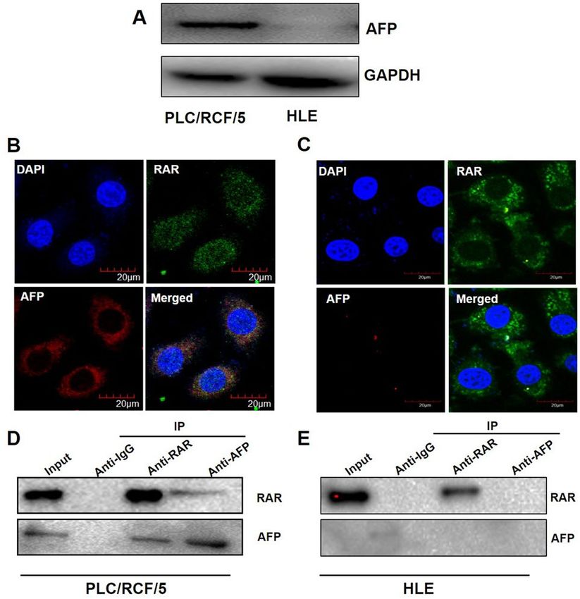
www.nature.com/scientificreports/
◂Figure 2. RAR signal cascade regulated transcription of ATG7 in HCC cells. (A) and (B). Various
concentrations of ATRA were used to treat HCC cells for 24 h. Expression of the ATG7 protein, p62/SQSTM1
and LC3 in PLC/PRF/5 (A) and HLE (A’) cells was detected using Western blotting. GAPDH was used as
loading control. (B) and (B’). Densitometry of the blots in Figure A and A’ were quantified and the ratio of
p62/SQSTM1 to GAPDH or LC3II to LC3I were calculated. *P < 0.05, **P < 0.01 compared with control group,
one-way ANOVA. (C) and (C’). The mRNA expression level of ATG7 gene in PLC/PRF/5 (C) and HLE (C’)
cells were analyzed with qRT-qPCR. *P < 0.05, **P < 0.01 compared with control group, one-way ANOVA. (D)
Schematic overview of the RAR binding sites on ATG7 promoter predicted with JASPAR website (http://jaspa
r.genereg.net/). (E) and (E’). ChIP assays for RAR binding onto ATG7 promoter were carried out in PLC/PRF/5
(E) and HLE (E’) cells.
co-localized in cytoplasm in PLC/PRF/5 cell (Fig. 4B), but not in HLE cells (Fig. 4C), which were further con-
firmed by Co-IP analysis (Fig. 4D,E).
AFP perturbed ATRA induced ATG7 expression in HCC cells. To further investigate if AFP was
involved in ATRA-RAR mediated ATG7 expression by interacting with RAR, AFP was first knockdown by
specific shRNA in PLC/PRF/5 cells. Immunofluorescence and confocal microscopy assays showed that AFP
expression was obviously depleted upon specific shRNA transfection compared with scramble shRNA (Fig. 5A).
Following AFP depletion, binding of AFP with RAR was significantly decreased as demonstrated by Co-IP assay
in PLC/PRF/5 cells (Fig. 5B). On the contrary, when AFP was introduced into HLE cells with pcDNA3.1-afp
vectors (Fig. 5A’), notable interaction between AFP and RAR was observed as shown by Co-IP results (Fig. 5B’),
accompanied with co-localization of AFP and RAR in the cytoplasm (Fig. 5A´). One intriguing phenomenon
was observed that alteration of the intracellular AFP level not only changed its interaction with RAR, but also
exhibit a negative regulation on the protein level of RAR itself (Fig. 5A,A’), which needs further investigation.
Interaction between AFP and RAR was able to disrupt the transcriptional regulation of RAR on its targets,
we wonder whether it was also the case in ATG7. Not surprisingly, when AFP was down regulated by shRNA
in PLC/PRF/5 cells, the ATG7 protein level was remarkably increased compared with the control in untreated
conditions (Fig. 5C). On the other hand, AFP expression in HLE cells resulted in an apparent reduction of ATG7
protein (Fig. 5C’). Similar results were also observed under ATRA treatment conditions, although to a less extent
in HLE cells (supplementary Figure 4A,B). However, knockdown of AFP in PLC/PRF/5 did not obviously alter
the effect of ATRA on p62/SQSTM1 degradation and LC3 conversion while ectopic expression of AFP in HLE
cells significantly attenuated ATRA induced alterations of p62/SQSTM1 and LC3II, possibly suggesting dose
dependence of AFP on ATRA induced autophagy as well as involvement of other regulators beyond ATG7 in
this process (see discussion).
Discussion
In the present study, ATRA treatment robustly induced autophagy in HCC cells through transcriptional up-
regulation of ATG7. Mechanistically, ATRA induced nuclear accumulation of RAR, which bound onto the pro-
moter region of ATG7 that harbors RAR binding motifs. Intracellular AFP interacted with RAR and exhibited an
inhibitory effect on nuclear accumulation of RAR, resulting in down-regulation of ATG7 of HCC cells. Functional
studies indicated a protective role of the induced expression of ATG7 and autophagy, and impairment of ATG7
induction or blockade of autophagy further aggravated ATRA induced cell apoptosis (supplementary Figure 5).
ATRA has long been used clinically to induce differentiation of APL cells, where the relationship between
ATRA and autophagy were mostly studied. An array of ATGs and important regulators of autophagy, includ-
ing ATG1, ATG5, Beclin1, mTOR, PI3KC3, WIPI and TFEB, DRAM etc., were implicated in ATRA induced
autophagy14,21–24. In other cell types, including several other solid tumor types, ATRA was also able to induce
autophagy15,25. In these studies, expression alterations of certain ATGs or signaling molecules were always
displayed as the underlying mechanisms, which seemingly was not powerful enough to establish direct links
between ATRA and autophagy, as the involvement and the function of RAR always lacked. For example, ATRA
induced autophagy in human B cells through mTOR inhibition26, and induced autophagy in APL cells via
potent up-regulation of T FEB23, how the inhibition or promotion occurred, directly through RAR or by other
alternative pathways? The present study directly linked ATRA and autophagy in HCC cells with RAR mediated
transcriptional activation of ATG7. Of course, as ATRA was able to elicit a number of other downstream signal-
ing pathways27, it still cannot rule out the possibility that other regulators were also involved in ATRA induced
autophagy in HCC cells.
Autophagy and apoptosis are generally discrete cellular processes mediated by distinct groups of m olecules28.
However, they often occurred in the same cell under stresses, and interactions among molecules involved in apop-
tosis and autophagy dictated the progression of each process. Autophagy under moderate levels of stress generally
exhibited a protective role and inhibited cell a poptosis29. For example, blockade of autophagy by knockdown
Atg1, Atg5 and PI3KC3 etc. or by specific autophagy inhibitors like 3-methyladenine (3-MA) impaired ATRA
induced differentiation of APL c ells14, suggesting the necessity of autophagy for the primary function of ATRA.
In breast cancer cells, autophagy was reported to be cell protective and inhibition of autophagy genetically or
pharmacologically resulted in robust apoptosis15. In the current study, as in most cases of chemotherapeutic drug
treatment, ATRA induced autophagy also played a protective role for the cancer cells, as ATG7 knockdown or CQ
treatment both potentiated ATRA induced apoptosis (Fig. 3 and Supplementary Figure 3), though overwhelmed
by the potency of ATRA. Besides the general protective role of autophagy, ATG7 was also able to directly bind
to p53 to modulate cell survival under metabolic stress independent of its E1-like enzymatic activity30. Both
Scientific Reports | (2021) 11:2146 | https://doi.org/10.1038/s41598-021-81678-7 5
Vol.:(0123456789)www.nature.com/scientificreports/
Figure 3. ATG7 prevented ATRA-induced HCC cell apoptosis. (A) and (A’). PLC/PRF/5 and HLE cells were
pre-treated with siRNAs (si-con and si-ATG7) for 24 h, and was then treated with ATRA at final concertation of
40 μM for further 24 h. PLC/PRF/5 cells (A) and HLE (A’) cell viability was determined by CCK-8 assays. * and
#
P < 0.05. **and ##P < 0.01, two-way ANOVA. (B) and (B’), Caspase-3 activity of PLC/PRF/5 cells (B) and HLE
(B’) cells were also detected following treatment as in (A) and (A’). * and #P < 0.05. ** and ##P < 0.01, two-way
ANOVA. (C) and (C’). Protein level for ATG7, p62/SQSTM1 and LC3 were detected upon ATG7 knockdown
and ATRA treatment. GAPDH was used as loading control. Densitometry of the blots were quantified, and
the ratio of ATG7 and p62/SQSTM1 to GAPDH, and LC3II to LC3I were calculated and marked under the
corresponding lanes. (D, D’) and (E, E’). Apoptosis rates of PLC/PRF/5 cells (D and E) and HLE (D’ and E’)
cells were determined with TUNEL assay. * and #P < 0.05. ** and ##P < 0.01, two-way ANOVA.
mechanisms may function in HCC cells under ATRA treatment. ATRA has been shown to induce differentiation
of tumor initiating cells in HCC, and potentiated the cytotoxicity of chemotherapeutic drugs like c isplatin31. It has
also been shown to enhance the anti-tumor activity of sorafenib through activation of A MPK32, a potent regulator
for autophagy induction. Together with the reports that most reagents for HCC treatment induced autophagy,
and autophagy was cell protective in most cases, it is plausible to consider combinational use of chemotherapeutic
drugs with ATRA and autophagy inhibitors like chloroquine to improve the efficacy of chemotherapy for HCC.
We recently reported that AFP was able to block basal level of autophagy in HCC cells through direct seques-
tration of PTEN, which leads to overactivation of PI3K-Akt-mTOR c ascade19. Together with current results,
AFP was thus able to disrupt both the basal and ATRA induced autophagy through interaction of different part-
ners (PTEN or RAR) and modulation of different autophagy regulators (PI3K/Akt/mTOR or ATG7). However,
whether autophagy and AFP played identical roles in these conditions still needs further illustration. Under
basal conditions, increased level of autophagy with AFP knockdown was accompanied with PTEN overactiva-
tion and increased cell apoptosis19. However, to what extent did autophagy contribute to increased apoptosis
could not be figured out with the evidence provided; In ATRA treated conditions, ATG7 played cell protective
roles and AFP perturbed ATG7 expression. From this point of view, AFP seemed to contribute to ATRA induced
cell death, which was obviously against its well-known tumor promoting function. The discrepancy could be
explained that PTEN and RAR were both able to elicit multiple important downstream signalings or effectors
to exert their potent anti-tumor functions. AFP, as an important oncoprotein for HCC, interacted with both
molecules to counteract their biological functions, with autophagy induction being one of them. Interestingly,
ATRA has been shown to induce expression of PTEN, which contributed its anticancer activity in APL c ells33.
Whether similar crosstalk exists, and how overexpression of AFP could orchestrate those signaling pathways in
HCC required further investigation.
Some discrepancies still exist in the current study. The first one is that impairment of ATG7 expression upon
ATRA significantly attenuated LC3 conversion but further promoted p62/SQSTM1 degradation (Fig. 3C,C’).
It was probable that blockade of autophagy with ATG7 knockdown enhanced proteotoxicity of ATRA in HCC
cells, activating alternative protein degradation pathways like the proteasome or endosomal microautophagy,
both of which has been involved in p62/SQSTM1 degradation34,35. The second is that the extent of expression
alteration for ATG7 was more obvious upon AFP knockdown in PLC/PRF/5 than its ectopic expression in HLE
cells while the inhibitory effect of AFP on autophagy was much better manifested in HLE cells (supplementary
Scientific Reports | (2021) 11:2146 | https://doi.org/10.1038/s41598-021-81678-7 6
Vol:.(1234567890)www.nature.com/scientificreports/
Figure 4. AFP interacted with RAR in cytoplasm of HCC cells. (A) Western blotting was used for analysis of
expression of AFP in PLC/PRF/5 and HLE cells. (B) and (C). Expression and localization of AFP and RAR were
analyzed with confocal microscopy in PLC/PRF/5 (B) and HLE (C) cells. (D) and (E). In PLC/PRF/5 (D) and
HLE (E) cells, Co-IP was employed to detect the interaction of AFP and RAR. The images captured by confocal
microscope are representatives of experiments that were repeated at least three times.
Figure 5. AFP attenuated RAR mediated of ATG7 expression in HCC cells. (A) and (A’). Localization of AFP
(red) and RAR (green) in AFP–shRNA923-transfected PLC/PRF/5 cells (A) and pcDNA3.1-afp-transfected
HLE cells (A’) were detected with immunofluorescence and observed with confocal microscopy. (B) and (B’).
Co-IP was carried out to detect the interaction between AFP and RAR in AFP knockdown or ectopic expression
in PLC/PRF/5 cells (B) or HLE cells (B’), respectively. (C) and (C’). Effects of AFP knockdown or ectopic
expression on ATG7 expression in PLC/PRF/5 cells (C) and HLE (C’) cells were analyzed by Western blotting.
GAPDH was used as loading control. Densitometry quantification were performed, and ratio of ATG7 to
GAPDH were calculated. *P < 0.05, Two tailed Student’s t test.
Scientific Reports | (2021) 11:2146 | https://doi.org/10.1038/s41598-021-81678-7 7
Vol.:(0123456789)www.nature.com/scientificreports/
Primary and sencondary antibodies
P62/SQSTM1 Cell Signaling Technology CST-5114S
LC3 Sigma-Aldrich L7543-100UL
ATG7 Cell Signaling Technology CST-8558S
RAR Santa Cruz Biotechnology sc773
AFP Santa Cruz Biotechnology sc8399
HDAC Cell Signaling Technology CST-3949
GAPDH Cell Signaling Technology CST-2118S
Goat anti-rabbit IgG-HRP Santa Cruz Biotechnology sc-2004
Goat anti-mouse IgG-HRP Santa Cruz Biotechnology sc-2354
Goat anti-Rabbit IgG Alexa Fluor 488 Invitrogen R37116
Goat anti-Mouse IgG Alexa Fluor 594 Invitrogen R37121
Oligonucleotides sequences
Primers for RT-qPCR
ATG7
Sense 5′-AAGAAATAATGGCGGCAGCT-3’
Antisense 5′-ACCCAACATCCAAGGCACTA-3’
GAPDH
Sense 5′-TGAAGGTCGGAGTCAACGGA-3’
Antisense 5′-CCTGGAAGATGGTGATGGGAT-3’
Primers for ChIP-qPCR
ATG7
Sense 5′-TTGGGTTGTTGCATTTCTGA-3’
Antisense 5′-CCATCCTAATTGGCCTGTGT-3’
AFP-shRNA
5′-CACCGAACGTGGTCAATGTATAATTCAAGAGATTATACATTGACCACGTTCTTT
Sense
TTTG-3’
5′-GATCCAAAAAAGAACGTGGTCAATGRATAATCTCTTGAATTATACATTGACCAC
Antisense
GTTC-3’
Table 1. Antibodies and Sequences of oligonucleotides.
Figure 4). Explanations for this might include: (1) the dosage alteration of AFP was much more prominent in the
ectopic expression model in HLE than in the knockdown model in PLC/PRF/5; (2) other regulators and path-
ways of autophagy like mTOR were affected by the higher dosage of AFP in HLE cells; (3) further up-regulation
of ATG7 upon AFP knockdown participated non-autophagic processes as previous s uggested36. More detailed
investigations are needed to be taken to address these issues.
Materials and methods
Cell lines. AFP-producing hepatocellular carcinoma cell line PLC/PRF/5 cells and AFP-non producing cell
line HLE were both maintained in a 5% C
O2 incubator and cultured in DMEM medium supplemented with
10% FCS.
Western blotting. For western blotting, total cell proteins from each sample were extracted with radio-
immunoprecipitation (RIPA, Thermofisher, USA) cell lysis buffer containing protease inhibitor cocktail (CST,
USA), 15 μg of which were then subjected to 12% SDS-PAGE. Electrophoretic transfer of proteins from gels onto
nitrocellulose membrane was carried out in a transblotting cell. Membranes were blocked by immersing in 5%
nonfat milk (w/v) /PBS for 1 h, and then incubated with primary antibodies at 4 °C for overnight. After rinsing
with PBS/0.1% Tween-20, membranes were incubated with horseradish peroxidase-conjugated secondary Ab.
The signals were visualized by incubation with the Enhanced Chemiluninescence kit and exposure on an X-ray
film. Densitometry quantification of the bands were performed with Image J software. Primary and secondary
antibodies used in this study are listed in Table 1.
Immunofluorescence staining. Cells were seeded on coated glass coverslips, then fixed with 4% para-
formaldehyde. After incubated with PBST buffer (containing 0.05% triton-100, 0.5% BSA in PBS) at room tem-
perature for 10 min, the cells were washed with PBS twice followed with blocking using 1% BSA at room tem-
perature for 10 min. The primary antibodies were then added and incubated at 4 °C overnight. After rinsing with
PBST for 10 min, secondary antibodies, Alexa Fluor 594 and 488 (Thermofisher, USA) were incubated with cells
for one hour in room temperature followed by addition of DAPI for counterstaining of the nuclei. Cells images
were captured with a Laser Confocal Microscope (Leica TCS STED-3X, Germany).
Scientific Reports | (2021) 11:2146 | https://doi.org/10.1038/s41598-021-81678-7 8
Vol:.(1234567890)www.nature.com/scientificreports/
Quantitative real‑time reverse transcription PCR (RT‑qPCR). Expression of ATG7 at the mRNA
level was evaluated by quantitative real-time reverse transcription PCR (RT-qPCR) assay. Briefly, total RNA
was extracted from HCC cells using the RNeasy Mini Kit (Qiagen, Germany) according to the manufacturer’s
instructions. cDNA was then synthesized by reverse transcription of the extracted RNA using a SuperScript II
First-stand Synthesis System (Invitrogen, USA). SYBR Green was used to detect the dsDNA products during the
real-time PCR reaction. The mRNA content was normalized to the housekeeping gene GAPDH and fold change
was calculated with the 2−ΔΔCT method. All primer sequences for RT-qPCR are listed in Table 1.
Co‑immunoprecipitation (Co‑IP). Co-IP experiments for evaluation of the interaction between AFP and
RAR were performed in PLC/PRF/5 and HLE cells. HCC cells were lysed in RAPI buffer containing 1% protease
inhibitor cocktails. The lysates (1 mg/500 μl) were then incubated with 1 μg anti-AFP, anti-IgG and anti-RAR
antibodys for overnight at 4 °C, then added 100 μl Protein A SepHarose agarose beads (CL-4B) (17-0780-01,
GE, USA) for further incubation for at least 8 h at 4 °C. Collected these beads and washed 3 times with 1 ml
PBS contain 1% protease inhibitor cocktails before boiling them in the loading buffer, and then Western blotting
analyses were performed.
Transient transfection. The AFP-expressing plasmid (pcDNA3.1-afp) was used to overexpress AFP in
HLE cells, and the AFP interference shRNA (AFP-shRNA923) was employed to knockdown endogenous AFP in
PLC/PRF/5 cells. ATG7 siRNA (sc-41447) and control siRNA (sc-37707) were used to silence the ATG7 mRNA
in the PLC/RCF/5 and HLE cells. In brief, HLE were transfected with pcDNA3.1-afp or control vector, and PLC/
PRF/5 cells were transfected with AFP-shRNA923 or scramble control shRNA with Lipofectamine 3000 (Invit-
rogen, USA) according to the manufacturer’s instructions. After 24 h of transfection, the expression of AFP and
ATG7 were evaluated by Western blotting and the interaction of AFP and RAR was examined by Co-IP. PLC/
RCF/5 and HLE cells were transfected with ATG7 siRNA or control siRNA for 24 h, followed with ATRA treat-
ment for another 24 h, and then subjected to functional evaluation.
Chromatin immunoprecipitation and PCR (ChIP‑PCR). ChIP was performed to verify the capacity
of RAR for binding to the 5′regulatory region of ATG7 gene. Briefly, HCC cells were cross-linked in 1% formal-
dehyde/PBS for 10 min at 37 °C and were then washed twice with ice-cold PBS prior to be lysed. Chromatin
fragments ranging from 200 to 1000 bp were obtained by sonication (SCIENTZ, China). The solution containing
chromatin fragments was then incubated overnight at 4 °C with anti-RAR or anti-IgG. The chromatin–antibody
complexes were then washed, eluted and reverse cross-linked at 65 °C for 6 h. The eluted DNA was purified with
the phenol–chloroform. Immunoprecipitated chromatin was analyzed with PCR using primers targeting the
human ATG7 promoter containing RAR binding motif. The primers used for ChIP-PCR are listed in Table 1.
Evaluation of fluorescent LC3 puncta. The tandem mRFP-GFP-LC3 adenoviruses construct was
obtained from Hanbio Inc (Shanghai, China) and was used to evaluate autophagy induction. Briefly, 5 × 10⁴
PLC/PRF/5 or HLE cells cultured on coverslips in 24- well microplates were transduced with mRFP-GFP-LC3
adenoviruses at 50 MOI. Two hours after transduction, removed the adenovirus by washing the cells with PBS,
and then maintained the cells in complete culture medium with 40 μM ATRA or vehicle for 24 h. The cells were
washed with PBS, fixed with 4% paraformaldehyde, and viewed under a fluorescence microscope. The number
of mRFP, GFP and GFP-mRFP (yellow) dots were determined by manual counting of fluorescent puncta in five
fields from three different cells. The number of dots per cell was obtained by dividing the total number of dots by
the number of cell in each microscopic field.
Determination of viability. Cell Counting Kit (CCK)-8 (CK04, Dojindo, Japan) was used to assess the
cells viability. PLC/PRF/5 and HLE cells were seeded into 96-well microplates, and transfected with Si-Con
or Si-ATG7. 24 h after transfection, the cells were subjected to 40 μM ATRA and solvent for another 24 h. The
absorbance at 570 nm wavelength was measured with a Universal Microplate Reader (EL × 800). The percentage
of cell viability was calculated as (A570sample-background)/ (A570control-backgroud).
Caspase‑3 activation and TUNEL assay. For caspase-3 activity assays, PLC/PRF/5 and HLE cells were
homogenized in lysis buffer. Thereafter, 30µL lysates were added to a white 96-well plate, and then mixed with
60µL assay buffer. 90µL assay buffer was added into the blank well. After incubation for 10minutes at 37 °C,
each well was added 10µL AC-DEVD-AFC at final concentration of 10 µg/mL, followed with further incuba-
tion for 1 h at 37 °C in the dark. The luminescence was measured using Imaging Multi-Mode Reader (BioTek).
The changes of caspase-3 activity was calculated as [(A400sample-blank)/Protein concentration]/[(A400control-blank)/
Protein concentration].
TUNEL assays of PLC/PRF/5 and HLE cells upon different treatment were carried out using a commercial
cell death detection kit (Roche Applied Science, Germany) and analyzed as previously described37.
Statistical analysis. The results of at least three separate experiments are presented as the mean ± s.d. Sta-
tistical significance was determined using the Two tailed Student’s t test, one-way and two-way ANOVA tests
(SPSS 16 software).
Scientific Reports | (2021) 11:2146 | https://doi.org/10.1038/s41598-021-81678-7 9
Vol.:(0123456789)www.nature.com/scientificreports/
Received: 28 April 2020; Accepted: 11 January 2021
References
1. Heimbach, J. K. et al. AASLD guidelines for the treatment of hepatocellular carcinoma. Hepatology 67, 358–380 (2018).
2. Wong, R. J., Ahmed, A. & Gish, R. G. Elevated alpha-fetoprotein: differential diagnosis—hepatocellular carcinoma and other
disorders. Clin. Liver Dis. 19, 309–323 (2015).
3. Li, M. et al. Cytoplasmic alpha-fetoprotein functions as a co-repressor in RA-RAR signaling to promote the growth of human
hepatoma Bel 7402 cells. Cancer Lett. 285, 190–199 (2009).
4. Wang, S. et al. Alpha-fetoprotein acts as a novel signal molecule and mediates transcription of Fn14 in human hepatocellular
carcinoma. J. Hepatol. 57, 322–329 (2012).
5. Zhang, C. et al. Alpha fetoprotein mediates HBx induced carcinogenesis in the hepatocyte cytoplasm. Int. J. Cancer 137, 1818–1829
(2015).
6. Li, C. et al. Impact of intracellular alpha fetoprotein on retinoic acid receptors-mediated expression of GADD153 in human
hepatoma cell lines. Int. J. Cancer 130, 754–764 (2012).
7. Okuno, M. et al. Retinoids in cancer chemoprevention. Curr. Cancer Drug Targets 4, 285–298 (2004).
8. Shi, J. et al. All-trans-retinoic acid (ATRA) plus oxaliplatin plus 5-fluorouracil/leucovorin (FOLFOX) versus FOLFOX alone as
palliative chemotherapy in patients with advanced hepatocellular carcinoma and extrahepatic metastasis: study protocol for a
randomized controlled trial. Trials 20, 245 (2019).
9. Levy, J., Towers, C. G. & Thorburn, A. Targeting autophagy in cancer. Nat. Rev. Cancer 17, 528–542 (2017).
10. Levine, B. & Kroemer, G. Biological functions of autophagy genes: a disease perspective. Cell 176, 11–42 (2019).
11. Yang, S. et al. New insights into autophagy in hepatocellular carcinoma: mechanisms and therapeutic strategies. Am. J. Cancer Res.
9, 1329–1353 (2019).
12. Kim, J., Kundu, M., Viollet, B. & Guan, K. L. AMPK and mTOR regulate autophagy through direct phosphorylation of Ulk1. Nat.
Cell Biol. 13, 132–141 (2011).
13. Isakson, P., Bjoras, M., Boe, S. O. & Simonsen, A. Autophagy contributes to therapy-induced degradation of the PML/RARA
oncoprotein. Blood 116, 2324–2331 (2010).
14. Wang, Z. et al. Autophagy regulates myeloid cell differentiation by p62/SQSTM1-mediated degradation of PML-RARalpha onco-
protein. Autophagy 7, 401–411 (2011).
15. Brigger, D., Schlafli, A. M., Garattini, E. & Tschan, M. P. Activation of RARalpha induces autophagy in SKBR3 breast cancer cells
and depletion of key autophagy genes enhances ATRA toxicity. Cell Death Dis. 6, e1861 (2015).
16. Fang, S. Y. et al. Changes in autophagy during maturation and differentiation of Hepa1-6 cells induced by all-trans retinoic acid.
Nan Fang Yi Ke Da Xue Xue Bao 38, 527–533 (2018).
17. Sheng, J., Qin, H., Zhang, K., Li, B. & Zhang, X. Targeting autophagy in chemotherapy-resistant of hepatocellular carcinoma. Am.
J. Cancer Res. 8, 354–365 (2018).
18. Niu, L. et al. New insights into sorafenib resistance in hepatocellular carcinoma: responsible mechanisms and promising strategies.
Biochim. Biophys. Acta Rev. Cancer 1868, 564–570 (2017).
19. Wang, S. et al. Alpha-fetoprotein inhibits autophagy to promote malignant behaviour in hepatocellular carcinoma cells by activat-
ing PI3K/AKT/mTOR signalling. Cell Death Dis. 9, 1027 (2018).
20. Shimizu, S. Biological roles of alternative autophagy. Mol. Cells 41, 50–54 (2018).
21. Humbert, M., Mueller, C., Fey, M. F. & Tschan, M. P. Inhibition of damage-regulated autophagy modulator-1 (DRAM-1) impairs
neutrophil differentiation of NB4 APL cells. Leuk Res. 36, 1552–1556 (2012).
22. Brigger, D., Proikas-Cezanne, T. & Tschan, M. P. WIPI-dependent autophagy during neutrophil differentiation of NB4 acute
promyelocytic leukemia cells. Cell Death Dis. 5, e1315 (2014).
23. Orfali, N. et al. All-trans retinoic acid (ATRA)-induced TFEB expression is required for myeloid differentiation in acute promye-
locytic leukemia (APL). Eur. J. Haematol. 104, 236–250 (2020).
24. Trocoli, A. et al. ATRA-induced upregulation of Beclin 1 prolongs the life span of differentiated acute promyelocytic leukemia
cells. Autophagy 7, 1108–1114 (2011).
25. Patties, I., Kortmann, R. D., Menzel, F. & Glasow, A. Enhanced inhibition of clonogenic survival of human medulloblastoma cells
by multimodal treatment with ionizing irradiation, epigenetic modifiers, and differentiation-inducing drugs. J. Exp. Clin. Cancer
Res. 35, 94 (2016).
26. Eriksen, A. B. et al. Retinoic acid-induced IgG production in TLR-activated human primary B cells involves ULK1-mediated
autophagy. Autophagy 11, 460–471 (2015).
27. Costantini, L., Molinari, R., Farinon, B. & Merendino, N. Retinoic acids in the treatment of most lethal solid cancers. J. Clin. Med.
9, 360 https://doi.org/10.3390/jcm9020360(2020).
28. Mukhopadhyay, S., Panda, P. K., Sinha, N., Das, D. N. & Bhutia, S. K. Autophagy and apoptosis: where do they meet?. Apoptosis
19, 555–566 (2014).
29. Marino, G., Niso-Santano, M., Baehrecke, E. H. & Kroemer, G. Self-consumption: the interplay of autophagy and apoptosis. Nat.
Rev. Mol. Cell Biol. 15, 81–94 (2014).
30. Lee, I. H. et al. Atg7 modulates p53 activity to regulate cell cycle and survival during metabolic stress. Science 336, 225–228 (2012).
31. Zhang, Y. et al. All-trans retinoic acid potentiates the chemotherapeutic effect of cisplatin by inducing differentiation of tumor
initiating cells in liver cancer. J. Hepatol. 59, 1255–1263 (2013).
32. Ishijima, N., Kanki, K., Shimizu, H. & Shiota, G. Activation of AMP-activated protein kinase by retinoic acid sensitizes hepatocel-
lular carcinoma cells to apoptosis induced by sorafenib. Cancer Sci. 106, 567–575 (2015).
33. Lee, Y. R. et al. Peroxisome proliferator-activated receptor gamma and retinoic acid receptor synergistically up-regulate the tumor
suppressor PTEN in human promyeloid leukemia cells. Int. J. Hematol. 85, 231–237 (2007).
34. Song, P. et al. Parkin promotes proteasomal degradation of p62: implication of selective vulnerability of neuronal cells in the
pathogenesis of Parkinson’s disease. Protein Cell 7, 114–129 (2016).
35. Mejlvang, J. et al. Starvation induces rapid degradation of selective autophagy receptors by endosomal microautophagy. J. Cell
Biol. 217, 3640–3655 (2018).
36. Subramani, S. & Malhotra, V. Non-autophagic roles of autophagy-related proteins. Embo Rep. 14, 143–151 (2013).
37. Xing, S. Q. et al. Adiponectin induces apoptosis in hepatocellular carcinoma through differential modulation of thioredoxin
proteins. Biochem. Pharmacol. 93, 221–231 (2015).
Acknowledgements
W.S. was supported by funds from the Chinese Nature Science Foundation (81870424). D.H was supported by the
Chinese Nature Science Foundation (81672725 and 81970525) and Beijing Nature Science Foundation Program
and Scientific Research Key Program of Beijing Municipal Commission of Education (KZ201810025037). S.Y
Scientific Reports | (2021) 11:2146 | https://doi.org/10.1038/s41598-021-81678-7 10
Vol:.(1234567890)www.nature.com/scientificreports/
was supported by the Beijing Nature Science Foundation (7182072).The funding bodies did not influence the
content of this article.
Author contributions
All authors contributed to the study conception and design. S.W. and C.Z. designed this study. Material prepara-
tion, data collection and analysis were performed by S.W., R.F., Y.S. and D.C. The first draft of the manuscript
was written by S.W., H.W. and H.D. C.Z. reviewed the manuscript.
Funding
This study was funded by the National Natural Science Foundation of China (No. 81870424; 81672725 and
81970525), the Beijing Nature Science Foundation (7182072) and the Beijing Natural Science Foundation Pro-
gram and Scientific Research Key Program of Beijing Municipal Commission of Education (KZ201810025037).
Competing interests
The authors declare no competing interests.
Additional information
Supplementary Information The online version contains supplementary material available at https://doi.
org/10.1038/s41598-021-81678-7.
Correspondence and requests for materials should be addressed to C.Z.
Reprints and permissions information is available at www.nature.com/reprints.
Publisher’s note Springer Nature remains neutral with regard to jurisdictional claims in published maps and
institutional affiliations.
Open Access This article is licensed under a Creative Commons Attribution 4.0 International
License, which permits use, sharing, adaptation, distribution and reproduction in any medium or
format, as long as you give appropriate credit to the original author(s) and the source, provide a link to the
Creative Commons licence, and indicate if changes were made. The images or other third party material in this
article are included in the article’s Creative Commons licence, unless indicated otherwise in a credit line to the
material. If material is not included in the article’s Creative Commons licence and your intended use is not
permitted by statutory regulation or exceeds the permitted use, you will need to obtain permission directly from
the copyright holder. To view a copy of this licence, visit http://creativecommons.org/licenses/by/4.0/.
© The Author(s) 2021
Scientific Reports | (2021) 11:2146 | https://doi.org/10.1038/s41598-021-81678-7 11
Vol.:(0123456789)You can also read